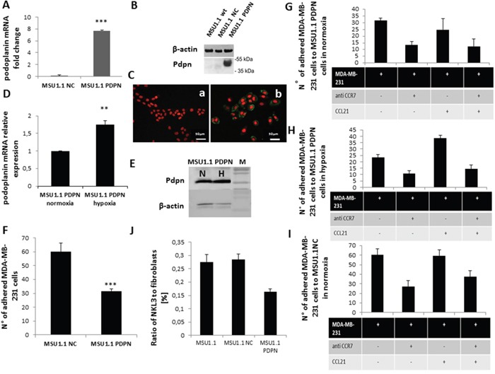
Figure 5. Impact of podoplanin/CCL21 interaction on MDA-MB-231 cells adhesion to MSU1.1 and MSU1.1 PDPN cells surface in normoxia and hypoxia.

A. Quantification of podoplanin expression at mRNA level in hypoxia vs normoxia. B. PDPN protein detection by immunoblotting. C. PDPN protein detection (b) in cells by immunocytochemistry with AlexaFuor-murine IgG anti human PDPN (green), nuclei are labelled with DRAG-5 (red). Control (a) is MSU1.1 NC cells (transduced with empty vector). D. PDPN mRNA expression level in hypoxia vs normoxia in MSU1.1 PDPN. E. PDPN protein expression level in hypoxia vs normoxia in MSU1.1 PDPN. F. Quantification of the MDA-MB-231 cells adhesion to MSU1.1 and MSU1.1 PDPN cells surface. MDA-MB-231 cells were counted on the surface of MSU1.1 NC or MSU1.1 PDPN cells (ten representative fields were counted) after flow adhesion in normoxia. * p<0.05, N=3 (mean from ten representative fields). G. Effect of CCL21/CCR7 interaction on MDA-MB-231 cell adhesion onto MSU 1.1 PDPN cells in normoxia. H. Effect of CCL21/CCR7 interaction on MDA-MB-231 cell adhesion on MSU1.1 PDPN cells in hypoxia. I. Effect of CCL21/CCR7 interaction on MDA-MB-231 cell adhesion on MSU1.1 NC cells in normoxia. J. Reduction of adhesion of NKL3 cells to MSU1.1 PDPN vs MSU1.1 NC and MSU 1.1.
