

Abstract
Supplemental Digital Content is available in the text.
The orbicularis oris muscle (OOM) has a unique form in that its muscle fibers cross the midline of the upper lip from both of the modioli. These crossing fibers are considered to form the distinctive structure of the philtrum.1 The objective of this study was to visualize the functional contraction and insertion area of the crossing fibers of the OOM using ultrasonography.
Five healthy adult volunteers were recruited. The subjects had no history of facial trauma, neuromuscular disorder, or any other congenital disorder. All examinations were performed using a diagnostic ultrasound system (Aplio500; Toshiba Medical Systems, Japan). A 12 MHz linear-array transducer was applied with ultrasound gel (Aquasonic 100; Parker Laboratories, N.J.), a polymer gel pad (Sonagel; Takiron, Japan) as a coupling medium, and cling film (New Kure Wrap; Kureha, Japan). The transducer was positioned under the surface of the oral mucosa, perpendicular to the skin surface and the philtrum at the middle of the upper lip. Videos of the repetitive motion between resting and protrusion of the upper lip were recorded as an audio video interleave file. The ethical committees of Kansai Electric Power Hospital approved the study protocol before it was performed.
The bundles of the crossing fibers of the OOM and the morphological changes of the philtrum were clearly visualized without pressure to the philtrum skin (Fig. 1). During lip protrusion, the OOM was observed to spread from both sides into trumpet-like shapes crossing the midline of the contralateral lip skin, which extends to approximately 7–10 mm outside the philtrum ridge. The skin was pulled most tightly inward at the outer end of the insertions. Although muscle fibers were not inserted into the skin at the philtrum hollow, there was a marked increase in the thickness of the muscle and the subcutaneous tissue. The thickening of the deep muscle fibers, which are regarded as muscle crossing between both of the modioli, could not be confirmed (see video, Supplemental Digital Content 1, which demonstrates lip protrusion, http://links.lww.com/PRSGO/A438).
Fig. 1.

A, Resting. B, Lip protrusion. CF, crossing fiber; DCT, deep connective tissue; Epi, epithelium; LCT, loose connective tissue; PhR, philtrum ridge.
Video Graphic 1.
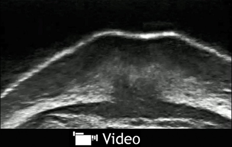
. See video, Supplemental Digital Content 1, which demonstrates lip protrusion, http://links.lww.com/PRSGO/A438.
Although histological studies have been performed, there are several theories regarding the site of fiber attachment and the functional mechanism of the crossover structures of the OOM and a consensus opinion has not been obtained.2–4 The previous reports make inferences based on still images (such as computed tomography, magnetic resonance imaging) or tissue specimens obtained from cadaver dissection. In other words, no actual muscle movement could be verified. In recent years, much attention has been given to analyzing the contraction of the facial mimic muscles, which can be visualized with ultrasound5; however, the OOM is scarcely mentioned because of its complex three-dimensional structure.
The observation of the obtained images revealed that the peripheral portion of the OOM acts on the lateral lips rather than the philtrum ridges or dimple and increases the thickness of the philtrum at the time of lip protrusion. These results provide information that is useful for surgical treatments for conditions involving the lips, such as cleft lip or facial nerve paralysis.
Supplementary Material
Footnotes
Disclosure: The author has no financial interest to declare in relation to the content of this article. The Article Processing Charge was paid for by the author.
Supplemental digital content is available for this article. Clickable URL citations appear in the text.
REFERENCES
- 1.Nicolau PJ. The orbicularis oris muscle: a functional approach to its repair in the cleft lip. Br J Plast Surg. 1983;36:141–153.. [DOI] [PubMed] [Google Scholar]
- 2.Latham RA, Deaton TG. The structural basis of the philtrum and the contour of the vermilion border: a study of the musculature of the upper lip. J Anat. 1976;121:151–160.. [PMC free article] [PubMed] [Google Scholar]
- 3.Briedis J, Jackson IT. The anatomy of the philtrum: observations made on dissections in the normal lip. Br J Plast Surg. 1981;34:128–132.. [DOI] [PubMed] [Google Scholar]
- 4.Bo C, Ningbei Y. Reconstruction of upper lip muscle system by anatomy, magnetic resonance imaging, and serial histological sections. J Craniofac Surg. 2014;25:48–54.. [DOI] [PubMed] [Google Scholar]
- 5.Power SM, Matic DB, Zhong T, et al. Definition of the lateral bulge deformity after primary cleft lip repair using real-time high-resolution ultrasound. J Craniofac Surg. 2010;21:1493–1499.. [DOI] [PubMed] [Google Scholar]