Figure 1.
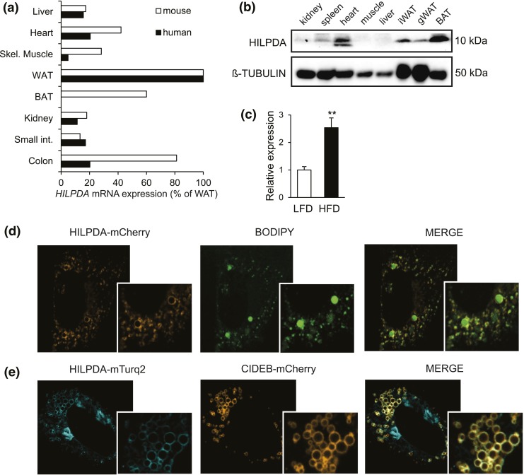
HILPDA is abundant in adipose tissue and localizes to lipid droplets. (a) HILPDA mRNA levels across various human (n = 1) and mouse tissues (n = 4). From top to bottom: liver, heart, skeletal muscle, WAT, BAT, kidney, small intestine, and colon. Expression levels in WAT were set at 100%. (b) Immunoblot for HILPDA in various mouse tissues (n = 1). (c) Hilpda mRNA in adipose tissue of C57BL/6 mice fed a low-fat diet (LFD) or high-fat diet (HFD) for 20 weeks. (d) The 3T3-L1 preadipocytes were transfected with HILPDA-mCherry plasmid, loaded with 400 μM oleic acid and, 48 hours posttransfection, stained with BODIPY and analyzed by confocal microscopy. (e) The 3T3-L1 preadipocytes were transfected with HILPDA-Turquoise2 plasmid (HILPDA-mTurq2) and CIDEB-mCherry plasmid, loaded with 400 μM oleic acid, and analyzed by confocal microscopy 48 hours after transfection. Asterisks indicate significant differences according to Student t test relative to (c) LFD; **P < 0.01. gWAT, gonadal white adipose tissue.

