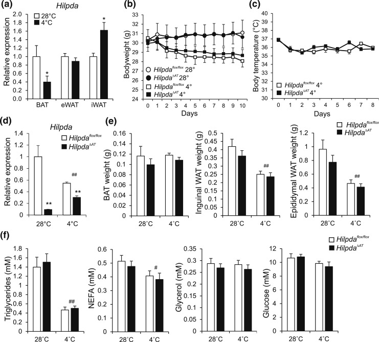
Figure 11.
Adipocyte-specific inactivation of Hilpda has no effect on tissue weights or plasma parameters in mice exposed to thermoneutral temperatures or cold. (a) Hilpda mRNA levels in BAT, eWAT, and iWAT of C57BL/6 mice exposed to 4°C or 28°C for 10 days (n = 8 to 10 animals per group). Gene expression levels of mice exposed to 28°C were set at one. (b) Body weight of Hilpdaflox/flox and HilpdaΔAT mice exposed to 4°C or 28°C for 10 days (n = 8 to 10 animals per group). (c) Body temperature of Hilpdaflox/flox and HilpdaΔAT mice exposed to 4°C or 28°C for 10 days (n = 10 animals per group). (d) Hilpda mRNA levels in BAT of Hilpdaflox/flox and HilpdaΔAT mice exposed to 4°C or 28°C for 10 days (n = 8 to 10 animals per group). Gene expression levels of Hilpdaflox/flox mice exposed to 28°C were set at one. (e) Tissue weights of BAT, eWAT, and iWAT of Hilpdaflox/flox and HilpdaΔAT mice exposed to 4°C or 28°C for 10 days (n = 8 to 10 animals per group). (f) Triglycerides, NEFA, glucose, and glycerol levels in plasma of Hilpdaflox/flox and HilpdaΔAT mice exposed to 4°C or 28°C for 10 days (n = 8 to 10 animals per group). Data are mean ± standard error of the mean. (a) Asterisks indicate significant differences according to Student t test (*) relative to mice exposed to thermoneutrality. Asterisks or hashtags indicate significant differences according to two-way analysis of variance (#), followed by Tukey’s honest significant difference post hoc test (*) relative to mice exposed to (d–h) thermoneutrality (#) and relative to (d–h) Hilpdaflox/flox mice (*); ** or ##P < 0.01; * or #P < 0.05.

