
Figure 6. Visualization of end-point aggregates of Aβ(M1-42), [Aβ(M1-42)]DiY and [Aβ(M1-42)]Q-K display distinct morphologies from one another.
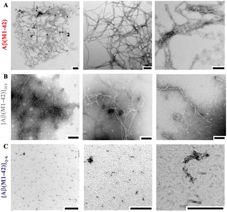
End-point samples of (A) Aβ(M1-42), (B) [Aβ(M1-42)]DiY and (C) [Aβ(M1-42)]Q-K were used for negative contrast EM. Electron micrographs are representative of 4 - 5 fields of view from at least two grids per sample. Size bars = 100 nm.