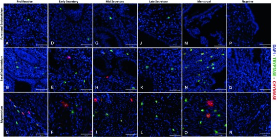
Figure 2. Comparison of immunoexpression of chymase and tryptase in “full thickness” (uterine lumen to endometrial/myometrial junction) tissue samples obtained from across the menstrual cycle.
Note that mast cells were less abundant in the functional layer and appeared to be exclusively tryptase+/chymase-. ( A– C) Functional, basal endometrium and myometrium during proliferative phase (P); ( D– F) Early secretory phase (ES); ( G– I) Mid secretory phase (MS); ( J– L) Late Secretory phase (LS); ( M– O) Menstrual phase (M); ( P– R) Negative control (omission of primary antibody). Double immunofluorescence has revealed the presence of three uterine mast cell subtypes, single tryptase, single chymase and double tryptase-chymase positive cells. (P n=4, ES n=4, MS n=2, LS n=3, M n=2): negative controls were included on all sections.

