Corrigendum to Toonen, Ma, and Gutman. Defining the temporal course of murine neurofibromatosis-1 optic gliomagenesis reveals a therapeutic window to attenuate retinal dysfunction. Neuro Oncol (doi:10.1093/neuonc/now267) first published online December 30, 2016.
The authors wish to convey that Figure 5 and Figure 6 were incorrectly displayed in the original manuscript. Figure 5 and Figure 6 were transposed. Please see the below for the correct Figure 5 and Figure 6.
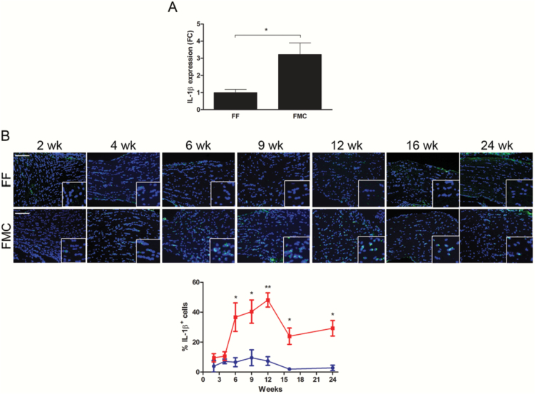
Fig. 5.
Nf1 optic glioma mice have more tumoral IL-1β expression. (A) The %IL-1β+ cells increased from 6 weeks to 12 weeks of age, but declined at 16 and 24 weeks of age. (B) Quantitative real-time PCR revealed 2.2-fold greater Il1b optic nerve RNA expression in 12-week-old FMC (n = 5) relative to FF (n = 3) mice. Data are represented as a fold change (FC) following normalization to H3f3a expression. *P ≤ .05.
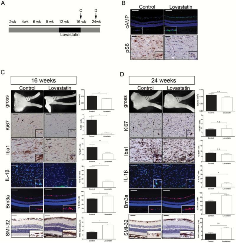
Fig. 6.
Lovastatin attenuates retinal dysfunction. (A) Schematic of treatment schedule. (B) cAMP immunostaining in the RGC layer revealed increased cAMP levels (top) and decreased optic nerve pS6Ser240/244 immunoreactivity (bottom) following lovastatin treatment compared with controls. (C) Lovastatin treatment decreased the volumes, %Ki67+ cells, %Iba1+ cells, and %IL-1β+ cells in FMC optic nerves compared with controls immediately following treatment cessation (16 wk of age). There was also an increase in the %Brn3a+ cells and RNFL thickness (SMI-32 immunohistochemistry). (D) Mice treated with lovastatin for 4 weeks and euthanized 8 weeks later showed no differences in optic nerve volumes, %Ki67+ cells, or %Iba1+ cells relative to controls; however, the %IL-1β+ cells was reduced, and the %Brn3a+ cells and RNFL thickness (SMI-32 immunohistochemistry) were increased. Black = vehicle, white = lovastatin, *P ≤ .05,**P ≤ .01, P ≤ .001. Scale bars = 50 μm.


