Abstract
Objective:
Breast cancer is a heterogeneous disease and very common malignancy in women worldwide. The efficacy of chemotherapy as an important part of breast cancer treatment is limited due to its side effects. While pharmaceutical companies are looking for better chemicals, research on traditional medicines that generally have fewer side effects is quite interesting. In this study, apoptosis and necrosis effect of Arctium lappa and doxorubicin was compared in MCF7, and MDA-MB-231 cell lines.
Materials and Methods:
MCF7 and MDA-MB-231 cells were cultured in RPMI 1640 containing 10% FBS and 100 U/ml penicillin/streptomycin. MTT assay and an annexin V/propidium iodide (AV/PI) kit were used respectively to compare the survival rate and apoptotic effects of different concentrations of doxorubicin and Arctium lappa root extract on MDA-MB-231 and MCF7 cells.
Results:
Arctium lappa root extract was able to reduce cell viability of the two cell lines in a dose and time dependent manner similar to doxorubicin. Flow cytometry results showed that similar to doxorubicin, Arctium Lappa root extract had a dose and time dependent apoptosis effect on both cell lines. 10µg/mL of Arctium lappa root extract and 5 µM of doxorubicin showed the highest anti-proliferative and apoptosis effect in MCF7 and MDA231 cells.
Conclusion:
The MCF7 (ER/PR-) and MDA-MB-231 (ER/PR+) cell lines represent two major breast cancer subtypes. The similar anti-proliferative and apoptotic effects of Arctium lappa root extract and doxorubicin (which is a conventional chemotherapy drug) on two different breast cancer cell lines strongly suggests its anticancer effects and further studies.
Keywords: Arctium lappa, Doxorubicin, breast cancer, apoptosis, cell lines
Introduction
Cancer is a disease, which starts with the accumulation of genomic alterations in DNA (Aghaei et al., 2014). Breast cancer is one of the most prevalent cancers among women as 12% of women are susceptible to it and one of eight will suffer from it at some stage in their lives. Doxorubicin (DOX) is an anthracyclin antibiotic used in chemotherapy. Its structure resembles a natural daunorubicin product and like all anthracyclin medicines binds to DNA (Yang et al., 2014). Unfortunately nonspecific toxicity of DOX causes severe side effects such as chronic cardiac toxicity, which limits the higher doses when it is necessary (Octavia et al., 2012).
Although, important progress in breast cancer diagnosis and treatment are in place but still extensive further studies are needed for the unsolved problems (Saba et al., 2017). Natural products have been used in the treatment of various mild and chronic human pathological conditions with believe that they are rich in antioxidants and have fewer side effects (Haddad-Kashani et al., 2012; Moghaddasi et al., 2012; Pal et al., 2003). Herbal medicine with different effective materials, vitamins, antioxidants and fewer side effects have always been considered in cancer treatment (Hosseini et al., 2012; Hosseyni et al., 2012; Zhang et al., 2012). Among these, Arctium lappa root extract has important components with anti-tumour activity (Chan et al., 2011). Arctium lappa root extract have been considered for its different properties, such as improving the immune system and metabolism. Their biological activities include anti-carcinogenic, anti-inflammatory, anti-diabetic, anti-infective and anti-viral activities (Chan et al., 2011).
Breast cancer cell lines have been used widely to investigate breast cancer pathobiology, and to screen and characterize new therapeutics (Vargo-Gogola and Rosen, 2007). Breast cancer cell lines present the main genetic and transcriptional features of breast tumors (Neve et al., 2006). Compared to cancer tissue, cell lines have the advantage of relative easiness of culture and pharmacologic manipulation. Furthermore cell lines are pure (free of stromal cells) and a variety of functional assays are available (Kao et al., 2009). In this study anti-proliferative effect of Arctium lappa is compared with DOX, a conventional chemotherapy drug on two different breast cancer cell lines (MCF7 and MDA-MB-231). MCF7 cells are ER-positive and have been used widely to develop anti-hormonal therapies in breast cancer (Fabbro et al., 1986; Hilliard et al., 2017). MDA-MB231 cell line is ER-negative and best resemble the recently characterized claudin-low tumor subtype of breast cancer (Heiser et al., 2012). From the current breast cancer subtypes, Luminal A (40.9%), Luminal B (21.5%) and Luminal HER2 (24.8%) can be ER/PR+ but the triple negative subtype is ER/PR (Liao, et al., 2015). Therefore, these cell lines are representative of two major breast cancer subtypes (ER/PR-positive and ER/PR-negative).
Materials and Methods
The human breast cell lines MCF7 and MDA-MB-231 were obtained from the Pasteur Institute of Iran. Doxorubicin, DMSO, MTT powder (3-(4,5-Dimethyl-2-thiazolyl)- 2,5-diphenyltetrazolium bromide) and penicillin/streptomycin were purchased from Sigma (St. Louis, MO, USA). DOX was dissolved in PBS as stock solution and stored at -20 °C. RPMI medium and Fetal Bovine Serum (FBS) were purchased from Gibco, UK. Annexin TACSTM kits and propidium iodide (PI) were obtained from Roche (Germany).
Aqueous extract of Arctium lappa L.
Arctium lappa root was supplied from Barij Essence Company (Kashan, Iran), powdered and mixed with 70% alcohol in a blender and incubated for 72 hours at room temperature. The solution was separated with a filter paper and dried in oven at 50 °C. To prepare stock solution the dried extract was resolved in PBS and was sterilized with 0.2 um syringe filter (Djeridane et al., 2006; Ferdosian et al., 2015; Kashani et al., 2015; Kashani et al., 2013; Nikzad et al., 2013).
Cell culture
MCF7 and MDA-MB-231 cells were cultured in RPMI-1640 medium supplemented with 10% FBS and 100 U/ml penicillin/streptomycin and grown at in a humidified incubator (37°C) with 5% CO2.
MTT assay
A colorimetric assay with tetrazolium salt was used to assess the anti-proliferative effects of DOX or Arctium lappa root extract. Briefly, 96-well plates were seeded with breast cancer cells (10000 cells/well) and allowed to grow for 24 hours. Cells were treated with different concentrations of DOX (0.1, 0.5,1, 2, 5 µM) or Acrium lappa extract (0, 0.1, 0.5, 1, 5, 10 µg/mL) for 24, 48 or 72 hours. Then 10 ul of MTT solution (5 mg/ml) in PBS was added to each well and incubated at 37 °C for 4 hours. The formazan crystals were resolved with DMSO and the absorbance of the wells for 570 nm was measured with a micro plate reader. All experiments were carried out more than three times, and the data is presented as means ± standard deviation. Statistical analysis of the data was performed using Excel program. IC50 (half maximal inhibitory concentration) values were estimated from in vitro dose-response curves using a linear regression analysis.
Flow cytometry
Annexin V-FITC Binding Assay (R&D USA, Cat No. TA4638) was used to assay apoptosis and necrosis in breast cancer cells after DOX or Arctium lappa root extract treatment. MCF-7 and MDA-MB-231 cells were cultured in small plates (5 mm). Upon reaching approximately 80% confluency, the cells were treated with DOX (0.1, 0.5, 1, 2 µM) or Arctium lappa root extract (0.1, 0.5, 1, 5 and 10 µg/ml) for 24, 48 or 72 hours. The cells were separated with 0.25% trypsin and 0.02% EDTA, centrifuged at 1000 rpm for 5 min. Then the cell pellet was washed and resuspended in binding buffer in a microfuge tube. 5 µl FITC-conjugated annexin V and 10 ul of PI was added to the tubes, mixed and incubated at r/t at dark for 10 min. The labeled cells were run in flow cytometry (Becton Dickinsonfacscalbur USA) and the percent of early apoptotic, late apoptotic and necrotic cells were determined.
Statistical Analysis
The Kolmogrov-Smirnov test was applied to determine the normal distribution of variables (Dehghani et al., 2016a; Dehghani et al., 2016b; Kashani et al., 2012; Lotfi et al., 2016; Sharif et al., 2016). These results were representative of three independent experiments. The results of the quantitative data analysis were expressed as mean ± standard deviation. Repeated measure Analysis of Variance (ANOVA) and 2-tailed independent samples Student’s t tests was used to compare the average body temperature at different study times. Differences among groups were stated to be statistically significant when P-values <0.05. All statistical analyses were done using the Statistical Package for Social Science version 19 (SPSS Inc., Chicago, Illinois, USA) (Dehghani 2016c; Hosseini et al., 2016; Jalali et al., 2016).
Results
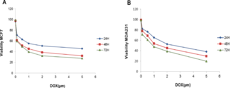
The effect of DOX on cell viability in MCF7 and MDA-MB-231 cells for different incubation times are shown in Figure 1. Our results showed that DOX reduced the viability of MCF7 and MDA-MB-231 cells in a dose and time-dependent manner (Figure 1).
Figure 1.

Viability of MCF7 (A) and MDA-MB-231 (B) Cells after Treatment with Different Concentrations and Incubation Times of Doxorubicin
Statistical analysis showed that by increasing treatment times (24, 48 and 72 hours) of DOX, the percent of viable MCF7 or MDA-MB-231 cells was decreased significantly (Table 1 and 2).
Table 1.
Statistical Analyses of Percent of Alive MCF7cells after Treatment with Different Concentrations of DOX for Different Incubation Times
| Doxorubicin | concentration (µM) | on MCF7 cells | ||||
|---|---|---|---|---|---|---|
| Treatment time | 0 | 0.1 | 0.5 | 1 | 2 | 5 |
| 24 H | 96.20±4.90 | 71.09±5.82 | 63.15±10.52 | 55.53±2.57 | 50.87±1.44 | 45.80±1.06 |
| 48 H | 98.00±2.88 | 61.53±3.06 | 51.74±3.60 | 45.52±3.81 | 38.84±2.16 | 32.60±2.58 |
| 72 h | 99.36±1.84 | 59.61±0.94 | 49.63±0.82 | 39.79±1.58 | 32.38±1.84 | 27.71±2.19 |
| P-value* | 0.0007 | 0.0006 | 0.0004 | 0.0004 | 0.0003 |
Statistical significance was attained when (P-value < 0.05).
Table 2.
Statistical Analyses of Percent of Alive MDA-MB-231 Cells after Treatment with Different Concentrations Of DOX for Different Incubation Times
| Doxorubicin | Concentrations | (µM) on MDA- | MB-231 cells | |||
|---|---|---|---|---|---|---|
| Treatment time | 0 | 0.1 | 0.5 | 1 | 2 | 5 |
| 24 H | 99.61±0.69 | 82.15±0.00 | 76.43±0.35 | 65.42±0.70 | 52.68±0.94 | 38.36±0.26 |
| 48 H | 99.47±0.90 | 77.93±0.70 | 68.92±1.16 | 53.68±0.42 | 45.00±0.63 | 29.62±0.57 |
| 72 H | 98.93±1.16 | 71.35±0.26 | 60.87±0.89 | 47.65±1.01 | 39.02±0.81 | 20.00±0.65 |
| P-value* | 0.002 | 0.002 | 0.001 | 0.0001 | 0.0001 |
Statistical significance was attained when (P-value < 0.05).
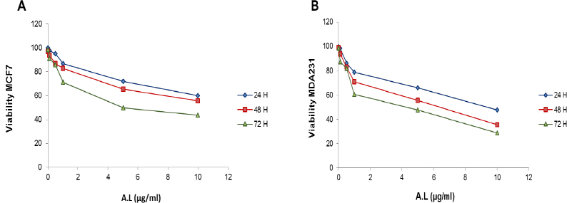
Arctium lappa root extract displayed cytotoxicity effect in a concentration and time-dependent manner in both cell lines (Figure 2). The percent of viable cells were decreased by increasing the dose and treatment time of Arctium lappa which is reflected in different curves in Figure 2.
Figure 2.

Viability of MCF7 (A) and MDA-MB-231 (B) Cells after 24, 48 and 72 H Incubation with Different Concentrations of Arctium Lappa Root Extract
Statistical analysis showed that the percent of viable MCF7 or MDA-MB-231 cells for each dose of Arctium lappa root extract was significantly different for different treatment times (24, 48 and 72 hours) (Table 3 and 4).
Table 3.
Statistical Analysis of the Percent of Viable MCF7cells for Different Concentrations of Arctium Lappa Root Extract after Different Incubation Times
| Arctium lappa | root extract | Concentrations | (µg/ml) on MCF7 cells | |||
|---|---|---|---|---|---|---|
| Treatment time | 0 | 0.1 | 0.5 | 1 | 5 | 10 |
| 24 H | 99.83±0.02 | 97.862±0.02 | 95.362±0.02 | 86.90±0.02 | 71.94±0.02 | 60.181±0.01 |
| 48 H | 97.22±0.03 | 93.86±0.03 | 87.06±0.03 | 82.96±0.0 | 65.39±0.02 | 55.70±0.01 |
| 72 H | 98.57±0.02 | 91.19±0.02 | 85.91±0.02 | 71.39±0.01 | 50.00±0.01 | 43.72±0.01 |
| P-value* | 0.071 | 0.04 | 0.015 | 0.004 | 0.0008 |
Statistical significance was attained when (P-value < 0.05).
Table 4.
Statistical Analysis of the Percent of Viable MDA-MB-231cells for Different Concentrations of Arctium Lappa Root Extract after Different Incubation Times
| Arctium lappa | root extract | Concentrations | (µg/ml) on | MDA-MB-231 cells | ||
|---|---|---|---|---|---|---|
| Treatment time | 0 | 0.1 | 0.5 | 1 | 5 | 10 |
| 24 H | 99.80±0.01 | 98.56±0.00 | 86.31±0.01 | 78.78±0.01 | 65.87±0.01 | 47.75±0.01 |
| 48 H | 99.22±0.03 | 93.86±0.03 | 83.06±0.04 | 70.96±0.03 | 55.39±0.02 | 35.70±0.02 |
| 72 H | 98.64±0.03 | 87.48±0.02 | 81.91±0.02 | 60.39±0.02 | 47.56±0.01 | 28.72±0.01 |
| P-value* | 0.14 | 0.0003 | 0.005 | 0.001 | 0.0003 |
Statistical significance was attained when (P-value < 0.05)
Apoptotic effect of DOX and Arctium lappa root extract
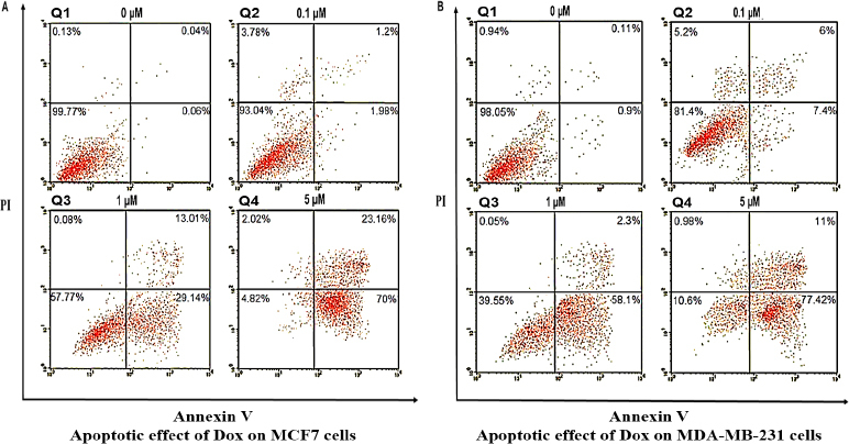
The breast cancer cells were treated with different concentrations of DOX or Arctium lappa root extract for 72 hours and after staining with Annexin v and PI, apoptosis rate was determined by flow cytometry and analyzed by FlowJo software. In the flow cytometry results, after each treatment, the cells were divided into 4 groups shown in quadrant 1 to 4 (Q1 to Q4). The cells in Q1 are necrotic cells which are labelled only with PI. Cells in the Q2 are late apoptotic cells, which are labelled with both PI and Annexin V. Normal cells are in Q3 that are not labelled with PI or Annexin V. In Q4 cells are labelled only with Annexin V and are early apoptotic cells. Figure 3 shows flow cytometric analysis of apoptosis in MCF7 and MDA-MB-231 cells induced by various concentrations of DOX (0, 0.1, 1 and 5 µM) for 72 h. For each cell lines four panels (group1 to group 4) are presented for different concentrations of DOX (0, 0.1, 1 and 5 µM respectively).
Figure 3.

Flow Cytometric Analysis of Apoptosis in MCF7 (A) and MDA-MB-231 (B) cells induced by various concentrations of DOX (0, 0.1, 1 and 5 µM) for 72 hours. In each scattergraph, cells in the Q1 are necrotic cells which are labelled only with PI. The cells in Q2 are late apoptotic cells, which are labelled with both PI and Annexin V. Normal cells are in Q3 that are not labelled with PI or Annexin V. In Q4 cells are labelled only with Annexin V and are early apoptotic cells
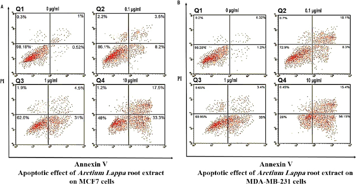
Figure 4 shows flow cytometric analysis of apoptosis in MCF7 and MDA-MB-231 cells induced by various concentrations of Arctium lappa root extract (0, 0.1, 1 and 10 µg/ml) for 72 h. For each cell lines four panels are presented for different concentrations of Arctium lappa root extract (0, 0.1, 1and 10 µg/ml respectively).
Figure 4.

Flow Cytometric Analysis of Apoptosis in MCF7 (A) and MDA-MB-231 (B) cells induced by various concentrations of Arctium lappa root extract (0, 0.1, 1 and 10 µg/ml) for 72 hours. The cells in Q1 are necrotic cells which are labelled only with PI. Cells in the Q2 are late apoptotic cells, which are labelled with both PI and Annexin V. Normal cells are in Q3 that are not labelled with PI or Annexin V. In Q4 cells are labelled only with Annexin V and are early apoptotic cells
In order to facilitate the comparison, the percentage of normal, early apoptotic, late apoptotic and necrotic cells after treating with DOX or Arctium lappa are shown in Table 5 and 6.
Table 5.
Shows the Percent of Normal, Early Apoptotic, Late Apoptotic and Necrotic Cells in MCF7 Cells after Treating with Different Concentrations of DOX Or Arctium Lappa
| Cell line | Drug | Dose | Normal (alive) cells | Early Apoptosis | Late Apoptosis | Necrotic |
|---|---|---|---|---|---|---|
| DOX | 0 μM | 99.77 | 0.06 | 0.04 | 0.13 | |
| 0.1 μM | 93.04 | 1.98 | 1.2 | 3.78 | ||
| 1 μM | 57.77 | 29.14 | 13.01 | 0.08 | ||
| MCF7 | 5 μM | 4.82 | 70 | 23.16 | 2.02 | |
| A. L. Extract | 0 µg/ml | 98.18 | 0.52 | 1 | 0.3 | |
| 0.1 µg/ml | 86.1 | 8.2 | 3.5 | 2.2 | ||
| 1 µg/ml | 62.6 | 31 | 4.5 | 1.9 | ||
| 10 µg/ml | 48 | 33.3 | 17.5 | 1.2 |
Table 6.
Shows the Percent of Normal, Early Apoptotic, Late Apoptotic and Necrotic Cells in MDA-MB-231 Cells after Treating with Different Concentrations of DOX Or Arctium Lappa
| Cell line | Drug | Dose | Normal (alive) cells | Early Apoptosis | Late Apoptosis | Necrotic |
|---|---|---|---|---|---|---|
| DOX | 0 μM | 98.5 | 0.9 | 0.1 | 0.94 | |
| 0.1 μM | 81.4 | 7.4 | 6.0 | 5.2 | ||
| 1 μM | 39.5 | 58.1 | 2.3 | 0.05 | ||
| MDA-231 | 5 μM | 10.6 | 77.4 | 0.9 | 11.0 | |
| A.L. Extract | 0 µg/ml | 98.2 | 1.2 | 0.3 | 0.2 | |
| 0.1 µg/ml | 72.9 | 8.3 | 16.1 | 0.7 | ||
| 1 µg/ml | 60.9 | 35.0 | 3.4 | 0.6 | ||
| 10 µg/ml | 20.0 | 54.1 | 15.4 | 0.4 |
These results showed that the Arctium lappa extract had a dose and time-dependent apoptotic effect on both cell lines similar to DOX (Table 5 and 6).
Two-color flow cytometry with annexin V and PI labeling showed that early apoptosis was the predominant mode of cell death in MCF-7 cells treated with different concentrations of DOX or AL extract for 72 hours (Table 5).
In both treatment the highest dose of DOX and AL (5 μM and 10 µg/ml respectively) resulted the highest percentage of early and late apoptotic cells. Regarding late apoptotic cells the results were dose dependent as well. However for necrotic cells a lower concentration of dox and AL (0.1 μM and 0.1 µg/ml respectively) resulted the highest rate of necrotic cells (Table 5).
For MDA-231 cells the results was almost similar to MCF7 cells. In both treatments the predominant form of cell defect was early apoptosis and it was dose dependent as well (Table 6). Regarding late apoptotic in MDA-231 cells, the dox or AL treatment did not result a dose dependent response. In MDA-231 cells, a lower dose (0.1 μM) of dox but in AL treatment a lower (0.1 µg/ml) and the highest dose (10 µg/ml) caused the highest rate of late apoptotic cells.
In MDA-MB-231 cells the 0.1 μM concentration of DOX caused a high rate of necrotic cells but AL treatment did not cause any significant change. Compared to AL, the DOX treatment in MDA-MB-231 cells caused a higher rate of early apoptosis and necrosis. Whereas compared to DOX, AL treatment in MDA-MB-231 cells lead to a higher late apoptotic cells (Table 6).
Discussion
The current study indicated that the alcoholic extract of Arctium lappa root extract was able to suppress the breast cancer cell lines similar to DOX. The MTT assay showed that the Arctium lappa extract had a dose dependent and time dependent anti-proliferative effect on MCF7 cells similar to the conventional chemotherapy drug DOX. Similar effect of Arctium lappa was detected on the ER-negative breast cancer cell line MDA-MB-231 as well.
The anti-proliferative effect of DOX has been documented in many studies. The dose dependent anti-proliferative effects of DOX on MCF-7 and MDA-MB-231 cells has been shown by the MTT assay and western blot analysis (Tang et al., 2014). From the different concentrations (0, 0.3, 0.6, 1.25, 2.5, and 5 μM) they used the 5 μM had the highest rate of cell death in both cell lines. The anti-proliferative effect of DOX and gemcitabine was examined on MDA-MB-231 and results showed that both drugs increased apoptosis of main and stem cell population of the cell line. Furthermore these drugs were able to inhibit tumor growth and improved the survival rate of mice in vivo (Zheng et al., 2014).
In a study the anti-proliferative and free radical scavenging activity of different preparations of Arctium lappa root (Hot and room temperature dichloromethanic, ethanolic and aqueous extracts; hydroethanolic and total aqueous extract of Arctium lappa roots) was compared and results showed that dichloromethane extract of Arctium lappa had the best anti-proliferative effect (Predes et al., 2011). Another Korean traditional herbal mixture (Hwang-Heuk-San) including Arctium lappa has been tested for anti-inflammatory effects using a lipopolysaccharide (LPS)-activated RAW 264.7 macrophage model. Results showed that a noncytotoxic concentration of the herbal remedy significantly reduced the production of NO, IL-1β and TNF-α in LPS-stimulated RAW 264.7 cells, suggesting that Hwang-Heuk-San may offer therapeutic potential for treating inflammatory diseases accompanied by macrophage activation (Kang et al., 2015). Arctium lappa has significant anti-inflammatory effect that is reflected in different activity like suppression of pro-inflammatory cytokine expression, inhibition of nuclear factor-kappa B (NF-κB) pathway and scavenging free radicals (Knipping et al., 2008). The excessive production of NO by iNOS is involved in various inflammatory diseases such as rheumatoid arthritis, autoimmune disease, chronic inflammation and atherosclerosis. Some of the compounds like Lappaol F, diarctignin and arctigenin found in the seeds or leaves of Arctium lappa can inhibit NO production (B-S Wang et al., 2007). Since the methanolic extract of Acrium lappa can inhibit the expression of COX-2 mRNA, therefore the anti-inflammatory effect of Acrium lappa is accredited to the lowered PGE2 release (B-S Wang et al., 2007). Since many scientists believe that inflammation is the main reason for cancer development (Lu et al., 2012) the anti-inflammatory effect of Arctium lappa could be accounted for its anti tumoric effect as well. Beside these several other benefits have been reported for Arctium lappa extract. A study reported that oral administration of Arctium lappa root extract was able to significantly improve the sexual behavior of male rats (JianFeng et al., 2012). Another study showed that different Arctium lappa extract (aqueous, ethanol, chloroform and flavone) was able to significantly revert the high fat diet-induced deteriorated lipid profile and antioxidant status in quail serum (Wang et al., 2016).
The benefit of using these herbs reported in different studies are most probably is contributed to the whole content of the extract. Therefore the safe dose of herbal extract should be adjusted in different experiments including human trial studies. The dose dependent activity of different herbal remedies has been reported in different studies. For instance, it’s been reported that the lower dose of Pumpkin seed extract (300 mg/kg) was better than the higher dose (600 mg/kg) to recover the Cisplatin-induced side effects in rat epididymis histology and sperm parameters (Aghaei et al., 2014). These results confirm the dose dependent activity of herbal remedies similar to conventional drugs as well. Therefore public awareness about possible adverse effect of herbal remedies should be enhanced.
Our flow cytometry results showed that Arctium lappa root extract treatment caused apoptosis in the two breast cancer cell lines similar to DOX. In the flow cytometry results, the percent of normal, early apoptosis, late apoptosis and necrotic cells are shown. Since the major part of defected cells after treatment with DOX or AL, in both cell lines were early apoptotic cells the difference between these cells are briefly discussed here.
Apoptosis and necrosis are two processes that can occur independently, sequentially, as well as simultaneously (Ouyang et al., 2012). Apoptosis is often energy-dependent process that involves the activation of a group of cysteine proteases called “caspases” and a complex cascade of events that link the initiating stimuli to the final death of the cell (Ouyang et al., 2012). Apoptotic ells consist of cytoplasm with tightly packed organelles with or without a nuclear fragment. Macrophages, parenchymal cells, or neoplastic cells phagocytise these bodies to degrade them within phagolysosomes. Therefore, since apoptotic cells do not release their cellular constituents into the surrounding interstitial tissue there is essentially no inflammatory reaction associated with the process of apoptosis (Ouyang et al., 2012). In contrast to apoptosis, necrosis is considered to be a toxic process because the loss of cell membrane integrity results in the release of the cytoplasmic contents into the surrounding tissue, sending chemotatic signals, leading to inflammatory response (Ouyang et al., 2012).
However the fact is that our results are based on cell culture, which is very different from in vivo environment. In whole organism there are other forms of cell death like stress-induced apoptosis-like cell death (SIaLCD) (Ouyang et al., 2012) that cannot happen in cell culture since there is no scavenging cell. The Different subtypes of breast cancer respond differently to cancer treatments including chemotherapy. MCF7 and MDA-MB-231 cells representing two major breast cancer subtypes (ER/PR- and ER/PR+ respectively) responded differently to doxorubicin or docetaxel regarding the expression of Erk, and phosph-Erk (Taherian and Mazoochi, 2012). The similar anti-proliferative effect of the Arctium lappa root extract on the two different breast cancer cell lines could be considered as a positive activity. This is the first study to examine the effect of Arctium lappa extract on two different breast cancer cell lines in one study.
our results showed that Arctium lappa root extract had a dose and time dependent anti-proliferative effect on MCF7 and MDA-MB-231 cells. The flow cytometry results showed that the Arctium lappa root extract was able to induce apoptosis (mostly early apoptosis) in both cell lines as well. Both anti-proliferative and apoptotic effect of Arctium lappa root extract was similar to DOX which is a conventional chemotherapy drug. Since MCF7 (ER/PR-) and MDA-MB-231 (ER/PR+) cell lines covers two major breast cancer subtypes, these results are strengthening.
Acknowledgments
The financial support for this research was provided by a grant no. 92135 from the Research Deputy of Kashan University of Medical Sciences and Kashan Anatomical Sciences Research Center. We wish to thank Dr. Maryam Anjomshoaa and the entire lab personal for their help.
Ethical Responsibilities of Authors
This paper is our original unpublished work and it has not been submitted to any other journal for reviews.
Compliance with ethical standards
This article does not contain any studies with human participants or animals performed by any of the authors.
Conflict of interest
The authors declared that they have no competing interests.
References
- Aghaei S, Nikzad H, Taghizadeh M, et al. Protective effect of Pumpkin seed extract on sperm characteristics, biochemical parameters and epididymal histology in adult male rats treated with Cyclophosphamide. Andrologia. 2014;46:927–35. doi: 10.1111/and.12175. [DOI] [PubMed] [Google Scholar]
- Chan Y-S, Cheng L-N, Wu J-H, et al. A review of the pharmacological effects of Arctium lappa (burdock) Inflammopharmacology. 2011;19:245–54. doi: 10.1007/s10787-010-0062-4. [DOI] [PubMed] [Google Scholar]
- Dehghani R, Sharif A, Assadi M A, et al. Fungal flora in the mouth of venomous and non-venomous snakes. Comp Clin Path. 2016a;25:1207–11. [Google Scholar]
- Dehghani R, Sharif A, Madani M, et al. Factors influencing animal bites in Iran: a descriptive study. Osong Public Health Res Perspect. 2016b;7:273–7. doi: 10.1016/j.phrp.2016.06.004. [DOI] [PMC free article] [PubMed] [Google Scholar]
- Dehghani R, Sharif MR, Moniri R, et al. The identification of bacterial flora in oral cavity of snakes. Comp Clin Path. 2016c;25:279–83. [Google Scholar]
- Djeridane A, Yousfi M, Nadjemi B, et al. Antioxidant activity of some Algerian medicinal plants extracts containing phenolic compounds. Food Chem. 2006;97:654–60. Fabbro D, Küng W, Roos W, et al (1986) Epidermal growth factor binding and protein kinase C activities in human breast cancer cell lines: possible quantitative relationship Cancer Res 46; 2720-5. [Google Scholar]
- Ferdosian M, Khatami MR, Malekshahi ZV, et al. Identification of immunotopes against Mycobacterium leprae as immune targets using PhDTm-12mer phage display peptide library. Trop J Pharm Res. 2015;14:1153–59. [Google Scholar]
- Kashani HH, Hoseini ES, Nikzad H, et al. Pharmacological properties of medicinal herbs by focus on secondary metabolites. Life Sci. 2012;9:509–20. [Google Scholar]
- Heiser LM, Sadanandam A, Kuo W-L, et al. Subtype and pathway specific responses to anticancer compounds in breast cancer. Proc Natl Acad Sci U S A. 2012;109:2724–9. doi: 10.1073/pnas.1018854108. [DOI] [PMC free article] [PubMed] [Google Scholar]
- Hilliard T, Miklossy G, Chock C, et al. 15-methoxypuupehenol induces antitumor effects in vitro and in vivo against human glioblastoma and breast cancer models. Mol Cancer Ther. 2017;16:0291. doi: 10.1158/1535-7163.MCT-16-0291. [DOI] [PMC free article] [PubMed] [Google Scholar]
- Hosseini ES, Moniri R, Goli YD, et al. Purification of antibacterial CHAPK protein using a self-cleaving Fusion tag and its activity against methicillin-resistant staphylococcus aureus. Probiotics Antimicrob Proteins. 2016;8:202–10. doi: 10.1007/s12602-016-9236-8. [DOI] [PubMed] [Google Scholar]
- Hosseini ES, Zeinoddini M, Kashani HH, et al. Intein as a Novel Strategy for Protein Purification. Life Sci. 2012;4:2187–95. [Google Scholar]
- Hosseyni ES, Kashani HH, Asadi MH. Mode of action of medicinal plants on diabetic disorders. Life Sci. 2012;4:2776–83. [Google Scholar]
- Jalali HK, Salamatzadeh A, Jalali AK, et al. Antagonistic activity of Nocardia brasiliensis PTCC 1422 against isolated Enterobacteriaceae from urinary tract infections. Probiotics Antimicrob Proteins. 2016;8:41–5. doi: 10.1007/s12602-016-9207-0. [DOI] [PubMed] [Google Scholar]
- JianFeng C, PengYing Z, ChengWei X, et al. Effect of aqueous extract of Arctium lappa L. (burdock) roots on the sexual behavior of male rats. BMC Complement Altern Med. 2012;12:8. doi: 10.1186/1472-6882-12-8. [DOI] [PMC free article] [PubMed] [Google Scholar]
- Kang HJ, Hong SH, Kang K-H, et al. Anti-inflammatory effects of Hwang-Heuk-San, a traditional Korean herbal formulation, on lipopolysaccharide-stimulated murine macrophages. BMC complementary and alternative medicine. 2015;15:447. doi: 10.1186/s12906-015-0971-2. [DOI] [PMC free article] [PubMed] [Google Scholar]
- Kao J, Salari K, Bocanegra M, et al. Molecular profiling of breast cancer cell lines defines relevant tumor models and provides a resource for cancer gene discovery. PloS One. 2009;4:e6146. doi: 10.1371/journal.pone.0006146. [DOI] [PMC free article] [PubMed] [Google Scholar]
- Kashani HH, Moniri R. Expression of recombinant pET22b-LysK-cysteine/histidine-dependent amidohydrolase/peptidase bacteriophage therapeutic protein in Escherichia coli BL21 (DE3) Osong Public Health Res Perspect. 2015;6:256–60. doi: 10.1016/j.phrp.2015.08.001. [DOI] [PMC free article] [PubMed] [Google Scholar]
- Kashani HH, Moshkdanian G, Atlasi MA, et al. Expression of galectin-3 as a testis inflammatory marker in vasectomised mice. Cell J. 2013;15:11. [PMC free article] [PubMed] [Google Scholar]
- Kashani HH, Nikzad H, Mobaseri S, et al. Synergism effect of Nisin peptide in reducing chemical preservatives in food industry. Life Sci J. 2012;9:496–501. [Google Scholar]
- Knipping K, van Esch EC, Wijering SC, et al. In vitro and in vivo anti-allergic effects of Arctium lappa L. Exp Biol Med. 2008;233:1469–77. doi: 10.3181/0803-RM-97. [DOI] [PubMed] [Google Scholar]
- Liao G-S, Chou Y-C, Hsu H-M, et al. The prognostic value of lymph node status among breast cancer subtypes. Am J Surg. 2015;209:717–24. doi: 10.1016/j.amjsurg.2014.05.029. [DOI] [PubMed] [Google Scholar]
- Lotfi A, Shiasi K, Amini R, et al. Comparing the effects of two feeding methods on metabolic bone disease in newborns with very low birth weights. Glob J Health Sci. 2016;8:249. doi: 10.5539/gjhs.v8n1p249. [DOI] [PMC free article] [PubMed] [Google Scholar]
- Lu H, Ouyang W, Huang C. Inflammation, a key event in cancer development. Mol Cancer Res. 2006;4:221–33. doi: 10.1158/1541-7786.MCR-05-0261. [DOI] [PubMed] [Google Scholar]
- Moghaddasi MS, Kashani HH, Azarbad Z. Capparis spinosa L. propagation and medicinal uses. Life Sci J. 2012;9:684–6. [Google Scholar]
- Morrison WB. Inflammation and cancer: a comparative view. J Vet Intern Med. 2012;26:18–31. doi: 10.1111/j.1939-1676.2011.00836.x. [DOI] [PubMed] [Google Scholar]
- Neve RM, Chin K, Fridlyand J, et al. A collection of breast cancer cell lines for the study of functionally distinct cancer subtypes. Cancer Cell. 2006;10:515–27. doi: 10.1016/j.ccr.2006.10.008. [DOI] [PMC free article] [PubMed] [Google Scholar]
- Nikzad H, Kashani HH, Kabir-Salmani M, et al. Expression of galectin-8 on human endometrium: Molecular and cellular aspects. Int J Reprod Biomed. 2013;11:65. [PMC free article] [PubMed] [Google Scholar]
- Octavia Y, Tocchetti CG, Gabrielson KL, et al. Doxorubicin-induced cardiomyopathy: from molecular mechanisms to therapeutic strategies. J Mol Cell Cardiol. 2012;52:1213–25. doi: 10.1016/j.yjmcc.2012.03.006. [DOI] [PubMed] [Google Scholar]
- Ouyang L, Shi Z, Zhao S, et al. Programmed cell death pathways in cancer: a review of apoptosis, autophagy and programmed necrosis. Cell Prolif. 2012;45:487–98. doi: 10.1111/j.1365-2184.2012.00845.x. [DOI] [PMC free article] [PubMed] [Google Scholar]
- Pal SK, Shukla Y. Herbal medicine: current status and the future. Asian Pac J Cancer Prev. 2003;4:281–8. [PubMed] [Google Scholar]
- Predes FS, Ruiz AL, Carvalho JE, et al. Antioxidative and in vitro antiproliferative activity of Arctium lappa root extracts. BMC Complement Altern Med. 2011;11:25. doi: 10.1186/1472-6882-11-25. [DOI] [PMC free article] [PubMed] [Google Scholar]
- Saba M, Valeh T, Ehteram H, et al. Diagnostic value of neuron-specific enolase (NSE) and cancer antigen 15-3 (CA 15-3) in the diagnosis of pleural effusions. Asian Pac J Cancer Prev. 2017;18:257. doi: 10.22034/APJCP.2017.18.1.257. [DOI] [PMC free article] [PubMed] [Google Scholar]
- Sharif MR, Kashani HH, Ardakani AT, et al. The effect of a yeast probiotic on acute diarrhea in children. Probiotics Antimicrob Proteins. 2016;8:211–14. doi: 10.1007/s12602-016-9221-2. [DOI] [PubMed] [Google Scholar]
- Taherian A, Mazoochi T. Different expression of extracellular signal-regulated kinases (ERK) 1/2 and phospho-Erk proteins in MBA-MB-231 and MCF-7 Cells after chemotherapy with doxorubicin or docetaxel. Iran J Basic Med Sci. 2012;15:669–77. [PMC free article] [PubMed] [Google Scholar]
- Tang Z-H, Li T, Gao H-W, et al. Platycodin D from platycodonis radix enhances the anti-proliferative effects of doxorubicin on breast cancer MCF-7 and MDA-MB-231 cells. Chin Med J. 2014;9:16. doi: 10.1186/1749-8546-9-16. [DOI] [PMC free article] [PubMed] [Google Scholar]
- Vargo-Gogola T, Rosen JM. Modelling breast cancer: one size does not fit all. Nat Rev Cancer. 2007;7:659–72. doi: 10.1038/nrc2193. [DOI] [PubMed] [Google Scholar]
- Wang B-S, Yen G-C, Chang L-W, et al. Protective effects of burdock (Arctium lappa Linne) on oxidation of low-density lipoprotein and oxidative stress in RAW 264.7 macrophages. Food Chem. 2007;101:729–38. [Google Scholar]
- Wang Z, Li P, Wang C, et al. Protective effects of Arctium lappa L. root extracts (AREs) on high fat diet induced quail atherosclerosis. BMC Complement Altern Med. 2016;16:6. doi: 10.1186/s12906-016-0987-2. [DOI] [PMC free article] [PubMed] [Google Scholar]
- Yang F, Teves SS, Kem CJ, et al. Doxorubicin, DNA torsion, and chromatin dynamics. Biochim Biophys Acta. 2014;1845:84–9. doi: 10.1016/j.bbcan.2013.12.002. [DOI] [PMC free article] [PubMed] [Google Scholar]
- Zhang J, Wider B, Shang H, et al. Quality of herbal medicines: challenges and solutions. Complement Ther Med. 2012;20:100–6. doi: 10.1016/j.ctim.2011.09.004. [DOI] [PubMed] [Google Scholar]
- Zheng R-R, Hu W, Sui C-G, et al. Effects of doxorubicin and gemcitabine on the induction of apoptosis in breast cancer cells. Oncol Rep. 2014;32:2719–25. doi: 10.3892/or.2014.3513. [DOI] [PubMed] [Google Scholar]


