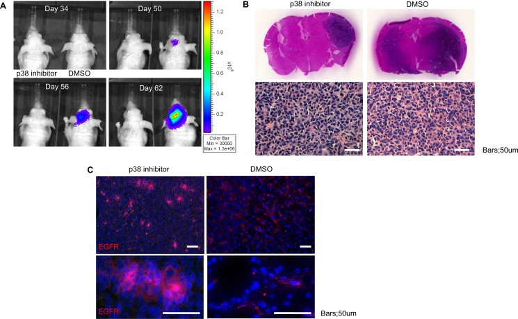
Figure 6. Inhibition of p38 delays in vivo tumor growth but increases EGFR expression.
(A) Tumor size was monitored via bioluminescence for mice treated with DMSO or p38 inhibitor demonstrating slow tumor growth (ten mice per group). (B) Post-mortem examination of coronal sections stained with hematoxylin and eosin (H&E) of mice treated with DMSO or p38 inhibitor shows larger tumor volume in the control group as well as more diffuse infiltration. Representative microscopic sections from control and p38 inhibitor treated groups demonstrates the typical histological appearance of glioblastoma with significant atypia and nuclear pleomorphism. (C) Immunofluorescent staining for EGFR (Alexa 555) counterstained with DAPI demonstrates increased EGFR expression in the p38 inhibitor group compared to control. Bars = 50 um.

