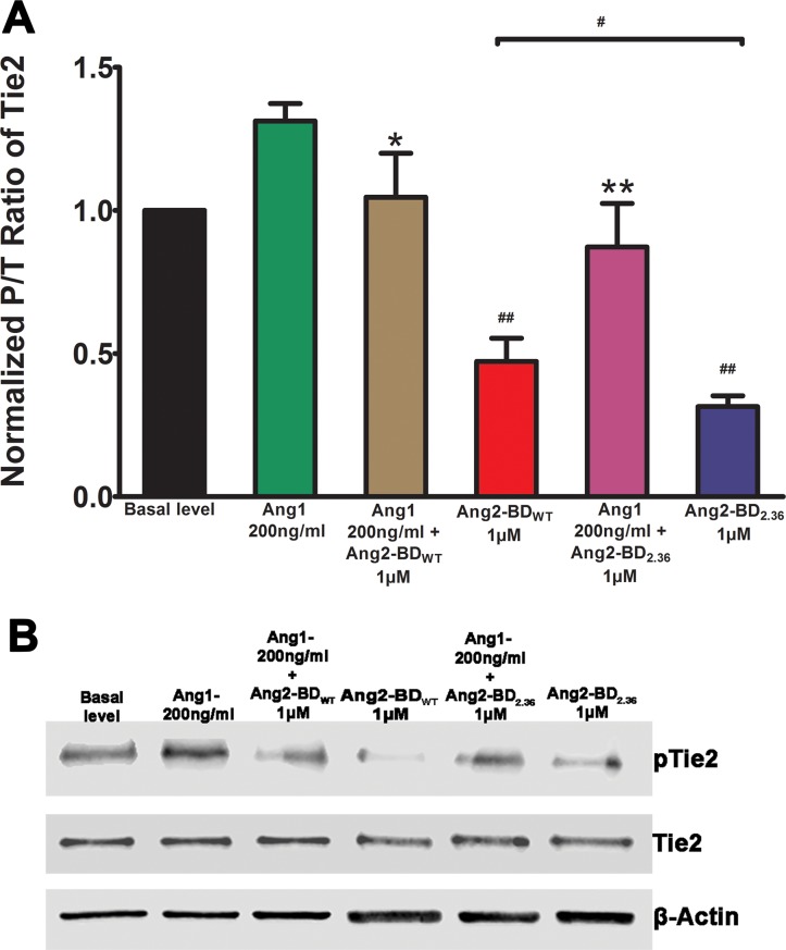
Figure 4. Inhibition of Tie2 phosphorylation by Ang2-BD variants.
(A) TIME cells were treated with control buffer (basal level, black), 200 ng/ml of Ang1 (green), 200 ng/ml of Ang1 and 1 μM Ang2-BDWT (brown), 1 μM Ang2-BDWT(red), 200 ng/ml of Ang1 and 1 μM Ang2-BDc2.36 (blue) and 1 μM Ang2-BDC2.36 (purple) for 15 min for Tie2 phosphorylation. (B) Cell lysates were analyzed by western blot using antibodies against pTie2, Tie2 and β-actin. # indicates P value < 0.05 for comparison of results between Ang2-BDWT and Ang2-BDC2.36; ## indicates P value < 0.01 for comparison of results between Ang1 + Ang2-BDWT and Ang2-BDWT and between Ang1 + Ang2-BDC2.36 and Ang2-BDC2.36; *indicates P value < 0.05 for comparison of results between Ang1 and Ang1 + Ang2-BDWT; **indicates P value < 0.01 for comparison of results between Ang1 and Ang1 + Ang2-BDC2.36. Data shown is the average of triplicate experiments, and error bars represent standard error of the mean.

