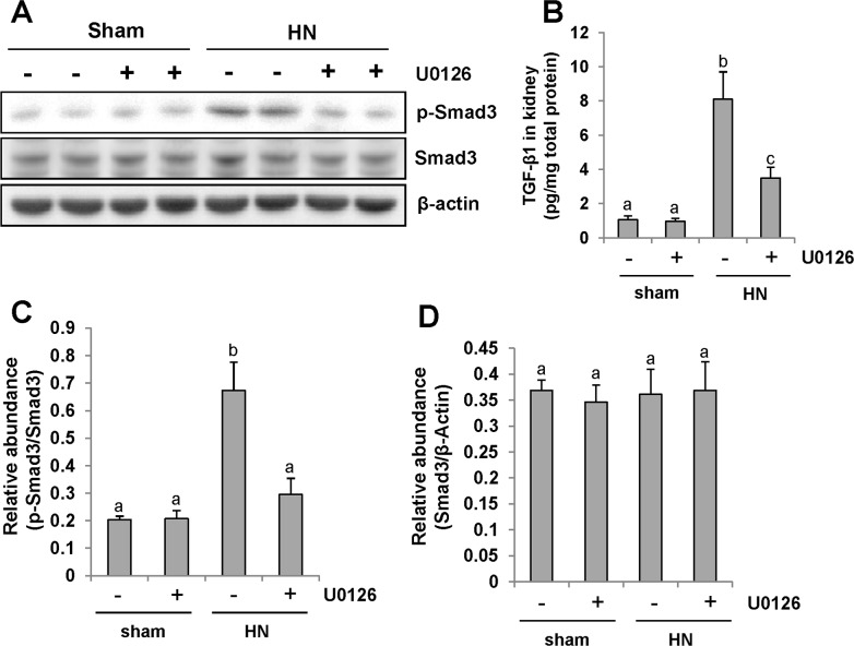
Figure 6. Pharmacologic blockade of ERK1/2 activity suppresses TGF-β1 signaling in the kidney of hyperuricemic rats.
(A) The kidneys were taken from immunoblot analysis of p-Smad3, Smad3, or β-actin. (B) Protein was extracted from the kidneys of rats after feeding of the mixture of adenine and potassium oxonate with or without U0126 and subjected to ELISA as described in the Concise Method section. Protein expression level of TGF-β1 was indicated. (C) Expression level of p-Smad3 was quantified by densitometry and normalized with total Smad3. (D) Expression level of Smad3 was quantified by densitometry and normalized with β-actin. Data are represented as the mean±SEM (n=6). Means with different letters are significantly different from one another (P<0.05).

