Abstract
Background:
Osteoporosis and hyperkyphosis could impose a considerable financial and therapeutic burden on the affected society. Thus, new strategies to prevent or manage such complications are of significant importance. Here we evaluate the effect of ‘Dynamic Hyperextension Brace’ (DHB) on bone density, and hyperkyphosis correction.
Methods:
Sixty postmenopausal women were randomly assigned to the case and control groups and followed for one year. DHB was applied in the case group according to the pre-designed protocol and the patients’ clinical and paraclinical parameters, including bone mineral density (BMD), kyphosis angle, osteoporotic fracture, and serum alkaline phosphatase (ALP) were evaluated in two groups.
Results:
Despite no significant difference in basic BMD and kyphosis between the case and control groups, BMD and kyphosis were significantly improved in the DHB treated group, at the end of the study (P=0.003 and P=0.001, respectively). Serum ALP level was significantly higher in cases compared to the controls (P=0.48). The vertebral fracture rate was also lower in the case group compared to the controls.
Conclusion:
The efficacy of bracing in osteoporosis and kyphosis management should be more emphasized. However, more detailed and controlled studies with more patients and a longer follow-up period is needed to adequately evaluate the long-term results of braces, including DHB.
Keywords: Dynamic hyperextension brace, Kyphosis, Osteoporosis
Introduction
Osteoporosis is a skeletal system disease defined by low bone mass and deterioration of bone structure leading to an increased risk of fracture, especially in the hip and spine (1, 2). Vertebral micro-fractures could result in anterior height reduction of vertebral bodies and hence thoracic hyperkyphotic (3). In addition to the surgical and postsurgical complications of corrective spinal surgeries in an already vulnerable osteoporotic patient, the high cost of such intervention imposes a significant financial burden on healthcare providers (4-6). Consequently, any intervention that may reduce the risk of fracture and kyphosis at either the individual or population level warrants critical appraisal (6). Several kinds of such interventions including pharmacologic, physical, and bracing approaches have already been introduced (7-10).
There is little consensus regarding the application of brace for the management of osteoporosis (11). Pfeifer et al. evaluated the six-month use of a thoracolumbar orthosis (TLO) brace for the management of osteoporotic compression fractures in a prospective randomized trial and found a significant improvement in the trunk muscle strength, posture, and body height amongst the treatment group, leading to a better quality of life and ability to perform activities of daily living (12).
Dynamic Hyperextension Brace (DHB), first introduced by Dr Sinaki, is an example of a regimen that focuses on strengthening the spinal extensors using a weighted kypho-orthosis and postural and proprioceptive training (13). It is based on the hypothesis that repetitive mechanical loading stimulates osteogenesis in osteoporotic patients (14). In this regard, the continuous pressure of the attached weight of DHB on the spine could induce osteogenesis and subsequently decrease the rate of osteoporosis and kyphosis through animprovement in bone quality. In addition, the extender base of the DHB stabilizes and off-loads the spine, which could correct the kyphosis through long-term extension force on the spine.
To the best of our knowledge, no study has been published on the efficacy of DHB in osteoporosis and kyphosis management. Considering bracing as a comfortable and affordable approach for osteoporosis and subsequent kyphosis management, we evaluated the effect of DHB on Bone Mineral Density (BMD) and kyphosis of osteoporotic postmenopausal women.
Materials and Methods
In an ethically approved randomized clinical trial (IRCT2016111430871N2) we compared two groups of osteoporotic patients treated with or without DHB.
After receiving an informed consent, 60 postmenopausal osteoporotic hyperkyphotic women (T-score < -2.5, Cobb angle of 50°–65°) were randomly divided into two equal groups of case and control. Patients were randomized in predesigned blocks of case and control groups, comprising 30 patients in each group. Block randomization was performed by a computer generated random number list prepared by an investigator with no clinical involvement in the study. Patients with metabolic or hormonal disorders affecting the skeletal structure; patients receiving medications with known effects on bone metabolism; patients currently receiving osteoporotic treatment; patients not able to perform hyperextension exercise; and malnourished patients were excluded from the study.
Since brace-wear compliance is considered as a major concern in brace evaluations, compliance monitoring and counselling were performed monthly, at each follow-up session, in order to improve bracing outcome (15-17). Patients were also asked to disclose any discomfort regarding brace-wear. Patients having trouble wearing a brace for any reason were planned to be excluded from the study as well. However, no such non-compliance was reported by any of our cases.
Anteroposterior and lateral thoracolumbar radiographs were obtained for each patient before starting the study and patients with vertebrate fracture were excluded.
Lumbar spine BMD, kyphosis angle, and alkaline phosphatase (ALP) were tested. A Dual-energy X-ray absorb ptiometry (DXA) system (Hologic Discovery DXA system, USA) was used to evaluate the BMD. Thoracic kyphosis was measured on standing lateral radiography of the spine.
A 600000 unit dose of intramuscular vitamin D was administered to patients in both groups at the start of the study (1-ml ampoule, 300000 IU/ml in sesame oil, Caspian Pharmaceutical, Iran). Patients in both groups received 1 gram calcium carbonate (Tehran Shimi Co, Iran)per day during the study, as well. A 35 mg weekly oraldose of Alendronate (Osteomed, Modava Company, Iran) was also administered to all patients.
Hyperextension exercise, three times a day, 20 minutes each session, was instructed to the patients in both groups.
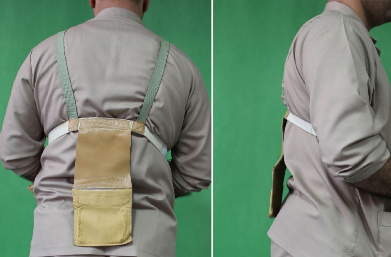
The case group was additionally intervened by the application of DHB (Handmade, Shafa Bracing Centre, Iran) containing one kilogram weight, and asked to use the brace 12 hours a day in their daily activities [Figure 1].
Figure 1.

Posterior and lateral view of Dynamic Hyperextentsion Brace on a patient.
The patients were informed that the brace should be detached in resting, lying, and sleeping positions.
Patient follow-up sessions were performed once a month in which the patients’ adherence to the study protocol was re-evaluated. The total follow-up time was 12 months. The patients’ characteristics including BMD, kyphosis angle and ALP were re-measured at the final follow-up and the results were compared with the primary results.
Loss of follow-up led to the exclusion of two patients in the control and three patients in the case group.
Central tendency and variability were measured using the mean and standard deviation, respectively. T-test was used for the analysis of results between the two groups. IBM SPSS Statistics 22 was used for all statistical analysis and a P value of <0.05 was considered as statistically significant.
Results
The mean age of the patients was 63 years, ranging from 51 to 76 years. The basic demographic and clinical characteristics of the patients are demonstrated in Table 1.
Table 1.
Patients baseline demographic, clinical and paraclinical characteristics have been demonstrated and compared. The results have been showed by P value. P<0.05 in considered as significant
| Variable | Case group | Control group | P Value |
|---|---|---|---|
| Mean Age (Year) | 63.1±11.1 | 63.3±10.8 | 0.31 |
| Mean±SD Lumbar spine BMD (mg/cm2) | 731± 87.11 | 733±84.47 | 0.54 |
| Mean±SD Lumbar spine T-score | -2.9 ± 0.4 | -2.9 ± 0.3 | 0.82 |
| Mean±SD Spinal Kyphosis (°) | 55.64±10.55 | 56.39±11.19 | 0.22 |
| Mean±SD Serum ALP (mg/dl) | 121.15±42.33 | 122±46.33 | 0.1 |
The mean increase in the lumbar spine BMD of case and control group was 23.57 mg/cm2 and 7.04 mg/cm2, respectively (P=0.003). In addition, the mean T-score was non-significantly higher in cases (-2.7±0.5) compared to the controls (-2.8±0.6) (P=0.23).
The mean decrease in spinal kyphosis significantly differed in the case (7.15°) and control (1.3°) groups (P=0.001). The final kyphosis value was 48.49±11.19 and 55.09±10.1 for the case and control group, respectively (P=0.001).
The mean increase in serum ALP was also significantly different in the case (6.2 mg/dl) and control (1.7 mg/dl) group (P=0.048). The detailed data of clinical and paraclinical evaluation of the patients is demonstrated in Table 2.
Table 2.
Patients clinical and paraclinical characteristics have been demonstrated and compared at the end of the study. The results have been showed by P value. P<0.05 is considered as significant
| Variable | Case group | Control group | P Value |
|---|---|---|---|
| Mean±SD Lumbar spine BMD (mg/cm2) | 754±79.11 | 740±81.42 | 0.003 |
| Mean±SD Lumbar spine T-score | -2.7 ± 0.5 | -2.8 ± 0.6 | 0.23 |
| Mean±SD Spinal Kyphosis (°) | 48.49±11.19 | 55.09±10.1 | 0.001 |
| Mean±SD Serum ALP(mg/dl) | 127.35±46.37 | 123.9±48.35 | 0.048 |
Interestingly, a total of eight vertebrate compression fractures were observed in our patients during the study period, of which six occurred in the control group (P=0.022).
Discussion
The prevention of osteoporosis and its complications is regarded as a preliminary health-care strategy. To this aim, new osteoporotic fracture prevention strategies including dietary, pharmacologic, physical, and so on are beneficial. In this study we evaluated the effect of DHB on BMD, kyphosis angle, and vertebra fracture of postmenopausal women.
The positive effects of bracing on osteoporotic spinal fractures have been evaluated in some earlier inves-tigations. Application of thoracolumbar orthosis (TLO) brace in patients with osteoporotic vertebral fractures has shown improvement in trunk muscle strength, posture, and body height as well as well-being and quality of life through pain reduction, decreased limitations of daily living. The use of an orthotic brace may represent an efficacious non-pharmacologic treatment option for spinal osteoporosis (12). This effect could be explained by the neutral spinal alignment and limited flexion imposed by bracing, thus reducing the axial loading on the fractured vertebra. Moreover, the brace allows for less paraspinal musculature fatigue and hence muscle spasm relief (18). It has been reported that brace treatment at its best though, can be only one step in the cascade of osteoporosis management, while general physiotherapy, analgesics and specific osteological drugs, and minerals essentially add to the treatment (19).
Our results showed a significant improvement in the spinal kyphosis of postmenopausal osteoporotic women and demonstrated that previous investigations might have underestimated the potential of bracing in the prevention of osteoporotic fractures. According to our results, DHB could simultaneously improve the spinal kyphosis and BMD in affected patients. Our paraclinical results also confirmed the increased osteogenic potential in patients of case group. Considering ALP as an important component of hard tissue formation and it shigher expression in mineralized tissuecells, the elevated serum ALP level in the case group could re-confirm the clinical results of osteogenes is improvement (20).
Bracing also has its own pitfalls including patient discomfort, which may decrease compliance. Moreover, prolonged periods of bracing may cause de-conditioning and atrophy of the trunk and paraspinal muscles. In conclusion, many authors have moved away from recommending rigid braces towards light-weight, soft braces, except in cases of severe deformity (21).
On the other hand, careful selection of the right brace for each given type of osteoporotic fracture is necessary. The overall brace-frames (Stagnara type) have been suggested to be used in only highly unstable or multiple osteoporotic fractures with impact on the spinal canal where surgery is not possible. Nevertheless, in the typical stable osteoporotic wedge fracture, light-weight constructions like the Jewett or Bähler-Vogt brace or, in less severe cases, dynamic braces (e.g. Torso Stretch brace or Spino Med Active brace) should be used to minimize muscle atrophy and demineralization (19). Such classification could also be employed in osteoporos is management. As an example, the attached weight of DHB might be optimized according to the kyphosis angle and BMD of the patients, which needs further evaluation in future investigations.
In addition, a longer follow-up period is needed to more adequately evaluate the effect of DHB on the management of osteoporosis and kyphosis in osteoporotic patients, which could also be regarded as the limitations of our study.
Application of DHB in postmenopausal women could improve osteoporosis and correct the kyphosis angle through elevated osteogenes is and extender capability of DHB. These conclusions have been supported by a significant difference in BMD, kyphosis angle, and serum ALP of the case group compared to the control group. However, evaluation of DHB with more patients and longer follow-up periods are needed to elucidate its long-term efficacy.
References
- 1.Nevitt CS, ODowd KJ. Epidemiology of osteoporosis and osteoporotic fractures. Epidemiol Rev. 1985;7(1):178–208. doi: 10.1093/oxfordjournals.epirev.a036281. [DOI] [PubMed] [Google Scholar]
- 2.Kanis JA. Diagnosis of osteoporosis and assessment of fracture risk. Lancet. 2002;359(9321):1929–36. doi: 10.1016/S0140-6736(02)08761-5. [DOI] [PubMed] [Google Scholar]
- 3.Katzman WB, Wanek L, Shepherd JA, Sellmeyer DE. Age-related hyperkyphosis:its causes, consequences, and management. J Orthop Sports Phys Ther. 2010;40(6):352–60. doi: 10.2519/jospt.2010.3099. [DOI] [PMC free article] [PubMed] [Google Scholar]
- 4.Papadakis M, Aggeliki L, Papadopoulos EC, Girardi FP. Common surgical complications in degenerative spinal surgery. World J Orthop. 2013;4(2):62–6. doi: 10.5312/wjo.v4.i2.62. [DOI] [PMC free article] [PubMed] [Google Scholar]
- 5.Nasser R, Yadla S, Maltenfort MG, Harrop JS, Anderson DG, Vaccaro AR, et al. Complications in spine surgery. J Neurosurg Spine. 2010;13(2):144–57. doi: 10.3171/2010.3.SPINE09369. [DOI] [PubMed] [Google Scholar]
- 6.Järvinen TL, Sievänen H, Khan KM, Heinonen A, Kannus P. Shifting the focus in fracture prevention from osteoporosis to falls. BMJ. 2008;336(7636):124–6. doi: 10.1136/bmj.39428.470752.AD. [DOI] [PMC free article] [PubMed] [Google Scholar]
- 7.Gass M, Dawson-Hughes B. Preventing osteoporosis-related fractures:an overview. Am J Med. 2006;119(4):S3–11. doi: 10.1016/j.amjmed.2005.12.017. [DOI] [PubMed] [Google Scholar]
- 8.Wilkins CH, Birge SJ. Prevention of osteoporotic fractures in the elderly. Am J Med. 2005;118(11):1190–5. doi: 10.1016/j.amjmed.2005.06.046. [DOI] [PubMed] [Google Scholar]
- 9.Cortet B. Osteoporosis:from early fracture prevention to better bone health with strontium ranelate. Rheumatology. 2009;48(Suppl 4):iv14–9. doi: 10.1093/rheumatology/kep275. [DOI] [PubMed] [Google Scholar]
- 10.Pisani P, Renna MD, Conversano F, Casciaro E, Di Paola M, Quarta E, et al. Major osteoporotic fragility fractures:risk factor updates and societal impact. World J Orthop. 2016;7(3):171–81. doi: 10.5312/wjo.v7.i3.171. [DOI] [PMC free article] [PubMed] [Google Scholar]
- 11.Wong CC, McGirt MJ. Vertebral compression fractures:a review of current management and multimodal therapy. J Multidiscip Healthc. 2013;6(1):205–14. doi: 10.2147/JMDH.S31659. [DOI] [PMC free article] [PubMed] [Google Scholar]
- 12.Pfeifer M, Begerow B, Minne HW. Effects of a new spinal orthosis on posture, trunk strength, and quality of life in women with postmenopausal osteoporosis:a randomized trial. Am J Phys Med Rehabil. 2004;83(3):177–86. doi: 10.1097/01.phm.0000113403.16617.93. [DOI] [PubMed] [Google Scholar]
- 13.Sinaki M. Exercise for patients with osteoporosis:management of vertebral compression fractures and trunk strengthening for fall prevention. PM R. 2012;4(11):882–8. doi: 10.1016/j.pmrj.2012.10.008. [DOI] [PubMed] [Google Scholar]
- 14.Chen JH, Liu C, You L, Simmons CA. Boning up on Wolff’;s Law:mechanical regulation of the cells that make and maintain bone. J Biomech. 2010;43(1):108–18. doi: 10.1016/j.jbiomech.2009.09.016. [DOI] [PubMed] [Google Scholar]
- 15.Karol LA, Virostek D, Felton K, Wheeler L. Effect of compliance counseling on brace use and success in patients with adolescent idiopathic scoliosis. J Bone Joint Surg Am. 2016;98(1):9–14. doi: 10.2106/JBJS.O.00359. [DOI] [PubMed] [Google Scholar]
- 16.DiRaimondo CV, Green NE. Brace-wear compliance in patients with adolescent idiopathic scoliosis. J Pediatr Orthop. 1988;8(2):143–6. [PubMed] [Google Scholar]
- 17.Reinker KA. Lori A, Karol MD, editors. Compliance counseling improves outcomes of bracing for patients with idiopathic scoliosis. Commentary on an article. “Effect of Compliance Counseling on Brace Use and Success in Patients with Adolescent Idiopathic Scoliosis”. J Bone Joint Surg Am. 2016;98(1):e4. doi: 10.2106/JBJS.O.01029. [DOI] [PubMed] [Google Scholar]
- 18.Longo UG, Loppini M, Denaro L, Maffulli N, Denaro V. Conservative management of patients with an osteoporotic vertebral fracture A review of the literature. J Bone Joint Surg Br. 2012;94(2):152–7. doi: 10.1302/0301-620X.94B2.26894. [DOI] [PubMed] [Google Scholar]
- 19.Matussek J, Boluki D, Füssel S, Grifka J. Orthotic methods for osteoporosis and osteoporotic vertebral fracture. Orthopade. 2010;39(4):387–96. doi: 10.1007/s00132-010-1596-2. [DOI] [PubMed] [Google Scholar]
- 20.Whyte MP. Hypophosphatasia and the role of alkaline phosphatase in skeletal mineralization. Endocr Rev. 1994;15(4):439–61. doi: 10.1210/edrv-15-4-439. [DOI] [PubMed] [Google Scholar]
- 21.Francis RM, Baillie SP, Chuck AJ, Crook PR, Dunn N, Fordham JN, et al. Acute and long-term management of patients with vertebral fractures. QJM. 2004;97(2):63–74. doi: 10.1093/qjmed/hch012. [DOI] [PubMed] [Google Scholar]


