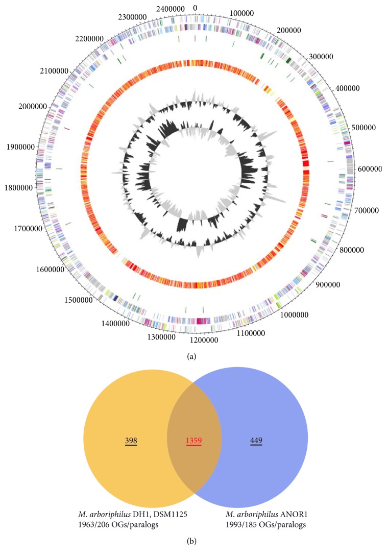
Figure 1.
Genomic features of Methanobrevibacter arboriphilus strain DH1 and ANOR1. A circular representation of the M. arboriphilus strain DH1 and comparison with strain ANOR1 is shown in (a). The two outer rings represent both strands of the strain DH1 genome with genes coloured by COGs. The orange/red ring shows genes present in the genome of strain ANOR1 as determined by BLAST. The two inner rings represent GC content and GC skew of strain DH1. The numbers of genes shared by and specific for the two Methanobrevibacter strains are shown in (b). Contigs of the strain DH1 genome were aligned to the genome of strain ANOR1. Ortholog detection was done with the Proteinortho software version 4.26 [33] (BLASTp) with an identity cutoff of 50% and an E value of 1e−10. Visualization was done using Proteinortho results and DNAPlotter [52]. COG categories of the genes were extracted from IMG database entries of M. arboriphilus DH1. Colour code according to E values of the BLASTp analysis performed using Proteinortho 4.26. Grey, 1e−20 to 1; light yellow, 1e−21 to 1e−50; gold, 1e−51 to 1e−90; light orange, 1e−91 to 1e−100; orange, 1e−101 to 1e−120; red, >1e−120.

