

Abstract
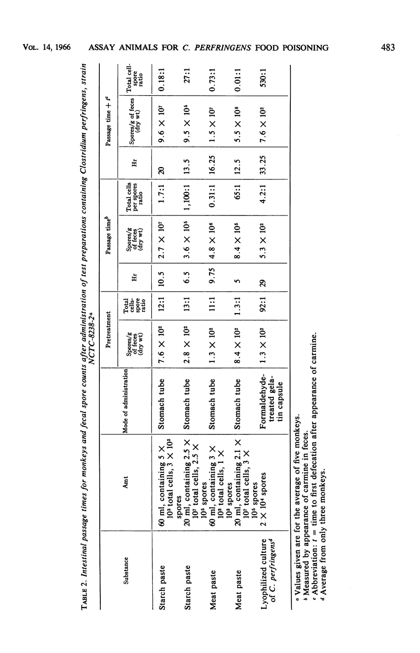
Spores and vegetative cells of Clostridium perfringens, in combination with meat or starch paste, sterile culture filtrates, lecithinase, and phosphorylcholine, were administered to mice and rhesus monkeys in an attempt both to evaluate the animals as test agents and, if possible, to elucidate the active factors producing food-poisoning symptoms caused by this organsim. Some of the preparations were administered to the monkeys by stomach tube; others, in gelatin capsules which were treated with formaldehyde so that the release of their contents was delayed and presumably reached the intestines of the animals. Any changes in intestinal passage times and in consistency of stools of the animals were observed, and the counts of C. perfringens in the feces of the monkeys previous and subsequent to treatment were recorded. The results obtained were inconclusive. Diarrhea occurred only relatively infrequently in both species, regardless of the substance fed or the mode of administration. The changes in intestinal passage times were not great, although in the monkeys there appeared to be a slight trend toward reduction as the magnitude of the bacterial load increased. Phosphorylcholine appeared to have little, if any, effect in reducing intestinal passage time of mice or monkeys. No procedures explored in these experiments could be said to be satisfactory as a means of animal assay for food poisoning strains of C. perfringens since typical symptoms did not appear with regularity.
Full text
PDF
Selected References
These references are in PubMed. This may not be the complete list of references from this article.
- ANGELOTTI R., HALL H. E., FOTER M. J., LEWIS K. H. Quantitation of Clostridium perfringens in foods. Appl Microbiol. 1962 May;10:193–199. doi: 10.1128/am.10.3.193-199.1962. [DOI] [PMC free article] [PubMed] [Google Scholar]
- Canada J. C., Strong D. H. Effects of animal alimentary passage on the heat resistance of Clostridium perfringens. Appl Microbiol. 1965 Sep;13(5):788–792. doi: 10.1128/am.13.5.788-792.1965. [DOI] [PMC free article] [PubMed] [Google Scholar]
- DACK G. M., SUGIYAMA H., OWENS F. J., KIRSNER J. B. Failure to produce illness in human volunteers fed Bacillus cereus and Clostridium perfringens. J Infect Dis. 1954 Jan-Feb;94(1):34–38. doi: 10.1093/infdis/94.1.34. [DOI] [PubMed] [Google Scholar]
- DISCHE F. E., ELEK S. D. Experimental food-poisoning by Clostridium welchii. Lancet. 1957 Jul 13;273(6985):71–74. doi: 10.1016/s0140-6736(57)92545-x. [DOI] [PubMed] [Google Scholar]
- ELLNER P. D. Fate of partially purified C-14-labeled toxin of Clostridium perfringens. J Bacteriol. 1961 Aug;82:275–283. doi: 10.1128/jb.82.2.275-283.1961. [DOI] [PMC free article] [PubMed] [Google Scholar]
- HALL H. E., ANGELOTTI R., LEWIS K. H., FOTER M. J. CHARACTERISTICS OF CLOSTRIDIUM PERFRINGENS STRAINS ASSOCIATED WITH FOOD AND FOOD-BORNE DISEASE. J Bacteriol. 1963 May;85:1094–1103. doi: 10.1128/jb.85.5.1094-1103.1963. [DOI] [PMC free article] [PubMed] [Google Scholar]
- HOBBS B. C., SMITH M. E., OAKLEY C. L., WARRACK G. H., CRUICKSHANK J. C. Clostridium welchii food poisoning. J Hyg (Lond) 1953 Mar;51(1):75–101. doi: 10.1017/s0022172400015515. [DOI] [PMC free article] [PubMed] [Google Scholar]
- NYGREN B. Phospholipase C-producing bacteria and food poisoning. An experimental study on Clostridium perfringens and Bacillus cereus. Acta Pathol Microbiol Scand Suppl. 1962;Suppl 160:1–88. [PubMed] [Google Scholar]
- YAMAMOTO R., SADLER W. W., ADLER H. E., STEWART G. F. Characterization of Clostridium perfringens (welchii) isolated from market poultry. Appl Microbiol. 1961 Jul;9:337–342. doi: 10.1128/am.9.4.337-342.1961. [DOI] [PMC free article] [PubMed] [Google Scholar]