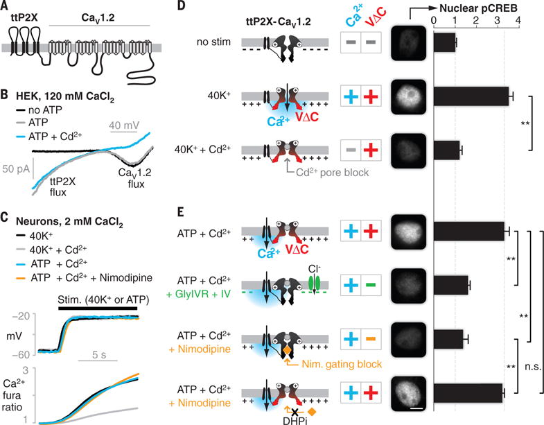
Fig. 1. Decoupling CaV1.2 Ca2+ and VΔC signals reveals dual requirement for CREB activation.

(A) The engineered chimeric construct, showing transmembrane domains and intra- and extracellular loops. (B) Ca2+ currents mediated by the chimeric channel in HEK cells. (C) Changes in voltage (top, current clamp recording) and Ca2+ influx (bottom, fura-2 imaging) under key stimulation conditions (N = 6 cells; fig. S2, A and B, shows SEM). (D and E) Nuclear pCREB in cultured cortical neurons assayed after 3 min of stimulation (conditions are indicated on the left; the cartoons represent the ttP2X-CaV1.2 chimera, with plus and minus signs indicating the presence and absence of the signals, respectively). pCREB was elevated only with dual Ca2+ and VΔC stimuli [second row in (D) and first and fourth rows in (E)]. Elimination of either Ca2+ [third row in (D)] or VΔC [second and third rows in (E)] abrogated signaling. Black bars show means ± SEM (normalized to no stimulation) of 50 cells from three independent cultures. Scale bar, 5 μm. **P < 0.01, determined by one-way analysis of variance (ANOVA) followed by Fisher’s least significant difference test.
