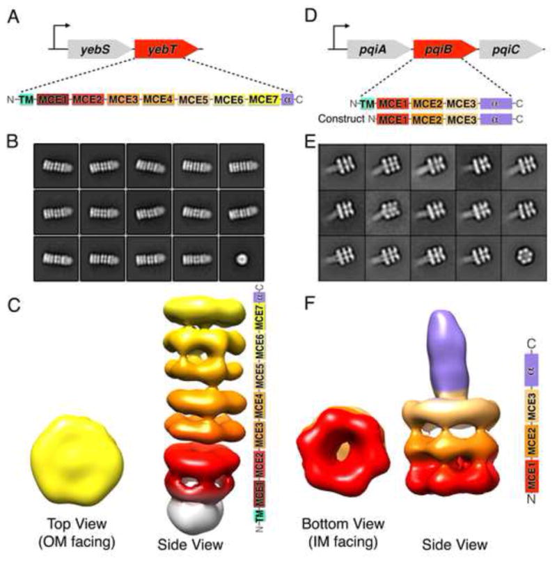
Figure 5. YebT and PqiB adopt elongated tube and syringe-like architectures.

(A) E. coli yeb operon and domain architecture of MCE protein YebT.
(B) Negative stain, 2D class averages for YebT.
(C) 3D reconstructions of YebT. Right, domain organization of YebT, color coded to show how the seven tandem MCE domains contribute to the seven stacked rings of EM density. White density corresponds to the TMs in the IM. Reconstruction is viewed from the side, IM at bottom and OM at top. Left, top view of YebT EM density map (OM towards IM).
(D) E. coli pqi operon and domain architecture of MCE protein PqiB and “periplasmic construct”.
(E) Negative stain, 2D class averages for PqiB.
(F) 3D reconstruction of PqiB. Right, side view of PqiB, with domain organization at right. Left, bottom view (IM towards OM).
