

A 75-year-old woman presented with hypoxemia, vomiting, and right facial weakness after falling. Head computed tomography (CT) demonstrated only a chronic left temporal infarct. Pelvis radiograph was negative for acute fracture. Within 24 hours, she developed bihemispheric coma and severe hypoxemic respiratory failure. Brain magnetic resonance imaging (MRI) demonstrated punctate acute infarcts scattered throughout both hemispheres. Fat embolism syndrome (FES) was suspected, despite a history of embolic stroke. Repeat brain MRI with susceptibility-weighted imaging (SWI) was consistent with FES and resolved diagnostic uncertainty (Figure 1). Subsequently, right hip CT revealed acute pelvic fractures. Petechiae emerged on the neck and conjunctiva 1 day later.
Figure 1.
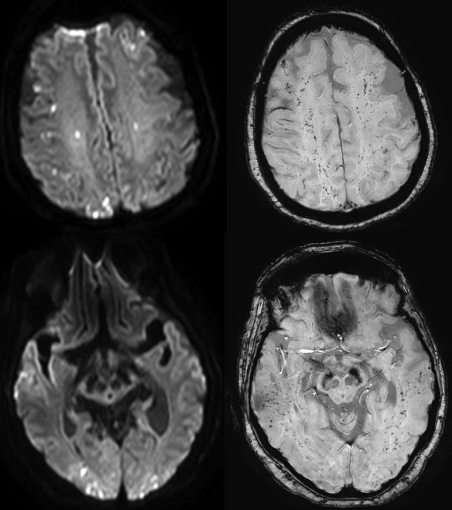
Diffusion-weighted (left) and susceptibility-weighted (right) magnetic resonance images of the brain at the level of centrum semiovale (top) and midbrain (bottom) demonstrate numerous cortical and subcortical diffusion abnormalities consistent with acute infarcts, as well as innumerable susceptibility foci within the white matter consistent with petechial hemorrhages of fat embolism syndrome (FES).
While clinical criteria remain the gold standard for FES diagnosis, validated diagnostic criteria do not exist.1 Further, because many of the elements of proposed diagnostic criteria are common in critically ill patients, their interpretation might be especially challenging in this population. Diffuse white matter microhemorrhages demonstrated by SWI may be a highly specific indicator of cerebral fat embolism.2 Susceptibility-weighted imaging of the brain should be considered in patients with suspected FES. Focused repeat imaging may reveal an initially undiagnosed culprit fracture.
Footnotes
Declaration of Conflicting Interests: The authors declared no potential conflicts of interest with respect to the research, authorship, and/or publication of this article.
Funding: The authors received no financial support for the research, authorship, and/or publication of this article.
References
- 1. Kosova E, Bergmark B, Piazza G. Fat embolism syndrome. Circulation. 2015;131(3):317–320. [DOI] [PubMed] [Google Scholar]
- 2. Kuo KH, Pan YJ, Lai YJ, Cheung WK, Chang FC, Jarosz J. Dynamic MRI patterns of cerebral fat embolism: a systematic review with illustrative cases. AJNR Am J Neuroradiol. 2014;35(6):1052–1057. [DOI] [PMC free article] [PubMed] [Google Scholar]