Abstract
Although H5N1 influenza viruses have been responsible for hundreds of human infections, these avian influenza viruses have not fully adapted to the human host. The lack of sustained transmission in humans may be due, in part, to their avian-like receptor preference. Here, we have introduced receptor binding domain mutations within the hemagglutinin (HA) gene of two H5N1 viruses and evaluated changes in receptor binding specificity by glycan microarray analysis. The impact of these mutations on replication efficiency was assessed in vitro and in vivo. Although certain mutations switched the receptor binding preference of the H5 HA, the rescued mutant viruses displayed reduced replication in vitro and delayed peak virus shedding in ferrets. An improvement in transmission efficiency was not observed with any of the mutants compared to the parental viruses, indicating that alternative molecular changes are required for H5N1 viruses to fully adapt to humans and to acquire pandemic capability.
Keywords: Influenza virus, Transmission, Ferrets, H5N1 virus
Introduction
Highly pathogenic avian influenza H5N1 viruses have caused not only catastrophic outbreaks in poultry and wild birds in many countries in Asia, the Middle East, Europe and Africa, but have also resulted in over 500 human infections with approximately 60% of those being fatal (http://www.who.int/en). Human H5N1 virus infections have mainly resulted from direct avian-to-human transmission of this virus, but despite repeated exposure of humans to infected birds during these outbreaks, H5N1 viruses have not adapted to the human host and developed the ability to transmit efficiently in a sustained manner from person to person, an essential property of a pandemic strain. The genetic changes necessary for H5N1 influenza viruses to adapt to humans and acquire efficient and sustained transmissibility are poorly understood. Due to the ongoing occurrence of H5N1 outbreaks and a continuing genetic evolution of these viruses in avian species, there is an urgent public health need to address this question.
To initiate influenza virus infection, the hemagglutinin (HA), which is the major surface glycoprotein of influenza viruses, binds to host cell surface complex glycans via a terminal sialic acid (SA). The preference of HA for particular SA moieties on host cells is a key determinant of host range and tissue tropism (Connor et al., 1994; Ito et al., 1998; Matrosovich et al., 2000). Avian influenza viruses preferentially bind SA in α2–3 linkage with the vicinal galactose (α2–3 SA), which predominate in the intestinal tracts of waterfowl, the natural reservoir of influenza viruses. In contrast, human-adapted influenza viruses preferentially bind to SA in α2–6 linkage (α2–6 SA), the major linkage of human respiratory epithelia (Shinya et al., 2006). Of the 16 HA subtypes known to exist in waterfowl, only H1, H2 and H3 subtypes have adapted to humans, causing the pandemics of 1918, 1957 and 1968, respectively. Each of them acquired a preferential α2–6 SA binding specificity (Matrosovich et al., 2000) that most likely contributed to the ability of these viruses to transmit efficiently among humans. While the origins of the 1918 H1N1 pandemic virus remain unclear, the H2N2 and H3N2 pandemic viruses were avian–human influenza reassortant viruses that acquired the PB1, HA and NA genes from an avian virus, in the case of H2N2, and PB1 and HA avian virus genes in the case of the H3N2 pandemic strain. The particular mutations within the HA that contributed to the adaptation of avian influenza viruses to human hosts have been elucidated for the previous pandemic subtypes. H2 and H3 glycoproteins were able to switch between α2–3 SA and α2–6 SA binding with two changes (Q226L and G228S) (Connor et al., 1994; Naeve et al., 1984) while two different mutations (E190D, G225D) in the H1 of the 1918 pandemic virus resulted in a switch from α2–3 SA to α2–6 SA binding preference (Glaser et al., 2005). When these sets of mutations were introduced into the H5 HA from A/Vietnam/1203/ 2004, H1 mutations (E190D and G225D) abolished binding to all glycans tested while the H2/H3 mutations (Q226L and G228S) caused a moderate shift in binding yet an equivalent, complete shift from α2–3 SA to α2–6 SA binding preference, similar to the shift observed with previous pandemic strains, was not observed (Stevens et al., 2006). These results indicate that a different combination of mutations in HA would be required for this H5N1 virus to shift from avian-like to human-like receptor binding preference.
The binding preference of H5N1 viruses for α2–3 SA receptors and the predominance of α2–6 SA receptors on the upper respiratory tract (URT) epithelium of humans has been suggested as a reason for the lack of efficient spread of avian influenza viruses among humans (Shinya et al., 2006). However, more recently it was demonstrated that H5N1 virus is able to replicate in human URT tissues ex vivo (Nicholls et al., 2007). In addition, we observed that an avian influenza H5N1 virus isolated from a human in 2004 displayed a high-growth phenotype in polarized oropharyngeal cells derived from the human upper respiratory tract (Zeng et al., 2007). We have observed similar efficient H5N1 virus replication in the URT tissues of ferrets (Maines et al., 2006; Maines et al., 2005), which like humans, have a predominance of α2–6 SA receptors in their airway epithelia (Leigh et al., 1995) and exhibit similar H5N1 virus attachment patterns within their respiratory tracts as those observed for human respiratory tissue (van Riel et al., 2006). Characterizing the impact of receptor binding preference on the efficiency of replication and transmission of H5N1 viruses is an important step in understanding the pandemic risk posed by H5N1 viruses. In the present investigation, we identified amino acid changes in the H5 HA that caused a shift in the receptor binding specificity. These receptor binding domain mutations were introduced into the HA of two H5N1 viruses [A/ Hong Kong/486/97 (HK486) and A/Vietnam/1203/04 (VN1203)] in order to examine the role of H5 HA receptor binding specificity on replication, pathogenicity, and transmissibility of this avian virus subtype in mammalian systems.
Results
Receptor binding preference of parental and mutant H5N1 viruses
Different sets of mutations in glycan receptor-binding site (RBS) of H5N1 HA were designed based on analyses of RBS of human-adapted H1N1 (PDB ID: 2WRG), H2N2 (PDB ID: 2WR7) and H3N2 (PDB IDs: 1MQN, 1HGG) HAs complexed with α2–6 SA receptors. The common feature of RBS of H2N2 and H3N2 HAs was the presence of residues Leu at 226 position and Ser at 228 position. Therefore the first set of mutations of H5 HA RBS included Q226L and G228S. In the context of the more recent genetic clades of H5 HA, a characteristic, natural change in the RBS is substitution of K193→R. This change was shown to increase α2,6 glycan binding of VN1203 virus (Stevens et al., 2008). To accommodate this substitution, the second set of mutations included K193R in addition to the Q226L/G228S mutations.
One of the characteristic differences between RBS of avian-adapted and human-adapted H1N1 HAs is the substitution of E190→D and G225→D. In human-adapted H1N1 HA, D225 is positioned to interact with penultimate Gal sugar of α2–6 SA receptor while D190 is positioned to interact with sugars beyond (at the reducing end) the penultimate Gal sugar. Therefore in the context of Q226L and G228S mutations in the H5 HA, it is preferable to have E190D substitution since L226 and S228 already provide sufficient contacts with SAα2–6Gal-motif. Unlike any of the other human-adapted HAs (H1, H2 and H3) which have Ser or Thr or Ala at 193, the RBS of H5 HA has a Lys at 193 (or Arg in the more recent genetic clades). The bulkier side chains of Arg or Lys at 193 in H5 HA in comparison to Ser/Thr/Ala in H1, H2 and H3 HAs could potentially have unfavorable steric contacts with the sugars beyond (at the reducing end) the penultimate Gal sugar of the α2–6 SA receptor. Based on this analysis, the third set of mutations included E190D/K193S in addition to Q226L/G228S mutations.
H5 HA mutant viruses, rHK486-LS (HA: Q226L, G228S), rHK486-DSLS (HA: E190D, K193S, Q226L, G228S), rHK486-GDSLS (HA: D187G, E190D, K193S, Q226L, G228S), rVN1203-DSLS (HA: E190D, K193S, Q226L, G228S) and rVN1203-RLS (HA: K193R, Q226L, G228S) were generated by site-directed mutagenesis and the influenza virus reverse genetics system (Table 1). rHK486-DSLS and rVN1203-DSLS viruses propagated in MDCK cells also contained a mixed population (D and N) at amino acid 151 in the NA protein. Mutant virus rHK486-GDSLS was a natural variant that arose in cell culture while generating the rHK486-DSLS mutant. Interestingly, when the DSLS and GDSLS mutant viruses were passaged in eggs, a reversion of the E190D HA mutation was detected. To avoid this reversion, experimental virus stocks for these mutants were generated in MDCK cells, while all others were grown in eggs (Table 1). Both parental and mutant viruses were assessed for their ability to agglutinate horse (HRBC) or turkey red blood cells (TRBC), which have been demonstrated to be agglutinated by viruses that recognize α2–3 SA receptors and viruses that recognize both α2–3 SA and α2–6 SA receptors, respectively (Ito et al., 1997). HA titers of the mutant HK486 viruses were reduced by 32-fold when titers were measured with HRBC versus TRBC. In contrast, the HA titers of the parental HK486 virus were identical regardless of the species of red blood cells used for detection (Table 1). HA titers measured with HRBC were reduced by 64-fold and 4-fold for rVN1203-DSLS and rVN1203-RLS, respectively, and the parental rVN1203 virus binding was reduced by 2-fold compared to TRBC. Compared to parental viruses, rHK486-LS, rHK486-GDSLS and rVN1203-RLS viruses exhibited increased binding to red blood cells re-sialylated with α2,6 SA (Table 1). Together these results indicate a change in receptor binding preference of the mutant viruses.
Table 1.
Summary of reverse genetics-derived (r) H5N1 viruses.
| HA amino acida | Stock passage/infectivity (log10)b | Stock HA titer | Hemagglutination using modified RBCsc | |||||||
|---|---|---|---|---|---|---|---|---|---|---|
|
|
|
|
||||||||
| 187 | 190 | 193 | 226 | 228 | Horse RBCs | Turkey RBCs | α2,3 | α2,6 | ||
| rHK486 | E | E | K | Q | G | E3/9.7 | 256 | 256 | + | − |
| rHK486-LS | E | E | K | L | S | E1/8.0 | 4 | 128 | + | + |
| rHK486-DSLS | E | D | S | L | S | C2/6.3 | 0 | 32 | + | − |
| rHK486-GDSLS | G | D | S | L | S | C1/6.6 | 0 | 32 | + | + |
| rVN1203 | E | E | K | Q | G | E3/9.8 | 64 | 128 | + | − |
| rVN1203-DSLS | E | D | S | L | S | C2/7.0 | 0 | 64 | + | − |
| rVN1203-RLS | E | E | R | L | S | E2/8.7 | 64 | 256 | − | + |
H3 numbering is shown; introduced mutations are in bold.
Infectivity of stocks were determined in indicated passage host (50% egg infectious dose or MDCK plaque forming units).
HA assay was performed using desialylated turkey RBCs that were re-sialylated as shown.
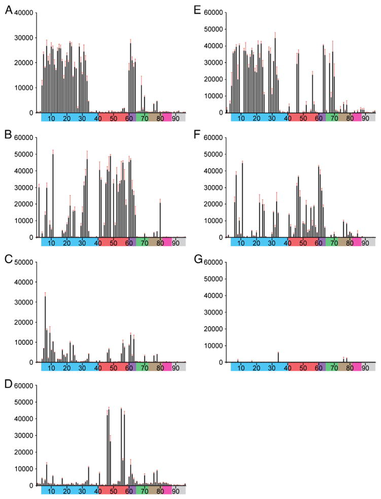
Receptor binding preference was more precisely assessed for each virus using glycan microarray analysis (Blixt et al., 2004; Stevens et al., 2008). The α2–3 and α2–6 SA binding preference of whole, inactivated parental (rHK486 and rVN1203) and mutant (rHK486-LS, rHK486-DSLS, rHK486-GDSLS, rVN1203-RLS and rVN1203-DSLS) viruses was determined (Fig. 1). The parental virus, rHK486, consistently bound to all of the α2–3 sialosides included in the array except for the α2–3 internal sialosides and bound none of the α2–6 sialosides (Fig. 1A). The rVN1203 parental virus bound all α2–3 glycan categories plus a few α2–6 sialosides (#45, 46 and 56; Fig. 1E). In contrast, the mutant viruses exhibited very different binding patterns. Mutant virus rHK486-LS bound nearly all of the α2–6 glycans included in the panel at varying degrees and reduced binding was observed with several of the linear and sulfated α2–3 glycans (Fig. 1B). rVN1203-RLS virus bound all categories of α2–3 and α2–6 SA glycans, except α2–3 internal sialosides, although overall binding intensity was reduced with the mutant virus compared to the rVN1203 parental virus (Figs. 1E and F). Binding to almost all of the α2–3 and α2–6 sialosides was substantially reduced with rHK486-DSLS and nearly eliminated with rVN1203-DSLS viruses (Figs. 1C and G). However, with the additional N187G spontaneously derived mutation present in the rHK486-GDSLS virus, binding was restored with four of the α2–6 sialoside glycans (#46–48 and 56–58; Table 2) and binding was only detected with two of the α2–3 sialosides (#4, 34; Fig. 1D). Influenza viruses that have flourished in the human population not only have increased binding preference for α2–6 SA glycans but also have reduced binding preference for α2–3 SA glycans (Matrosovich and Klenk, 2003).
Fig. 1.
Comparison of parental and mutant H5N1 receptor binding preference using a glycan microarray. Glycan specificity was assessed for H5N1 viruses; rHK486 (A), rHK486-LS (B), rHK486-DSLS (C), rHK486-GDSLS (D), rVN1203 (E), rVN1203-RLS (F) and rVN1203-DSLS (G) using glycan microarrays. Viruses, rHK486, rHK486-LS, rHK486-DSLS, rVN1203 and rVN1203-RLS were assessed using 256 HA units while rHK486-GDSLS was assessed using 128 HA units and VN1203-DSLS was assessed using 309 HA units. Due to different imprinting between the chips used for these glycan microarray studies, glycans # 10, 11, 26, 27, 42–45, 48, 49, 60–62, 79–81, 84, and 99 are absent for VN1203 and VN1203-DSLS. Colored bars highlight glycans that contain α2–3 SA (blue) and α2–6 SA (red), α2–6/α2–3 mixed SA (purple), N-glycolyl SA (green), α2–8 SA (brown), β2–6 and 9-O-acetyl SA, and non-SA (gray). Error bars reflect the standard error in the signal for six independent replicates on the array. Structures of each of the numbered glycans are listed in Table 2.
Table 2.
Sialic acid linked glycans represented on the glycan microarray.
| Chart number | Chart color | Structure |
|---|---|---|
| 1 | α-Neu5Ac–Sp8 | |
| 2 | α-Neu5Ac–Sp11 | |
| 3 | b-Neu5Ac-Sp8 | |
| 4 | Neu5Acα2–3(6-O-Su)Gal β1–4(Fucα1–3)GlcNAcβ-Sp8 | |
| 5 | Neu5Acα2–3Galβ1–3[6OSO3]GalNAcα-Sp8 | |
| 6 | Neu5Acα2–3Galβ1–4[6OSO3]GlcNAc β-Sp8 | |
| 7 | Neu5Acα2–3Galβ1–4(Fucα1–3)(6OSO3)GlcNAcβ-Sp8 | |
| 8 | Neu5Acα2–3Galβ1–3(6OSO3)GlcNAc-Sp8 | |
| 9 | Neu5Acα2–3Galβ1–3(Neu5Acα2–3Galβ1–4)GlcNAcβ-Sp8 | |
| 10 | Neu5Acα2–3Galβ1–3(Neu5Acα2–3Galβ1–4GlcNAcβ1–6)GalNAc-Sp14 | |
| 11 | Neu5Acα2–3Galβ1–4GlcNAcβ1–2Manα1–3(Neu5Acα2–3Galβ1–4GlcNAcβ1–2Manα1–6)Manβ1–4GlcNAcβ1–4GlcNAcβ-Sp12 | |
| 12 | Neu5Acα2–3Galβ1-Sp8 | |
| 13 | Neu5Acα2–3GalNAcα–Sp8 | |
| 14 | Neu5Acα2–3Galβ1–3GalNAcα-Sp8 | |
| 15 | Neu5Acα2–3Galβ1–3GlcNAcβ-Sp0 | |
| 16 | Neu5Acα2–3Galβ1–3GlcNAcβ-Sp8 | |
| 17 | Neu5Acα2–3Galβ1–4Glcβ-Sp0 | |
| 18 | Neu5Acα2–3Galβ1–4Glcβ-Sp8 | |
| 19 | Neu5Acα2–3Galβ1–4GlcNAcβ-Sp0 | |
| 20 | Neu5Acα2–3Galβ1–4GlcNAcβ-Sp8 | |
| 21 | Neu5Acα2–3GalNAcβ1–4GlcNAcβ-Sp0 | |
| 22 | Neu5Acα2–3Galβ1–4GlcNAcβ1–3Galβ1–4GlcNAcβ-Sp0 | |
| 23 | Neu5Acα2–3Galβ1–3GlcNAcβ1–3Galβ1–3GlcNAcβ-Sp0 | |
| 24 | Neu5Acα2–3Galβ1–4GlcNAcβ1–3Galβ1–4GlcNAcβ1–3Galβ1–4GlcNAcβ-Sp0 | |
| 25 | Neu5Acα2–3Galβ1–4GlcNAc β1–3Galβ1–3GlcNAc β-Sp0 | |
| 26 | Neu5Acα2–3Galβ1–3GalNAc-Sp14 | |
| 27 | Galβ1–3(Neu5Ac α2–3Galβ1–4(Fucα1–3)GlcNAc β1–6)GalNAc-Sp14 | |
| 28 | Neu5Acα2–3Galβ1–3(Fucα1–4)GlcNAcβ-Sp8 | |
| 29 | Neu5Acα2–3Galβ1–4(Fucα1–3)GlcNAcβ-Sp0 | |
| 30 | Neu5Acα2–3Galβ1–4(Fucα1–3)GlcNAcβ-Sp8 | |
| 31 | Neu5Acα2–3Galβ1–4(Fucα1–3)GlcNAcβ1–3Galβ-Sp8 | |
| 32 | Neu5Acα2–3Galβ1–4(Fucα1–3)GlcNAcβ1–3Galβ1–4GlcNAcβ-Sp8 | |
| 33 | Neu5Acα2–3Galβ1–4(Fucα1–3)GlcNAcβ1–3Galβ1–4(Fucα1–3)GlcNAcβ1–3Galβ1–4(Fucα1–3)GlcNAcβ-Sp0 | |
| 34 | Neu5Acα2–3Galβ1–4GlcNAcβ1–3Galβ1–4(Fucα1–3)GlcNAc-Sp0 | |
| 35 | Neu5Acα2–3(GalNAcβ1–4)Galβ1–4GlcNAcβ-Sp0 | |
| 36 | Neu5Acα2–3(GalNAcβ1–4)Galβ1–4GlcNAcβ-Sp8 | |
| 37 | Neu5Acα2–3(GalNAcβ1–4)Galβ1–4Glcβ–Sp0 | |
| 38 | Galβ1–3GalNAcβ1–4(Neu5Acα2–3)Galβ1–4Glcβ-Sp0 | |
| 39 | Fucα1–2Galβ1–3GalNAcβ1–4(Neu5Acα2–3)Galβ1–4Glcβ-Sp0 | |
| 40 | Fucα1–2Galβ1–3GalNAcβ1–4(Neu5Acα2–3)Galβ1–4Glcβ-Sp9 | |
| 41 | Neu5Acα2–6Galβ1–4[6OSO3]GlcNAcβ-Sp8 | |
| 42 | Galβ1–4GlcNAcβ1–2Manα1–3(Neu5Acα2–6Galβ1–4GlcNAcβ1–2Manα1–6)Manβ1–4GlcNAcβ1–4GlcNAcβ-Sp12 | |
| 43 | GlcNAcβ1–2Manα1–3(Neu5Acα2–6Galβ1–4GlcNAcβ1–2Manα1–6)Manβ1–4GlcNAcβ1–4GlcNAcβ-Sp12 | |
| 44 | Galβ1–4GlcNAcβ1–2Manα1–3(Neu5Acα2–6Galβ1–4GlcNAcβ1–2Manα1–6)Manβ1–4GlcNAcβ1–4GlcNAcβ-Sp21 | |
| 45 | Neu5Acα2–6Galβ1–4GlcNAcβ1–2Manα1–3(GlcNAcβ1–2Manα1–6)Manβ1–4GlcNAcβ1–4GlcNAcβ-Sp12 | |
| 46 | Neu5Acα2–6Galβ1–4GlcNAcβ1–2Manα1–3(Neu5Acα2–6Galβ1–4GlcNAcβ1–2Manα1–6)Manβ1–4GlcNAcβ1–4GlcNAcβ-Sp13 | |
| 47 | Neu5Acα2–6Galβ1–4GlcNAcβ1–2Manα1–3(Neu5Acα2–6Galβ1–4GlcNAcβ1–2Manα1–6)Manβ1–4GlcNAcβ1–4GlcNAcβ-Sp8 | |
| 48 | Neu5Acα2–6Galβ1–4GlcNAcβ1–2Manα1–3(Neu5Acα2–6Galβ1–4GlcNAcβ1–2Manα1–6)Manβ1–4GlcNAcβ1–4GlcNAcβ-Sp12 | |
| 49 | Neu5Acα2–6Galβ1–4GlcNAcβ1–2Manα1–3(Galβ1–4GlcNAcβ1–2Manα1–6)Manβ1–4GlcNAcβ1–4GlcNAcβ-Sp12 | |
| 50 | Neu5Acα2–6GalNAcα–Sp8 | |
| 51 | Neu5Acα2–6Galβ–Sp8 | |
| 52 | Neu5Acα2–6Galβ1–4Glcβ–Sp8 | |
| 53 | Neu5Acα2–6Galβ1–4GlcNAcβ–Sp0 | |
| 54 | Neu5Acα2–6Galβ1–4GlcNAcβ–Sp8 | |
| 55 | Neu5Acα2–6GalNAcβ1–4GlcNAcβ-Sp0 | |
| 56 | Neu5Acα2–6Galβ1–4GlcNAcβ1–3Galβ1–4GlcNAcβ-Sp0 | |
| 57 | Neu5Acα2–6Galβ1–4GlcNAcβ1–3Galβ1–4(Fucα1–3)GlcNAcβ1–3Galβ1–4(Fucα1–3)GlcNAcβ-Sp0 | |
| 58 | Galβ1–3(Neu5Acα2–6)GlcNAcβ1–3Galβ1–4Glcβ-Sp10 | |
| 59 | Galβ1–3(Neu5Acα2–6)GalNAcα-Sp8 | |
| 60 | Neu5Acα2–3Galβ1–4GlcNAcβ1–2Manα1–3(Neu5Acα2–6Galβ1–4GlcNAcβ1–2Manα1–6)Manβ1–4GlcNAcβ1–4GlcNAcβ-Sp12 | |
| 61 | Neu5Acα2–6Galβ1–4GlcNAcβ1–2Manα1–3(Neu5Acα2–3Galβ1–4GlcNAcβ1–2Manα1–6)Manβ1–4GlcNAcβ1–4GlcNAcβ-Sp12 | |
| 62 | Neu5Acα2–3Galβ1–3(Neu5Acα2–6)GalNAc-Sp14 | |
| 63 | Neu5Acα2–3Galβ1–3(Neu5Acα2–6)GalNAcα–Sp8 | |
| 64 | Neu5Acα2–3(Neu5Acα2–6)GalNAcα–Sp8 | |
| 65 | Neu5Gcα–Sp8 | |
| 66 | Neu5Gcα2–3Galβ1–3(Fucα1–4)GlcNAcβ-Sp0 | |
| 67 | Neu5Gca2–3Galβ1–3GlcNAcβ-Sp0 | |
| 68 | Neu5Gcα2–3Galβ1–4(Fucα1–3)GlcNAcβ-Sp0 | |
| 69 | Neu5Gcα2–3Galβ1–4GlcNAcβ–Sp0 | |
| 70 | Neu5Gcα2–3Galβ1–4Glcβ–Sp0α2–6GalNAcα–Sp0 | |
| 71 | Neu5Gcα2–6GalNAcα–Sp0 | |
| 72 | Neu5Gcα2–6Galβ1–4GlcNAcβ–Sp0 α2–8Neu5Acα-Sp8 | |
| 73 | Neu5Acα2–8Neu5Acα-Sp8 | |
| 74 | Neu5Acα2–8Neu5Acα2–8Neu5Acα-Sp8 | |
| 75 | Neu5Acα2–8Neu5Acα2–3(GalNAcβ1–4)Galβ1–4Glcβ–Sp0 | |
| 76 | Neu5Acα2–8Neu5Acα2–3Galβ1–4Glcβ–Sp0 | |
| 77 | Neu5Acα2–8Neu5Acα2–8Neu5Acα2–3(GalNAcβ1–4)Galβ1–4Glcβ-Sp0 | |
| 78 | Neu5Acα2–8Neu5Acα2–8Neu5Acα2–3Galβ1–4Glcβ–Sp0 | |
| 79 | Neu5Acα2–8Neu5Acα-Sp8 | |
| 80 | Neu5Acα2–8Neu5Acβ-Sp17 | |
| 81 | Neu5Acα2–8Neu5Acα2–8Neu5Acβ-Sp8 | |
| 82 | Neu5Acβ2–6GalNAcα–Sp8 | |
| 83 | Neu5Acβ2–6Galβ1–4GlcNAcβ-Sp8 | |
| 84 | Neu5Gcβ2–6Galβ1–4GlcNAc-Sp8 | |
| 85 | Galβ1–3(Neu5Acβ2–6)GalNAcα-Sp8 | |
| 86 | 9NAcNeu5Aca-Sp8 | |
| 87 | 9NAcNeu5Acα2–6Galβ1–4GlcNAcβ-Sp8 | |
| 88 | Galβ1–4GlcNAcβ1–3Galβ1–4GlcNAcβ1–3Galβ1–4GlcNAcβ–Sp0 | |
| 89 | Galβ1–3GlcNAcβ1–3Galβ1–3GlcNAcβ-Sp0 | |
| 90 | Fucα1–2Galβ1–3GlcNAcβ1–3Galβ1–4Glcβ–Sp8 | |
| 91 | Fucα1–2Galβ1–4(Fucα1–3)GlcNAcβ1–3Galβ1–4(Fucα1–3)GlcNAcβ-Sp0 | |
| 92 | GalNAcα1–3(Fucα1–2)Galβ1–3GlcNAcβ-Sp0 | |
| 93 | GalNAcα1–3(Fucα1–2)Galβ1–4GlcNAcβ–Sp8 | |
| 94 | Galα1–3(Fucα1–2)Galβ1–3GlcNAcβ-Sp0 | |
| 95 | Galα1–3(Fucα1–2)Galβ1–4(Fucα1–3)GlcNAcβ-Sp0 | |
| 96 | Galβ1–3GalNAc-Sp14 |
Replication of parental and mutant H5N1 viruses in cell culture
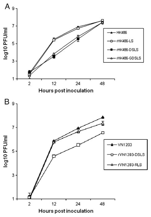
The effects of the introduced HA mutations on the replication efficiencies of HK486 and VN1203 viruses were evaluated in Calu-3 cells, a polarized cell line derived from human bronchial epithelium that express both α2,3 and α2,6 SA receptors (Zeng et al., 2007). Calu-3 cells were grown to confluency on transwell membrane inserts, infected with parental or mutant viruses at an MOI of 0.01 and maintained in either the presence or absence of trypsin. Virus titers in apical supernatants collected 2 to 48 h post inoculation (p.i.) were measured by standard plaque assays using MDCK cells. All viruses replicated with similar kinetics and to similar titers in Calu-3 cells with (not shown) or without (Fig. 2) the addition of exogeneous trypsin. At 12 and 24 h p.i., titers of the HK486 and rHK486-LS viruses were at least 14-fold higher than those of rHK486-DSLS and rHK486-GDSLS (p<0.05) (Fig. 2A). However, by 48 h p.i., all HK486 viruses had reached titers of greater than 107 PFU/ml. Similarly, titers of VN1203 virus were at least 25-fold higher than that of the mutant rVN1203-DSLS virus at 12 and 24 h p.i. (p<0.001), and by 48 h p.i. the mutant virus titers were still significantly reduced compared to the parental virus (p<0.05; Fig. 2B). However, rVN1203-RLS virus replicated on par with parental VN1203 virus up to 12 h p.i. and was reduced only by 2- and 3-fold at 24 and 48 h, respectively. Therefore, only the LS mutations introduced into the HK486 HA and the RLS mutation introduced into the VN1203 HA did not significantly reduce replication efficiency in the human bronchial epithelial cell line.
Fig. 2.
Replication of parental and mutant H5N1 viruses in Calu-3 cells. Calu-3 cells were grown to confluency on transwell inserts. Cells were infected with wild-type or mutant H5N1 viruses at an MOI of 0.01 for 1 h at 37°C. Unbound virus was removed by washing the cells 3 times and infected cells were cultured in DMEM medium supplemented with 0.3% bovine serum albumin. Calu-3 cells and virus were cultured in the presence (not shown) or absence of trypsin (1 μg/ml; Sigma, St. Louis, MO). Apical supernatants were collected at the indicated times and virus content was determined in a standard plaque assay using MDCK cells. The values shown represent the mean virus titer of fluids from three replicate infected cultures. A. HK486 parental and mutant viruses are shown. B. VN1203 parental and mutant viruses are shown. Error bars represent the standard deviation of three independent wells.
Replication and transmission of parental and mutant H5N1 viruses in ferrets
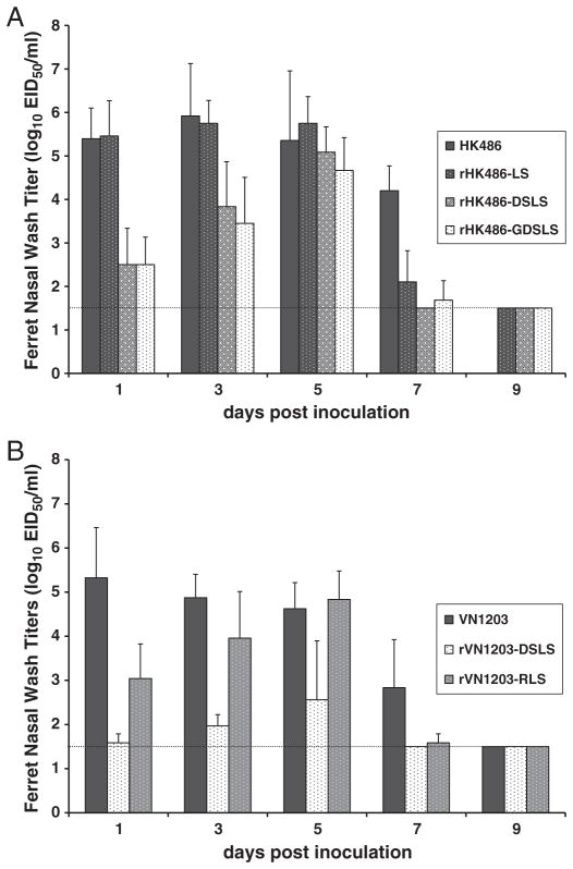
Replication efficiencies of these mutant H5N1 viruses were evaluated in vivo using the ferret model. A 106 50% egg infectious dose (EID50) or plaque forming unit (PFU) dose of each virus was administered intranasally (i.n.) to at least six ferrets and virus shedding was monitored in nasal washes collected on alternating days for 9 days p.i. and 3 days p.i. three ferrets were sacrificed for virus titration in ferret respiratory tract tissues. Consistent with the replication efficiencies observed in Calu-3 cells, HK486 and rHK486-LS viruses replicated to similar titers and with similar kinetics over the course of infection in ferrets, maintaining titers in nasal washes of greater than 105 EID50/ml for the first 5 days p.i. with clearance by day 9 p.i. (Fig. 3A). Similar levels of virulence were also observed between this virus pair. Ferrets exhibited a maximum mean weight loss of >10%, substantial lethargy and mortality reached approximately 30% (data not shown). In contrast, rHK486-DSLS and rHK486-GDSLS viruses exhibited delayed replication kinetics and did not reach comparable titers until day 5 p.i. when peak mean titers of 105.1 or 104.7 EID50/ml, respectively were attained (Fig. 3A). Lethargy and weight loss were minimal in rHK486-DSLS- and rHK486-GDSLS-infected ferrets and all ferrets survived infection (data not shown). Mean virus titers in lung tissues of ferrets inoculated with HK486 or rHK486-LS were similar, at least 104.9 EID50/g, 25 times greater than rHK486-DSLS or rHK486-GDSLS and no significant increase in virus titers in the upper respiratory tract of ferrets was detected with any of the HK486 mutant viruses (Table 3). Replication of mutant virus, rVN1203-DSLS, was significantly reduced compared to VN1203 with titers in nasal washes that were 18–12,000 fold lower through day 7 p. i.; a peak mean titer of 102.6 EID50/ml was detected in nasal washes 5 days p.i. (p<0.001) and virus was cleared by 7 days p.i. (Fig. 3B). In contrast, rVN1203-RLS virus replicated to titers similar to VN1203 by 5 days but was cleared in 5 of 6 animals by 7 days p.i. No notable clinical signs were observed in rVN1203-DSLS-infected ferrets, and only modest lethargy and weight loss were observed in a single animal infected with rVN1203-RLS virus, whereas VN1203-infected ferrets exhibited severe lethargy, weight loss and 100% mortality by day 9 p.i. (data not shown). Lung titers were similar for all of the VN1203 viruses while virus titers in nasal turbinates were similar for wild-type VN1203 and rVN1203-RLS but none was detected for rVN1203-DSLS virus (Table 3). These data indicate that the DSLS and GDSLS but not the LS HA mutations attenuated HK486 virus and that despite similar virus titers in lung tissues at 3 days p.i., the DSLS and RLS HA mutations attenuated VN1203 virus virulence in ferrets.
Fig. 3.
Replication of parental and mutant H5N1 viruses in ferrets. At least 3 ferrets each were inoculated i.n. with a 106 EID50 or PFU dose of virus. Virus titers in nasal washes collected every other day for 9 days were measured in eggs. A. Mean titers for HK486 parental (solid bar) and mutant (dotted bar) viruses are shown. B. Mean titers for VN1203 parental (solid bar) and mutant (dotted bar) viruses are shown. The limit of detection was 101.5 EID50/ml and is indicated by a dotted line.
Table 3.
Virus titers in tissues of inoculated ferrets and detection of transmission to contact ferrets.
| Inoculated ferrets | HK486 viruses
|
VN1203 viruses
|
||||||||||||
|---|---|---|---|---|---|---|---|---|---|---|---|---|---|---|
| Wildtype HK486 |
LS | DSLS | GDSLS | Wildtype VN1203 |
DSLS | RLS | ||||||||
| Nasal turbinates | 3.6±0.2a | 3.8±0.7 | 2.8±0.6 | 1.7±0.3 | 4.8±0.4 | ≤1.5±0.0 | 4.9±1.5 | |||||||
| Lungs | 4.9±0.7 | 5.0±1.0 | 2.5±0.2 | 3.5±0.9 | 5.3±0.7 | 5.4±0.1 | 6.1±1.4 | |||||||
|
| ||||||||||||||
| Contact ferrets | VDb | SCc | VD | SC | VD | SC | VD | SC | VD | SC | VD | SC | VD | SC |
| Respiratory droplet | 0/3 | 2/3 | 0/3 | 0/3 | 0/3 | 0/3 | 0/3 | 0/3 | 0/3 | 0/3 | 0/3 | 0/3 | 0/3 | 0/3 |
| Direct contact | 1/2 | 2/2 | 0/3 | 1/3 | 2/3 | 2/3 | 0/3 | 1/3 | 0/3 | 1/3 | 0/3 | 0/3 | 0/3 | 0/3 |
Virus titers in tissues collected 3 days p.i. are presented as log10 EID50/g for lungs and EID50/ml for nasal turbinate homogenates±standard deviation.
VD, virus detection in nasal washes was conducted in eggs; limit of detection was 1.5 EID50/ml.
SC, seroconversion was detected by hemagglutination inhibition assays that were performed using homologous virus and horse red blood cells (RBCs) for parental viruses or turkey RBCs for each of the mutant viruses.
To assess the impact of these HA mutations on the transmission efficiencies of the H5N1 viruses, both respiratory droplet and direct contact transmission experiments using the ferret transmission model were performed as previously described (Maines et al., 2006) (Table 3). Four or six ferrets each were inoculated i.n. with 106 EID50 or PFU of virus. A day later, two or three of the inoculated ferrets were each co-housed with a naïve ferret and the remaining inoculated ferrets were each housed in modified cages with a perforated side wall adjacent to a naïve ferret so that there was no direct contact between the ferrets or indirect contact with food and bedding, such that any transfer of virus occurred through the air. Transmission was assessed by virus detection in nasal washes of contact ferrets collected 2–10 days post exposure to infected animals, and seroconversion against the homologous virus detected by hemagglutination inhibition assay in sera collected 16 or more days post exposure. Transmission of parental HK486 virus was detected by seroconversion in 2 out of 2 ferrets co-housed with infected ferrets and 2 out of 3 ferrets in adjacent respiratory droplet cages (Table 3). However, during the course of infection, virus was detected in the nasal washes of only 1 ferret from the contact experiment and none from the respiratory droplet experiment. This level of transmissibility is similar to all previously reported results for this virus and is considered to be inefficient compared to the efficient transmission of human influenza H3N2 viruses in this model (Maines et al., 2006) in which all H3N2 virus contact ferrets seroconverted and shed virus regardless of the type of exposure to infected animals. Transmission of parental VN1203 virus was essentially undetectable with only 1 ferret from the contact transmission experiment seroconverting and no virus detected in any of the nasal washes of contact animals despite exposure to infected animals shedding as much as 106.5 EID50/ml at the peak of infection (Fig. 3B). None of the mutant viruses (rHK486-LS, rHK486-DSLS, rHK486-GDSLS, rVN1203-DSLS or rVN1203-RLS) transmitted more efficiently than their parental counterparts indicating that the introduced mutations did not confer properties that enhance either contact or respiratory droplet transmission in the ferret model.
Discussion
Efficient transmission of influenza viruses among humans is determined by multiple factors that are not well understood, but the ability to infect and replicate efficiently in the cells of the human respiratory tract is undoubtedly a prerequisite for transmission. In addition, the receptor binding preference of influenza viruses for receptors on cells of the airway epithelium is likely a contributing determinant. Here we describe HA mutations that, when introduced into H5N1 viruses, impacted receptor binding preference but that almost always attenuated the replication efficiency in vitro and in vivo. Compared to parental H5N1 viruses, the LS, RLS and GDSLS mutations resulted in increased binding to α2–6 SA glycans and reduced binding to some of the α2–3 SA glycans included in the microarray. This was most evident with the rHK486-GDSLS virus. The DSLS mutations resulted in reduced binding to both α2–6 and α2–3 SA glycans, a finding also observed by others (Yang et al., 2007). The greater binding of α2–6 and α2–3 SA glycans by rHK486-DSLS as compared to rVN1203-DSLS could be mediated by a subpopulation of viruses with the N187G substitution. Although it was not detected by consensus sequencing, it was previously detected in MDCK-grown stocks of rHK486-DSLS. Virus replication in human bronchial epithelial cells was significantly reduced by the DSLS and GDSLS HA mutations while the LS and RLS mutations had little impact on replication efficiency in vitro. In ferrets, peak replication of mutant viruses was delayed compared to parental strains, except for the LS mutation in HK486. None of the mutant viruses replicated to significantly higher titers in the ferret upper respiratory tract and most importantly, none of the introduced mutations improved the transmission efficiency of the H5N1 viruses.
Previous studies showed that the earliest isolates of the H2 and H3 pandemic strains already exhibited an α2–6 SA binding preference, but additional adaptive changes in the HA increased the affinity for α2–6 SA binding, a change that is proposed to have engendered efficient replication and transmission among humans (Matrosovich et al., 2000). Furthermore, when the H1N1 1918 pandemic virus was mutated to change receptor binding preference from α2–6 SA to the avian-like α2–3 SA, transmission in ferrets was abolished (Tumpey et al., 2007). Some H5N1 viruses isolated from humans have acquired changes in their HAs that increase their avidity for binding to α2–6 SA receptors while maintaining a moderate avidity for α2–3 SA glycans. A/HK/213/03 and A/Turkey/65–596/06 have an S227N HA change and were shown to bind both α2–6 SA and α2–3 SA glycans (Gambaryan et al., 2006; Yen et al., 2007) but efficient transmission of these viruses was not observed in ferrets (Maines et al., 2006; Yen et al., 2007). Furthermore, when two mutations (N182K, and Q192R) identified in H5N1 human isolates were introduced into an avian virus, again a moderate degree of binding to both α2–3 SA and α2–6 SA was observed (Yamada et al., 2006) and when mutations N182K, Q22L, and G224S were introduced into A/Indonesia/5/05 virus, binding patterns to ferret and human respiratory tract tissues resembled those of a human seasonal influenza virus (Chutinimitkul et al., 2010) but the transmissibility of these viruses were not tested. The human airway epithelium is a dynamic environment, rich in secreted sialic acid-containing mucins that act as a barrier between pathogens and epithelial cells (Lamblin et al., 2001). These mucins primarily contain α2–3 glycans and can bind virus and prevent them from reaching the cell surface (Couceiro et al., 1993). Thus, one consideration is that in order for an H5N1 virus to gain pandemic capability, it will likely need to not only gain an increased avidity for α2–6 SA glycans but also reduce its avidity for α2–3 SA glycans to avoid becoming trapped in mucin. It is possible that the reduced binding to α2,3 SA of the mutant viruses account for the observed reduced virulence and the subtle increase in α2,6 SA binding observed with some of the mutants was not sufficient to facilitate efficient replication, thereby affecting infectivity and ultimately transmission.
The NA protein also plays an important role by cleaving sialic acid inhibitors in the respiratory tract allowing the virus to reach the cell surface (reviewed in (Matrosovich and Klenk, 2003)). When a single mutation (R292K) was introduced into the NA of H3N2 viruses, reduced enzymatic activity was observed (Yen et al., 2005) as well as reduced infectivity and transmissibility in ferrets (Herlocher et al., 2002). The HA and NA must work in concert to establish a balance between cleavage of SA in the respiratory tract and binding to airway cell surface SA receptors. Not surprisingly when mutations are introduced into one protein, compensatory changes can occur in the other protein to maintain a balance in receptor binding and cleaving. Here we introduced mutations into the HA of H5N1 viruses that shift virus binding preference toward α2–6 SA receptors. When the DSLS mutations were introduced into the HA of HK486 and VN1203, a spontaneous D151N change was acquired in the NA of both viruses during propagation in the laboratory, but whether or not this is a compensatory change affecting NA activity is currently not known. Moreover, DSLS and GDSLS mutations attenuated replication and none of the introduced mutations were sufficient to enhance transmission among ferrets. It is possible that this change in specificity failed to provide robust binding to receptors on ferret airway cells, or impaired the balance between the HA and NA. However, other factors unrelated to the HA and NA may also play a role. As a case in point, we reported that a reassortant virus possessing the HA and NA from a highly transmissible, human H3N2 virus and the internal genes from an avian H5N1 virus, replicated efficiently but that transmission was not observed (Maines et al., 2006).
Recent studies have highlighted increased complexity of the structural topology among α2–3 and α2–6 SA and suggest that conformational features that go beyond linkage specificity contribute to virus binding and could play a role in virus transmissibility. In particular, as exemplified by glycan analysis of a human epithelial cell line (Chandrasekaran et al., 2008), the human upper airway appears to express a diversity of α2–6 SA structures including expression of long oligosaccharides. Human adapted H1 and H3 HA demonstrate increased binding affinity and high specificity for long α2–6 glycans, and it has been postulated that HA specificity for α2–6 glycan receptors with distinct topology is the critical determinant for human adaptation of HA (Chandrasekaran et al., 2008). The diversity of oligosaccharide lengths in the ferret respiratory tract is currently not known but diversity of sialic acid receptors could in part contribute to the inefficient transmission observed here with the H5N1 mutants.
Collectively, these results demonstrate that although the introduction of receptor binding domain mutations to the H5 HA resulted in viruses that bound to human influenza virus type receptors, the mutant viruses were generally attenuated and were unable to spread to naïve contact animals. The data suggest that additional or alternative receptor binding domain mutations in the H5 HA are needed in order to confer efficient replication and transmission of this potentially pandemic strain. Additional molecular changes in one or more of the remaining seven viral gene segments are likely needed for H5N1 viruses to transmit efficiently and acquire pandemic capability. Identifying these determinants will lead to a better understanding of mechanisms involved in influenza virus transmission and will assist in pandemic preparedness.
Materials and methods
Viruses
Plasmid-based, reverse genetics derived H5N1 avian influenza viruses were used in this study. Mutations were introduced into the HA plasmid of each parental virus by site-directed mutagenesis (Stratagene, Cedar Creek, TX) to generate mutant viruses, rHK486-LS, rHK486-DSLS, rHK486-GDSLS, rVN1203-DSLS and rVN1203-RLS. Virus stocks were prepared in 10-day-old embryonated eggs as previously described (Maines et al., 2006). The rHK486-DSLS, rHK486-GDSLS and rVN1203-DSLS mutant viruses were propagated in Madin-Darby Canine Kidney (MDCK) cells (ATCC, Manassas, VA) containing Dulbecco’s modified Eagle medium (DMEM), 0.025 M HEPES, 0.3% BSA (Gibco Invitrogen, Carlsbad, CA). The genetic makeup of each virus was confirmed by sequencing as previously described (Maines et al., 2005). All research with H5N1 mutant and wildtype viruses was conducted under biosafety level 3 (BSL-3) containment, including enhancements required by the U.S. Department of Agriculture and the Select Agent Program (Richmond and McKinney, 2007).
Glycan microarray analysis
All of the VN1203 viruses used with the glycan microarrays were generated with internal genes from PR8/34 virus and the multi-basic cleavage site changed to a standard HA cleavage site. All of the HK486 viruses were generated with HK486 internal genes and the multi-basic cleavage site was not modified from the wildtype version. Influenza viruses were concentrated by centrifugation and inactivated by overnight treatment with β-propiolactone at 4°C. Confirmation of inactivation was obtained by two passages in eggs and one in MDCK cells. Glycan microarray slides were produced specifically for influenza research at the CDC using the Consortium for Functional Glycomics (CFG) glycan library (see Table 2 for a list of glycans). Receptor specificity of influenza viruses was conducted as previously described (Stevens et al., 2008). Calculations to determine dilutions used for microarray analysis were based on titers determined using turkey erythrocytes.
Cell culture infections
A human bronchial epithelial cell line (Calu-3) was cultured to confluency on transwell inserts as previously described (Zeng et al., 2007). In triplicate, Calu-3 monolayers were inoculated apically with wildtype or mutant virus at an MOI of 0.01 for 1 h at 37°C. Unbound virus was removed by washing three times and cells were cultured in DMEM supplemented with 0.3% BSA and in the presence or absence of trypsin (1 μg/ml; Sigma, St. Louis, MO). Virus titers in apical supernatants collected at designated time pointswere determined in MDCK cells using a standard plaque assay described previously (Zeng et al., 2007).
Ferret infections and transmission experiments
Male Fitch ferrets, 6–8 months of age (Triple F Farms, Sayre, PA), that were serologically negative by HI assay for currently circulating influenza viruses were used in this study. Throughout each experiment ferrets were housed in cages within a Duo-Flo Bioclean mobile clean room (Lab Products, Seaford, DE). Baseline serum, temperature, and weight measurements were obtained prior to infection. Temperatures were measured with a subcutaneous implantable temperature transponder (BioMedic Data Systems, Seaford, DE). Respiratory droplet and contact transmission experiments were conducted for each virus included in the study as previously described (Maines et al., 2006). Briefly, for the respiratory droplet transmission experiments, ferrets were housed in adjacent cages each with a perforated side wall to facilitate transfer of respiratory droplets through the air but prevent direct contact between the animals or indirect contact with bedding or food of neighboring ferrets. Six ferrets were used for each experiment. Three of these ferrets were inoculated with 106 EID50 or PFU, for egg-grown or cell-grown viruses, respectively, and each placed in a separate cage. Twenty-four hours later a naïve ferret was placed in each of three cages adjacent to the inoculated ferrets. Contact transmission experiments were conducted similarly except that unmodified, solid-wall cages were used and 24 h after inoculation, a naïve ferret was placed in each cage containing an inoculated animal. Clinical signs were monitored daily for at least 16 days while nasal washes were collected and analyzed on alternating days for 9 days as previously described (Maines et al., 2005). Three additional ferrets each were inoculated with the same dose of virus and sacrificed 3 days p.i. for determination of virus titers in lung and nasal turbinate tissues. All animal procedures were conducted with the approval of the CDC Institutional Animal Care and Use Committee (IACUC).
Hemagglutination and hemagglutination inhibition assays
Hemagglutination assays using resialylated turkey red blood cells were performed as previously described (Pappas et al., 2010). Assays were performed by using both 4 and 8 hemagglutination units of virus yielding identical results. Convalescent sera were collected from ferrets 16–20 days post exposure and analyzed for the presence of H5-specific antibodies by HI using homologous virus and 1% turkey erythrocytes as previously described (Maines et al., 2006).
Statistical analysis
Student’s t test was performed on virus titration data. A p value of ≤0.05 was considered significant.
Acknowledgments
The authors thank the CDC Animal Resources Branch for exceptional animal care. This work was supported in part by the Singapore-Massachusetts Institute of Technology Alliance for Research and Technology (SMART) (RS). K. Gustin is supported by Oak Ridge Institute for Science and Education. The authors declare that they have no competing financial interests. The findings and conclusions in this report are those of the authors and do not necessarily reflect the views of the funding agency.
Footnotes
The authors wish to report no competing interests associated with this manuscript.
References
- Blixt O, Head S, Mondala T, Scanlan C, Huflejt ME, Alvarez R, Bryan MC, Fazio F, Calarese D, Stevens J, Razi N, Stevens DJ, Skehel JJ, van Die I, Burton DR, Wilson IA, Cummings R, Bovin N, Wong CH, Paulson JC. Printed covalent glycan array for ligand profiling of diverse glycan binding proteins. Proc Natl Acad Sci USA. 2004;101(49):17033–17038. doi: 10.1073/pnas.0407902101. [DOI] [PMC free article] [PubMed] [Google Scholar]
- Chandrasekaran A, Srinivasan A, Raman R, Viswanathan K, Raguram S, Tumpey TM, Sasisekharan V, Sasisekharan R. Glycan topology determines human adaptation of avian H5N1 virus hemagglutinin. Nat Biotechnol. 2008;26(1):107–113. doi: 10.1038/nbt1375. [DOI] [PubMed] [Google Scholar]
- Chutinimitkul S, van Riel D, Munster VJ, van den Brand JM, Rimmelzwaan GF, Kuiken T, Osterhaus AD, Fouchier RA, de Wit E. In vitro assessment of attachment pattern and replication efficiency of H5N1 influenza A viruses with altered receptor specificity. J Virol. 2010;84(13):6825–6833. doi: 10.1128/JVI.02737-09. [DOI] [PMC free article] [PubMed] [Google Scholar]
- Connor RJ, Kawaoka Y, Webster RG, Paulson JC. Receptor specificity in human, avian, and equine H2 and H3 influenza virus isolates. Virology. 1994;205(1):17–23. doi: 10.1006/viro.1994.1615. [DOI] [PubMed] [Google Scholar]
- Couceiro JN, Paulson JC, Baum LG. Influenza virus strains selectively recognize sialyloligosaccharides on human respiratory epithelium; the role of the host cell in selection of hemagglutinin receptor specificity. Virus Res. 1993;29(2):155–165. doi: 10.1016/0168-1702(93)90056-s. [DOI] [PubMed] [Google Scholar]
- Gambaryan A, Tuzikov A, Pazynina G, Bovin N, Balish A, Klimov A. Evolution of the receptor binding phenotype of influenza A (H5) viruses. Virology. 2006;344(2):432–438. doi: 10.1016/j.virol.2005.08.035. [DOI] [PubMed] [Google Scholar]
- Glaser L, Stevens J, Zamarin D, Wilson IA, Garcia-Sastre A, Tumpey TM, Basler CF, Taubenberger JK, Palese P. A single amino acid substitution in 1918 influenza virus hemagglutinin changes receptor binding specificity. J Virol. 2005;79(17):11533–11536. doi: 10.1128/JVI.79.17.11533-11536.2005. [DOI] [PMC free article] [PubMed] [Google Scholar]
- Herlocher ML, Carr J, Ives J, Elias S, Truscon R, Roberts N, Monto AS. Influenza virus carrying an R292K mutation in the neuraminidase gene is not transmitted in ferrets. Antivir Res. 2002;54(2):99–111. doi: 10.1016/s0166-3542(01)00214-5. [DOI] [PubMed] [Google Scholar]
- Ito T, Suzuki Y, Mitnaul L, Vines A, Kida H, Kawaoka Y. Receptor specificity of influenza A viruses correlates with the agglutination of erythrocytes from different animal species. Virology. 1997;227(2):493–499. doi: 10.1006/viro.1996.8323. [DOI] [PubMed] [Google Scholar]
- Ito T, Couceiro JN, Kelm S, Baum LG, Krauss S, Castrucci MR, Donatelli I, Kida H, Paulson JC, Webster RG, Kawaoka Y. Molecular basis for the generation in pigs of influenza A viruses with pandemic potential. J Virol. 1998;72(9):7367–7373. doi: 10.1128/jvi.72.9.7367-7373.1998. [DOI] [PMC free article] [PubMed] [Google Scholar]
- Lamblin G, Degroote S, Perini JM, Delmotte P, Scharfman A, Davril M, Lo-Guidice JM, Houdret N, Dumur V, Klein A, Rousse P. Human airway mucin glycosylation: a combinatory of carbohydrate determinants which vary in cystic fibrosis. Glycoconj J. 2001;18(9):661–684. doi: 10.1023/a:1020867221861. [DOI] [PubMed] [Google Scholar]
- Leigh MW, Connor RJ, Kelm S, Baum LG, Paulson JC. Receptor specificity of influenza virus influences severity of illness in ferrets. Vaccine. 1995;13(15):1468–1473. doi: 10.1016/0264-410x(95)00004-k. [DOI] [PubMed] [Google Scholar]
- Maines TR, Lu XH, Erb SM, Edwards L, Guarner J, Greer PW, Nguyen DC, Szretter KJ, Chen LM, Thawatsupha P, Chittaganpitch M, Waicharoen S, Nguyen DT, Nguyen T, Nguyen HH, Kim JH, Hoang LT, Kang C, Phuong LS, Lim W, Zaki S, Donis RO, Cox NJ, Katz JM, Tumpey TM. Avian influenza (H5N1) viruses isolated from humans in Asia in 2004 exhibit increased virulence in mammals. J Virol. 2005;79(18):11788–11800. doi: 10.1128/JVI.79.18.11788-11800.2005. [DOI] [PMC free article] [PubMed] [Google Scholar]
- Maines TR, Chen LM, Matsuoka Y, Chen H, Rowe T, Ortin J, Falcon A, Nguyen TH, Mai le Q, Sedyaningsih ER, Harun S, Tumpey TM, Donis RO, Cox NJ, Subbarao K, Katz JM. Lack of transmission of H5N1 avian–human reassortant influenza viruses in a ferret model. Proc Natl Acad Sci USA. 2006;103(32):12121–12126. doi: 10.1073/pnas.0605134103. [DOI] [PMC free article] [PubMed] [Google Scholar]
- Matrosovich M, Klenk HD. Natural and synthetic sialic acid-containing inhibitors of influenza virus receptor binding. Rev Med Virol. 2003;13(2):85–97. doi: 10.1002/rmv.372. [DOI] [PubMed] [Google Scholar]
- Matrosovich M, Tuzikov A, Bovin N, Gambaryan A, Klimov A, Castrucci MR, Donatelli I, Kawaoka Y. Early alterations of the receptor-binding properties of H1, H2, and H3 avian influenza virus hemagglutinins after their introduction into mammals. J Virol. 2000;74(18):8502–8512. doi: 10.1128/jvi.74.18.8502-8512.2000. [DOI] [PMC free article] [PubMed] [Google Scholar]
- Naeve CW, Hinshaw VS, Webster RG. Mutations in the hemagglutinin receptor-binding site can change the biological properties of an influenza virus. J Virol. 1984;51(2):567–569. doi: 10.1128/jvi.51.2.567-569.1984. [DOI] [PMC free article] [PubMed] [Google Scholar]
- Nicholls JM, Chan MC, Chan WY, Wong HK, Cheung CY, Kwong DL, Wong MP, Chui WH, Poon LL, Tsao SW, Guan Y, Peiris JS. Tropism of avian influenza A (H5N1) in the upper and lower respiratory tract. Nat Med. 2007;13(2):147–149. doi: 10.1038/nm1529. [DOI] [PubMed] [Google Scholar]
- Pappas C, Viswanathan K, Chandrasekaran A, Raman R, Katz JM, Sasisekharan R, Tumpey TM. Receptor specificity and transmission of H2N2 subtype viruses isolated from the pandemic of 1957. PLoS ONE. 2010;5(6):e11158. doi: 10.1371/journal.pone.0011158. [DOI] [PMC free article] [PubMed] [Google Scholar]
- Richmond JY, McKinney RW. In: Biosafety in Microbiological and Biomedical Laboratories. 5. Richmond JY, McKinney RW, editors. Centers for Disease Control and Prevention; Atlanta: 2007. [Google Scholar]
- Shinya K, Ebina M, Yamada S, Ono M, Kasai N, Kawaoka Y. Avian flu: influenza virus receptors in the human airway. Nature. 2006;440(7083):435–436. doi: 10.1038/440435a. [DOI] [PubMed] [Google Scholar]
- Stevens J, Blixt O, Tumpey TM, Taubenberger JK, Paulson JC, Wilson IA. Structure and receptor specificity of the hemagglutinin from an H5N1 influenza virus. Science. 2006;312(5772):404–410. doi: 10.1126/science.1124513. [DOI] [PubMed] [Google Scholar]
- Stevens J, Blixt O, Chen LM, Donis RO, Paulson JC, Wilson IA. Recent avian H5N1 viruses exhibit increased propensity for acquiring human receptor specificity. J Mol Biol. 2008;381(5):1382–1394. doi: 10.1016/j.jmb.2008.04.016. [DOI] [PMC free article] [PubMed] [Google Scholar]
- Tumpey TM, Maines TR, Van Hoeven N, Glaser L, Solorzano A, Pappas C, Cox NJ, Swayne DE, Palese P, Katz JM, Garcia-Sastre A. A two-amino acid change in the hemagglutinin of the 1918 influenza virus abolishes transmission. Science. 2007;315(5812):655–659. doi: 10.1126/science.1136212. [DOI] [PubMed] [Google Scholar]
- van Riel D, Munster VJ, de Wit E, Rimmelzwaan GF, Fouchier RA, Osterhaus AD, Kuiken T. H5N1 virus attachment to lower respiratory tract. Science. 2006;312(5772):399. doi: 10.1126/science.1125548. [DOI] [PubMed] [Google Scholar]
- Yamada S, Suzuki Y, Suzuki T, Le MQ, Nidom CA, Sakai-Tagawa Y, Muramoto Y, Ito M, Kiso M, Horimoto T, Shinya K, Sawada T, Usui T, Murata T, Lin Y, Hay A, Haire LF, Stevens DJ, Russell RJ, Gamblin SJ, Skehel JJ, Kawaoka Y. Haemagglutinin mutations responsible for the binding of H5N1 influenza A viruses to human-type receptors. Nature. 2006;444(7117):378–382. doi: 10.1038/nature05264. [DOI] [PubMed] [Google Scholar]
- Yang ZY, Wei CJ, Kong WP, Wu L, Xu L, Smith DF, Nabel GJ. Immunization by avian H5 influenza hemagglutinin mutants with altered receptor binding specificity. Science. 2007;317(5839):825–828. doi: 10.1126/science.1135165. [DOI] [PMC free article] [PubMed] [Google Scholar]
- Yen HL, Herlocher LM, Hoffmann E, Matrosovich MN, Monto AS, Webster RG, Govorkova EA. Neuraminidase inhibitor-resistant influenza viruses may differ substantially in fitness and transmissibility. Antimicrob Agents Chemother. 2005;49(10):4075–4084. doi: 10.1128/AAC.49.10.4075-4084.2005. [DOI] [PMC free article] [PubMed] [Google Scholar]
- Yen HL, Lipatov AS, Ilyushina NA, Govorkova EA, Franks J, Yilmaz N, Douglas A, Hay A, Krauss S, Rehg JE, Hoffmann E, Webster RG. Inefficient transmission of H5N1 influenza viruses in a ferret contact model. J Virol. 2007;81(13):6890–6898. doi: 10.1128/JVI.00170-07. [DOI] [PMC free article] [PubMed] [Google Scholar]
- Zeng H, Goldsmith C, Thawatsupha P, Chittaganpitch M, Waicharoen S, Zaki S, Tumpey TM, Katz JM. Highly pathogenic avian influenza H5N1 viruses elicit an attenuated type I interferon response in polarized human bronchial epithelial cells. J Virol. 2007;81(22):12439–12449. doi: 10.1128/JVI.01134-07. [DOI] [PMC free article] [PubMed] [Google Scholar]


