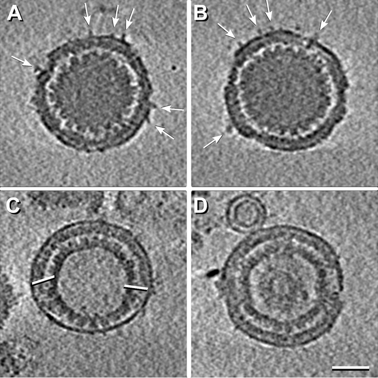
FIG 2 .

Central cryo-ET slices from reconstructions of four PEVs. The particles in panel A and B have a few protruding densities (white arrows). The PEV in panel C contains an A-capsid and has an unusually large fontanelle delimited by white bars, while the PEV in panel D contains a B-capsid. Scale bar = 50 nm.
