

Abstract
The presence and location of cellulose in different stages of the life cycles of the cellular slime molds can be demonstrated by use of the disodium salt of 4,4′-bis(4-anilino-6-bis (2-hydroxyethyl)-amino-s-triazin-2-ylamino)-2,2′ -stilbene disulfonic acid, a fluorescent brightener. It may be used successfully as a direct stain at a concentration of 0.1% in half-normal saline at pH 6; and it may be incorporated into growth media as a vital stain at a concentration of 0.0025% with no inhibitory effect at any developmental stage. Vegetative myxamoebae contain no cellulose and show no fluorescence in the presence of this brightener when viewed with ultraviolet light. In later stages of the life cycle, the time and sites of cellulose formation can be demonstrated with the brightener because of its fluorescence. e.g., in the slime covering of the pseudoplasmodia, in the sorophore sheath, in the walls of stalk cells and spores, in the walls of microcysts, and in the walls and sheath material of macrocysts. The brightener appears to be a very sensitive indicator for cellulose, and it has certain advantages over other cellulose stains, since the staining reaction (fluorescence) is very intense, long-lasting, and not obscured by unstained cellulose-free myxamoebae if such are present.
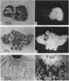
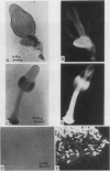
Full text
PDF


Images in this article


Selected References
These references are in PubMed. This may not be the complete list of references from this article.
- DARKEN M. A. Absorption and transport of fluorescent brighteners by microorganisms. Appl Microbiol. 1962 Sep;10:387–393. doi: 10.1128/am.10.5.387-393.1962. [DOI] [PMC free article] [PubMed] [Google Scholar]
- GEZELIUS K., RANBY B. G. Morphology and fine structure of the slime mold Dictyostelium discoideum. Exp Cell Res. 1957 Apr;12(2):265–289. doi: 10.1016/0014-4827(57)90141-6. [DOI] [PubMed] [Google Scholar]
- GEZELIUS K. The ultrastructure of cells and cellulose membranes in Acrasiae. Exp Cell Res. 1959 Nov;18:425–453. doi: 10.1016/0014-4827(59)90310-6. [DOI] [PubMed] [Google Scholar]
- Toama M. A., Raper K. B. Microcysts of the cellular slime mold Polysphondylium pallidum. I. Factors influencing microcyst formation. J Bacteriol. 1967 Oct;94(4):1143–1149. doi: 10.1128/jb.94.4.1143-1149.1967. [DOI] [PMC free article] [PubMed] [Google Scholar]