Abstract
The increasing prevalence of mild hypomineralization due to developmental defects on tooth surfaces poses a challenge for caries detection and caries risk assessment and reliable methods need to be developed to discriminate such lesions from active caries lesions that need intervention. Previous studies have demonstrated that areas of hypomineralization are typically covered with a relatively thick surface layer of highly mineralized and transparent enamel similar to arrested lesions. Seventy-six extracted human teeth with mild to moderate degrees of suspicious fluorosis were imaged using near-infrared reflectance and transillumination. Enamel hypomineralization was clearly visible in both modalities. However, it was difficult to distinguish hypomineralization due to developmental defects from caries lesions with contrast measurements alone. The location of the lesion on tooth coronal surface (i.e. generalized vs. localized) seems to be the most important indicator for the presence of enamel hypomineralization due to developmental defects.
Keywords: Fluorosis, Developmental defects of enamel, Dental Caries, Near-infrared imaging
1. INTRODUCTION
The developmental defects of enamel (DDEs) is a general term describing a wide range of enamel malformations due to many etiological factors including hereditary and environmental reasons.[1] Severe forms of DDEs such as enamel hypoplasia can be easily discerned, yet mild forms of DDEs such as enamel hypomineralization can be easily mistaken for caries lesions and this may lead to unnecessary intervention. [2]
Fluorescence imaging methods such as quantitative light-induced fluorescence (QLF) have been proposed as potential diagnostic tools for the assessment of early caries lesions. [3] QLF has also been used for the assessment of hypomineralization due to dental fluorosis because the subsurface porosities scatter light in a similar manner to demineralized carious lesions. [4]
In a previous study, we showed that the hypomineralization due to developmental defects manifests higher scattering at near-infrared (NIR) wavelengths similar to caries lesions. [5] However, in the same study, NIR transillumination imaging showed different optical behavior of hypomineralization due to developmental defects at 1310 nm compared to caries lesions due to the different geometric location of the hypomineralization. [5] Fejerskov et al. described the histological sections of permanent teeth with dental fluorosis as areas of diffuse hypomineralization, or porosity in the subsurface enamel in varying depth and severity with a well-mineralized surface layer, and the authors suggested that the superficial “remineralized” layer forms as the surface gets exposed to the oral cavity. [6] This presentation of the uniform hypomineralization with a highly mineralized surface layer was also observed with the PS-OCT of developmental defects. [5] We hypothesize that NIR imaging modalities are capable of detecting the subtle differences of caries lesions and hypomineralization due to developmental defects.
2. MATERIALS AND METHODS
2.1. Sample Selection
Teeth extracted from patients in the San Francisco Bay area were collected, cleaned and sterilized with gamma radiation. Seventy six teeth with suspected mild enamel hypomineralization due to developmental defects were screened. Enamel hypomineralization was identified as ranging from a few irregular white flecks to white opaque areas in the enamel covering as much as 50 percent of the tooth. Teeth were mounted on 1.2 cm × 1.2 cm × 3 cm pink translucent acrylic blocks. Samples were air-dried for 5 seconds prior to all image acquisition.
2.2. QLF Imaging
A Dino-Lite digital microscope AM4115TW (AnMo Electronics, Hsinchu, Taiwan) with 8 LEDs operating at 480 nm excitation wavelength and a 510 nm long-pass filter was used to acquire QLF images.
2.3. NIR Transillumination Imaging
Two iris diaphragms, a 60 nm NIR achromatic doublet AC254-060-C and 150-nm NIR achromatic doublet AC254-150-C (Thor Labs, Newton, New Jersey) lenses projected the captured light onto a high sensitivity NIR camera with an InGaAs focal plane array, Model GA1280J (Sensors Unlimited, Princeton, NJ) with a 1280 × 1024 pixel format and 15 μm pixel pitch. A band-pass filter BP1300-90 from (Spectrogon, Parsippany, NJ) was used for the transillumination imaging setup.
Light from a 150 W fiber-optic illuminator FOI-1 (E Licht Company, Denver, CO) was equally split into 4 line lights and directed at the cemento-enamel junction (CEJ) of each line angle (mesio-buccal, mesio-lingual, disto-buccal and disto-lingual).
2.4. NIR Reflectance Imaging
An Indigo Alpha NIR InGaAs focal plane array (FLIR Systems, Wilsonville, OR) with a spectral sensitivity range from 900 nm to 1750 nm, a resolution of 320 × 256 pixels, with an InfiniMite™ lens (Infinity, Boulder, CO) was used to acquire NIR reflectance images. Light from a 150 W fiber-optic illuminator FOI-1 (E Licht Company, Denver, CO) was directed at the sample with crossed polarizers and a long-pass filter at 1400 nm (1400 – 1700 nm).
2.5. Polarized Light Microscopy (PLM)
After sample imaging was completed, 200 μm thick serial sections were cut using an Isomet 5000 saw (Buehler, Lake Bluff, IL) for PLM. PLM was carried out using a Meiji Techno Model RZT microscope (Meiji Techno Co., LTD, Saitama, Japan) with an integrated digital camera, Canon EOS Digital Rebel XT (Canon Inc., Tokyo, Japan). The sample sections were imbibed in water and examined in the brightfield mode with crossed polarizers and a red I plate with 500 nm retardation. PLM images were acquired at 35x magnification and had a resolution of 3264 × 2448 pixels.
3. RESULTS AND DISCUSSION
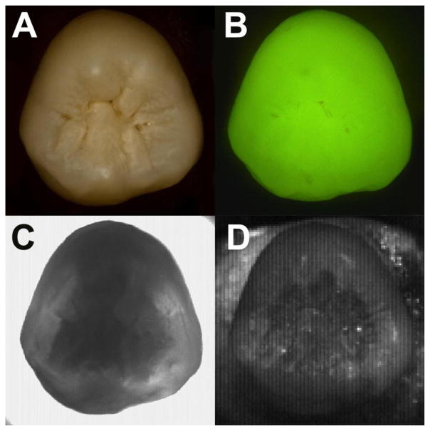
Figures 1–3 show visible, fluorescence and NIR images of the occlusal surfaces of three teeth. Figure 1 shows an occlusal view of a sound tooth. A visible image (Fig. 1A) shows no detectable caries lesions or hypomineralization, and the QLF image (Fig. 1B) shows localized decreased fluorescence intensity in the pits and fissures. In both transillumination and reflectance images (Figs. 1C and 1D), a lighter and translucent rim of enamel surrounding underlying dentin can be seen. In NIR occlusal transillumination of a sound tooth, the NIR light propagates up through more transparent enamel while it is blocked by the less transparent dentin. The cusps of the crowns appear darker than other surrounding enamel due to the increased thickness that the NIR light must travel. However, in NIR occlusal reflectance images of a sound tooth, the high contrast between the inner dentin and outer rim of enamel is most likely due to the high water content in dentin and the increased water absorption at NIR wavelengths.
Fig. 1.
Visible (A), QLF (B), NIR transillumination (C) and NIR reflectance images of a sound tooth (D).
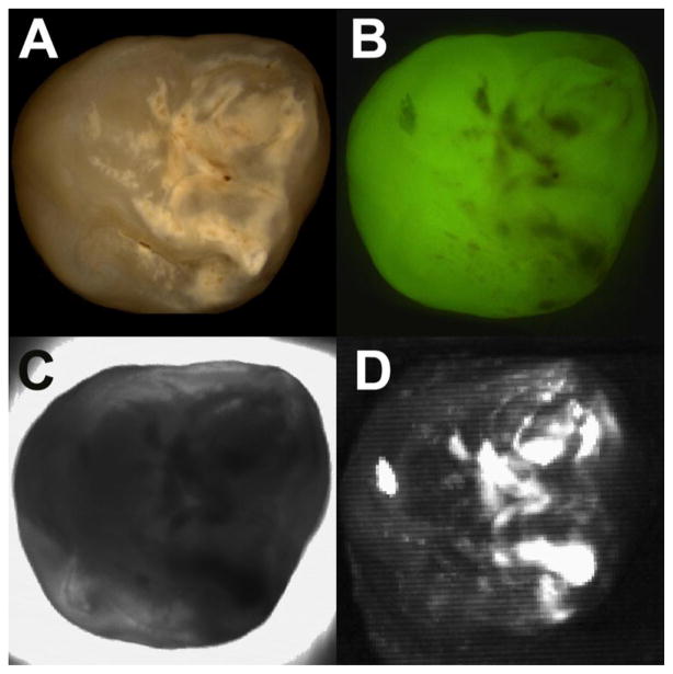
Fig. 3.
Visible (A), QLF (B), NIR transillumination (C) and NIR reflectance imaging of a tooth with mild enamel hypomineralization due to developmental defect (D).
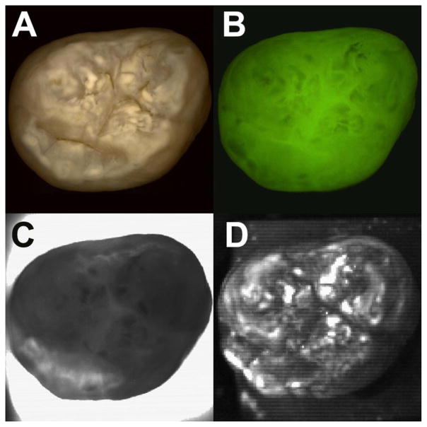
Figure 2 shows a tooth with caries lesion on the occlusal surface. The caries lesion manifests itself as generalized surface demineralization. The visible image (Fig. 2A) clearly shows localized white and brown opaque caries lesions around the pits and fissures with a clear demarcation of the lesion boundaries, and the decreased fluorescence intensity in the QLF image (Fig. 2B), decreased intensity in the NIR transillumination image (Fig. 2C) and increased intensity in the NIR reflectacnce image (Fig. 2D) match the locations of the highly lesion areas. The localized caries activity in both the NIR transillumination and reflectance images do not interfere with the contrast created by the surrounding lighter and translucent rim of enamel and the more opaque inner dentin.
Fig. 2.
Visible (A), QLF (B), NIR transillumination (C) and NIR reflectance images of a tooth with caries lesions (D).
Figure 3 shows a tooth with mild enamel hypomineralization due to developmental defects. Most of the samples are extracted impacted third molars, which have not been exposed to oral cavity, thus these specimens do not have any caries activity or surface remineralization. The visible image (Fig. 3A) shows generalized white hypomineralization around cusps and ridges of the crown, and there is decreased intensity in the fluorescence (Fig. 3B) and NIR transillumination images (Fig. 3C. There is increased intensity in the NIR reflectance image (Fig. 3D) around the cusps and ridges of the crown. However, the pits and fissures are not affected by the hypomineralization in all three imaging modalities, and this suggests that this hypomineralization is not likely to be related to caries activity. The generalized surface hypomineralization of the crown causes general opacity of the clear rim of enamel in the NIR transillumination image and a complete absence of contrast between the surrounding lighter rim of enamel and the inner more opaque dentin due in the NIR reflectance image.
The location of the lesion on tooth coronal surface seems to be the most important indicator for the presence of enamel hypomineralization due to developmental defects. The contrast produced by the surrounding lighter rim of enamel and inner more opaque dentin in multimodal NIR imaging methods may potentially be used to distinguish between generalized hypomineralization and localized caries lesions. Statistical analysis and further comparison with clinical methods such as visual and tactile will be discussed in full paper.
Acknowledgments
The authors acknowledge the support of NIH/NIDCR Grants F30-DE023278 and R01-DE14698.
References
- 1.Suckling GW. Developmental defects of enamel: Historical and present-day perspectives of their pathogenesis. Adv Dent Res. 1989;3(2):87–94. doi: 10.1177/08959374890030022901. [DOI] [PubMed] [Google Scholar]
- 2.Seow WK. Clinical diagnosis of enamel defects: pitfalls and practical guidelines. Int Dent J. 1997;47(3):173–82. doi: 10.1002/j.1875-595x.1997.tb00783.x. [DOI] [PubMed] [Google Scholar]
- 3.Pretty IA. Caries detection and diagnosis: novel technologies. J Dent. 2006;34(10):727–39. doi: 10.1016/j.jdent.2006.06.001. [DOI] [PubMed] [Google Scholar]
- 4.Pretty IA, McGrady M, Zakian C, Ellwood RP, Taylor A, Sharif MQ, Iafolla T, Martinez-Mier EA, Srisilapanan P, Korwanich N, Goodwin M, Dye BA. Quantitative light fluorescence (QLF) and polarized white light (PWL) assessments of dental fluorosis in an epidemiological setting. BMC Public Health. 2012;12:366. doi: 10.1186/1471-2458-12-366. [DOI] [PMC free article] [PubMed] [Google Scholar]
- 5.Hirasuna K, Fried D, Darling CL. Near-infrared imaging of developmental defects in dental enamel. Journal of Biomedical Optics. 044011;13(4):2008. doi: 10.1117/1.2956374. [DOI] [PubMed] [Google Scholar]
- 6.Fejerskov O, Thylstrup A, Larsen MJ. Clinical and structural features and possible pathogenic mechanisms of dental fluorosis. Scand J Dent Res. 1977;85(7):510–34. doi: 10.1111/j.1600-0722.1977.tb02110.x. [DOI] [PubMed] [Google Scholar]


