

Abstract
Purpose
Tibial fracture is the most common long bone fracture. Distal third tibial fractures are challenging though open reduction and plating can result in anatomical reduction and rigid fixation. This paper aimed to evaluate and compare the results of medial and lateral locking compression plates for distal third tibial fractures.
Methods
This prospective clinical study involved 36 patients with distal tibial fractures admitted in Department of Orthopaedics, Sawai Mansingh Medical College & Affiliated Hospital, Jaipur, India, from June 2011 to May 2012, including 29 closed fractures and 7 open fractures at the mean age of 38.9 years. Thirty-six patients were divided equally into two groups based on treatment method, including medial plating group (18 patients) and lateral plating group (18 patients). They were followed up for at least 5 months after discharge. The functional outcomes were evaluated using Tenny and Wiss clinical assessment criteria.
Results
Malunion was found in 3 cases of medial plating group and in 1 case of lateral plating group. In the medial plating group, there were 5 cases of superficial infections, 1 deep infection, 1 nonunion and 3 wound dehiscence. In the lateral plating group, there was 1 case of superficial infections, 1 deep infection and 1 nonunion. In the lateral plating group, 4 patients reported feeling the plates and screws but none of them asked to remove the hardware. In the medial plating group, 9 patients reported symptomatic hardware problems and 7 asked to remove the hardware. The number of cases graded as excellent/good/fair was 1/8/7 in the medial plating group and 3/7/7 in the lateral plating group respectively. In the medial plating group, the final range of motion was 17.2° in ankle dorsiflexion and 30.7° in ankle plantar flexion. In the lateral plating group, the final range of motion was 19° in ankle dorsiflexion and 34.2° in ankle plantar flexion.
Conclusion
Lateral plating of distal tibia is safe and feasible, which can provide biological fixation and prevent the soft tissue complications associated with medial plating.
Keywords: Tibial fractures, Bone plates, Open fracture reduction
Introduction
Distal third tibial fractures remain challenging due to peculiar soft tissue features and myriad treatment options. Surgical treatment itself has controversies such as difficulty in achieving and retaining good reduction by nailing methods, propensity to infection and nonunion due to dissecting the fracture site during the procedure of inserting the plate.1, 2 Open reduction and plating can result in rigid fixation and retention of the anatomical reduction. Traditionally popular method of medial plating offers good exposure to the tibia. However, it is at high risk of wound dehiscence, infection and hardware problems.3, 4 Recently, minimally invasive percutaneous medial plating has been devised.5, 6, 7, 8 However, this method is technically demanding, and it is often difficult to achieve anatomic reduction of the fracture site. Also in medial plating, if fixation of the fibula is required, an additional incision should be made on the lateral side of the shin. Single lateral plating for distal tibial and fibular fractures was reported to yield good results, but most of these studies were small series.9, 10 In their biomechanical study, Yenna et al11 pointed out that the patients with distal tibial extra-articular fractures demonstrated no statistically significant difference between anterolateral and medial locking plate in biomechanical stiffness in compression and torsion testing. There were few prospective studies comparing medial and lateral plating for distal tibial fractures. Whether the recently devised precontoured anterolateral distal tibial locking compression plate (LCP) shares problems of metaphyseal distal tibial LCP or overcomes them? To solve this question, our prospective study was conducted in the Department of Orthopaedics, Sawai Mansingh Medical College & Affiliated Hospital, Jaipur, India.
Materials and methods
During this study, the patients with distal tibial fractures admitted into our department from June 2011 to May 2012 were included according to the following criteria. Inclusion criteria were skeletally mature patients with distal third tibial fractures with or without concomitant fibular fractures, closed fractures and open fractures in which soft tissue injury was healed and skin condition was good enough for definitive treatment. Exclusion criteria were patients with open fractures in which soft tissue injury was not healed or skin condition was poor and patients with concomitant vascular injury. Totally 36 patients at the mean age of 38.9 years were included in this study and were followed up for at least 5 months after discharge, including 29 closed fractures and 7 open fractures. All open fractures were initially treated by irrigation, thorough debridement and appropriate intravenous antibiotics. Calcaneal pin traction was applied in 2 cases and spanning external fixator in one case as temporary fixation. With healed soft tissues and good skin condition, definitive management was undertaken in all patients. Thirty-six patients were divided equally into two groups based on treatment method.
The medial plating group used medial approach (Fig. 1), including 18 patients (mean 36.2 years). Fifteen patients (83.33%) suffered from vehicular trauma. Three patients had open fractures and 16 had tibial fractures concurrent with ipsilateral fibular fracture. The mean follow-up was 6.7 months.
Fig. 1.

X-ray showed tibial plating by medial approach.
The lateral plating group used a lateral approach (Fig. 2), including 18 patients (mean 41.6 years). Thirteen patients (72.22%) suffered from vehicle-related trauma. Four had open fractures and 14 had tibial fractures concurrent with ipsilateral fibular fracture. The mean follow-up was 6.2 months. In the medial plating group, the medial plating was performed as described by Clifford et al.3 If the distal fibula was fractured, it was fixed using separate incisions. In the lateral plating group, the lateral approach was performed as described by Manninen et al.9 A vertical skin incision was made from distal fibula to the anterior margin of the fibula. The dorsal cutaneous branches of the superficial peroneal nerve were retracted and protected. The fibula was exposed, if fractured, it was fixed firstly to obtain the reference length of the tibia. The anterior muscles and neurovascular tissue were bluntly dissected from the interosseous membrane and retracted anteriorly.
Fig. 2.
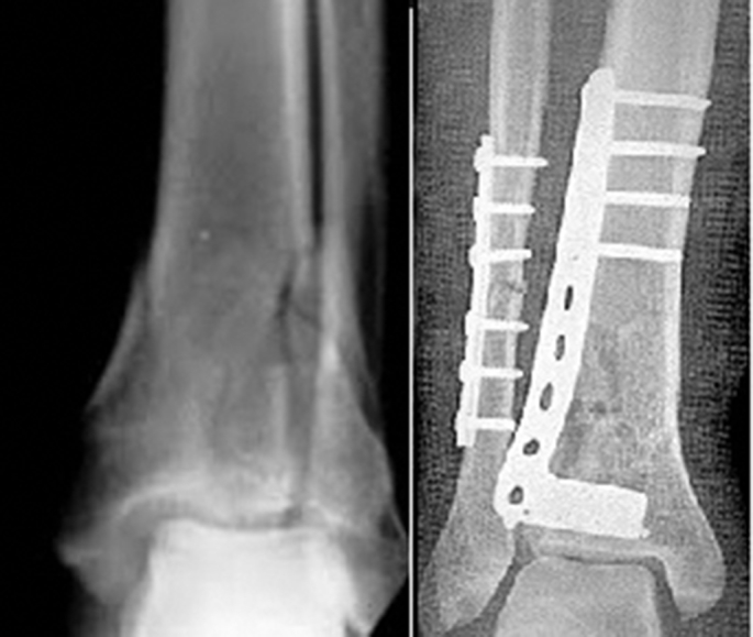
X-ray showed tibial plating by lateral approach.
After the reduction of tibial fracture, precontoured metaphyseal plate was applied in medial plating group and precontoured anterolateral distal tibial plate was applied in lateral plating group. The distal end of the plate could nearly reach the joint line. At least 6 cortex fixations proximal and distal to the fracture site were done. Operating time was recorded from the beginning of surgery to skin closure. Below-knee slab with the ankle in a neutral position was applied for 3 postoperative weeks for soft tissue healing. For patients with comminuted tibial fracture, a patellar tendon-bearing cast or removable brace was recommended for 6–8 weeks. The postoperative rehabilitation process consisted of partial weight bearing if patients had radiological signs of union. Full weight bearing was permitted when union was radiographically confirmed.
Radiographs taken immediately and postoperatively were reviewed for adequacy of fracture reduction in all patients. Anteroposterior alignment was determined by measuring the angle between a line paralleled to the proximal fragment and a line paralleled to the distal fragment on lateral radiographs. Varus–valgus alignment was determined by measuring the angle between the line perpendicular to and bisecting the tibial plateau and proximal medullary canal and the line bisecting the distal medullary canal and tibial plafond on anteroposterior radiographs. If both the fracture gap <2 mm and angular deformity ≤5° in any plane (valgus/varus, or anterior/posterior) were present, it was considered as excellent reduction. The fracture gap of 2–5 mm and angular deformity of ≤5° in any plane were regarded as good reduction. Adequate reduction included excellent and good reductions. Bony union was defined as evidence of bridging callus across the fracture sites or the obliteration of the fracture lines based on radiographic findings. Malunion was defined as angular deformity >5° in any plane, or internal rotation ≥10°, external rotation >15°, or shortening ≥2 cm. Nonunion was defined as no evidence of healing after 6 months.
Final evaluation was done for distal tibial fractures using Tenny and Wiss clinical assessment criteria based on 100 points system.12 The Student's t test was used to compare the outcomes of the two groups. p values less than 0.05 were considered significantly different.
Results
There were 3 cases of malunion in the medial plating group and 1 in the lateral plating group. In the medial plating group, there were 5 cases of superficial infections, 1 deep infection, 1 nonunion and 3 wound dehiscence. In the lateral plating group, there was 1 case of superficial infection, 1 deep infection and 1 nonunion.
Most of superficial infections were diagnosed clinically in the first follow-up of postoperative 7–10 days. After oral antibiotics treatment for 7–14 days, the wounds healed uneventfully. Both cases of deep infection presented in the first follow-up of postoperative 7–10 days as discharge around suture line. They were admitted and given intravenous antibiotics according to the results of drug sensitivity test. There were no signs of chronic osteomyelitis in the last follow-up.
In the lateral plating group, 4 out of 18 patients reported feeling the plates and screws but none of them asked to remove the hardware. In the medial plating group, 9 of 18 patients reported symptomatic hardware problems and 7 asked to have the hardware removed. Therefore, the lateral plating group experienced fewer hardware problems and hardware removals.
In the final follow-up, functional outcomes were evaluated using Tenny and Wiss clinical assessment criteria. The number of cases graded as excellent/good/fair was 1/8/7 in the medial plating group and 3/7/7 in the lateral plating group respectively. In the medial plating group, the final range of motion was 17.2° in ankle dorsiflexion and 30.7° in ankle plantar flexion. In the lateral plating group, the final range of motion was 19° in ankle dorsiflexion and 34.2° in ankle plantar flexion.
According to Table 1, it was seen that there was a significant difference in the sex distribution between two groups whereas there was no significant difference in the mechanism of injury and fracture characteristics. Table 2 showed significantly less operative time in the lateral plating group than in the medial plating group whereas there was no significant difference seen in the union rate, healing time, symptomatic hardware, superficial infection, functional score and range of ankle motion (p > 0.05).
Table 1.
Patients' characteristics in both medial plating group and lateral plating group.
| Parameters | Median plating group (n = 18) | Lateral plating group (n = 18) | t value | p value |
|---|---|---|---|---|
| Gender | 9.84 | 0.001 | ||
| Male | 1 | 4 | ||
| Female | 17 | 14 | ||
| Average age (years) | 36.3 | 41.6 | ||
| Average follow-up (months) | 6.7 | 6.2 | ||
| Mechanism of injury | 1.14 | 0.56 | ||
| Road traffic accidents | 15 | 13 | ||
| Fall from height | 2 | 2 | ||
| Others | 1 | 3 | ||
| Fracture type | 0.17 | 0.67 | ||
| Open | 3 | 4 | ||
| Closed | 15 | 14 | ||
| Distance from fracture site to tibial plafond (cm) | 5.1 | 5.6 | ||
| Concomitant fibular fracture | 16 | 14 | ||
Table 2.
Results of the medial and lateral plating in distal tibial fractures.
| Parameters | Median plating group (n = 18) | Lateral plating group (n = 18) | t value | p value |
|---|---|---|---|---|
| Operative time (min) | 95.6 ± 13.3 | 86.4 ± 12.7 | 2.12 | 0.04 |
| Union rate | 16/18 | 17/18 | 0.36 | 0.54 |
| Healing time (weeks) | 19.2 ± 4.2 | 18.6 ± 5.1 | 0.38 | 0.70 |
| Symptomatic hardware | 9 | 4 | 3.01 | 0.08 |
| Hardware removal | 7 | 0 | – | – |
| Superficial infection | 5 | 1 | 3.20 | 0.07 |
| Wound dehiscence | 2 | 0 | – | – |
| Result grading | 1.40 | 0.71 | ||
| Excellent | 1 | 3 | ||
| Good | 8 | 7 | ||
| Fair | 7 | 7 | ||
| Poor | 2 | 1 | ||
| Ankle dorsiflexion (°) | 17.2 ± 7.8 | 19.3 ± 8.5 | −0.77 | 0.45 |
| Ankle plantar flexion (°) | 30.7 ± 8.6 | 34.2 ± 9.1 | −1.19 | 0.24 |
Discussion
Nonsurgical treatment of tibial fractures can increase the incidence of malalignment with unacceptable shortening as Hooper et al13 concluded that nonoperative treatment resulted in more malunion and shortening. The most common surgical methods for distal tibial fractures are intramedullary nailing or medial plating.1, 2 However, malalignment of the distal tibia may develop after nailing.1 Vallier et al2 reported 113 cases of extra-articular distal tibial fractures treated with an intramedullary nail (n = 76) or a medial plate (n = 37) and found that the plating led to fewer malunions compared with the nailing (5.4% vs 38%). In our 36 patients, 4 (11.1%) reported malunion.
In our study, symptomatic hardware was a common problem in the medial plating group but was unusual in the lateral plating group. Totally 39% of the patients treated with medial plating requested a secondary operation to remove the implants because of the discomfort produced by the medial plate placed under the skin over anteromedial tibia. In contrast, in the lateral plating group, a lateral plate was placed beneath the anterior compartment muscles with thick soft tissue coverage.
Six cases had superficial infections in our study, including 5 in the medial plating group and 1 in the lateral plating group. No clear risk factors were found but we attributed to insufficient circulation at wound edges after closure. Wound necrosis and symptomatic hardware were more common in the medial plating group than in the lateral plating group. Theoretically, medial plating increases skin tension of the anteromedial tibia. In addition, medial plating often requires a separate incision for the accompanied distal fibular fracture, leading to double skin incisions around the ankle which may disturb the skin circulation between incisions. This may develop poor blood supply to the region and cause wound necrosis. Borrelli et al14 evaluated extraosseous blood supply of the tibia and the efficacy of different plating techniques in a cadaveric study. In their study, an anastomotic network of arteries from the anterior tibial artery and posterior tibial artery formed the rich extraosseous blood supply of the medial distal aspect of the tibia while open plating of the medial aspect of the distal tibia caused massive disruption of the extraosseous blood supply in the metaphyseal region. Disruption of the extraosseous vessels following fracture and subsequent operative stabilization may also increase the risk of nonunion.
Previous studies have reported that lateral plating for the distal tibia is technically demanding.9 However, in our study, operative time in the lateral approach was not significantly longer than that in the anterior approach. We experienced no difficulties using the lateral approach. Therefore, we concluded that the lateral plating is a safe and feasible technique.
Our result was consistent with that of a retrospective study conducted by Lee et al15 in Lin Shin Hospital, Taichung City. They retrospectively reviewed 88 patients with distal tibial fractures treated with medial or lateral plating and concluded that both methods achieved good functional outcomes with a low malunion rate; however, the lateral plating group had a lower complication rate and fewer hardware problems (p < 0.001). Our study was a prospective one, but with small sample size. Lateral plating of distal tibia can provide biological fixation and prevent the soft tissue complications associated with medial plating.
Footnotes
Peer review under responsibility of Daping Hospital and the Research Institute of Surgery of the Third Military Medical University.
References
- 1.Janssen K.W., Biert J., van Kampen A. Treatment of distal tibial fractures: plate versus nail: a retrospective outcome analysis of matched pairs of patients. Int Orthop. 2007;31:709–714. doi: 10.1007/s00264-006-0237-1. [DOI] [PMC free article] [PubMed] [Google Scholar]
- 2.Vallier H.A., Le T.T., Bedi A. Radiographic and clinical comparisons of distal tibia shaft fractures (4 to 11 cm proximal to the plafond): plating versus intramedullary nailing. J Orthop Trauma. 2008;22:307–311. doi: 10.1097/BOT.0b013e31816ed974. [DOI] [PubMed] [Google Scholar]
- 3.Clifford R.P., Beauchamp C.G., Kellam J.F. Plate fixation of open fractures of the tibia. J Bone Joint Surg Br. 1988;70:644–648. doi: 10.1302/0301-620X.70B4.3403616. [DOI] [PubMed] [Google Scholar]
- 4.Jensen J.S., Hansen F.W., Johansen J. Tibial shaft fractures. A comparison of conservative treatment and internal fixation with conventional plates or AO compression plates. Acta Orthop Scand. 1977;48:204–212. doi: 10.3109/17453677708985136. [DOI] [PubMed] [Google Scholar]
- 5.Helfet D.L., Shonnard P.Y., Levine D. Minimally invasive plate osteosynthesis of distal fractures of the tibia. Injury. 1997;28:A42–A47. doi: 10.1016/s0020-1383(97)90114-5. discussion A47–8. [DOI] [PubMed] [Google Scholar]
- 6.Maffulli N., Toms A.D., McMurtie A. Percutaneous plating of distal tibial fractures. Int Orthop. 2004;28:159–162. doi: 10.1007/s00264-004-0541-6. [DOI] [PMC free article] [PubMed] [Google Scholar]
- 7.Oh C.W., Kyung H.S., Park I.H. Distal tibia metaphyseal fractures treated by percutaneous plate osteosynthesis. Clin Orthop Relat Res. 2003;408:286–291. doi: 10.1097/00003086-200303000-00038. [DOI] [PubMed] [Google Scholar]
- 8.Pai V., Coulter G., Pai V. Minimally invasive plate fixation of the tibia. Int Orthop. 2007;31:491–496. doi: 10.1007/s00264-006-0228-2. [DOI] [PMC free article] [PubMed] [Google Scholar]
- 9.Manninen M.J., Lindahl J., Kankare J. Lateral approach for fixation of the distal tibia. Outcome of 20 patients. Technical note. Arch Orthop Trauma Surg. 2007;127:349–353. doi: 10.1007/s00402-006-0278-3. [DOI] [PubMed] [Google Scholar]
- 10.Shantharam S.S., Naeni F., Wilson E.P. Single-incision technique for internal fixation of distal tibia and fibula fractures. Orthopedics. 2000;23:429–431. doi: 10.3928/0147-7447-20000501-10. [DOI] [PubMed] [Google Scholar]
- 11.Yenna Z.C., Bhadra A.K. Anterolateral and medial locking plate stiffness in distal tibial fracture model. Foot Ankle Int. 2011;32:630–637. doi: 10.3113/FAI.2011.0630. [DOI] [PubMed] [Google Scholar]
- 12.Mahajan N. Minimally invasive techniques in distal tibial fractures. JK Sci. 2008;2:78–80. [Google Scholar]
- 13.Hooper G.J., Keddell R.G., Penny I.D. Conservative management or closed nailing for tibial shaft fractures. A randomised prospective trial. J Bone Joint Surg Br. 1991;73:83–85. doi: 10.1302/0301-620X.73B1.1991783. [DOI] [PubMed] [Google Scholar]
- 14.Borrelli J., Jr., Prickett W., Song E. Extraosseous blood supply of the tibia and the effects of different plating techniques: a human cadaveric study. J Orthop Trauma. 2002;16:691–695. doi: 10.1097/00005131-200211000-00002. [DOI] [PubMed] [Google Scholar]
- 15.Lee Y.S., Chen S.H., Lin J.C. Surgical treatment of distal tibia fractures: a comparison of medial and lateral plating. Orthopedics. 2009;32:163. [PubMed] [Google Scholar]