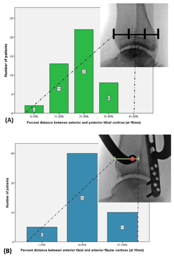
Figure 3.
Using three-dimensional reconstructions, the lateral tine of a reduction forceps was positioned on the fibular ridge, and, with the clamp positioned along the TSA, the location of the medial tine on a simulated lateral fluoroscopy image was noted with respect to its position (A) from anterior to posterior along the tibia and (B) in the space between the anterior cortices of the tibia and fibula.

