ABSTRACT
Botulinum neurotoxin (BoNT), produced by neurotoxigenic clostridia, is the most potent biological toxin known and the causative agent of the paralytic disease botulism. The nutritional, environmental, and genetic regulation of BoNT synthesis, activation, stability, and toxin complex (TC) formation is not well studied. Previous studies indicated that growth and BoNT formation were affected by arginine and glucose in Clostridium botulinum types A and B. In the present study, C. botulinum ATCC 3502 was grown in toxin production medium (TPM) with different levels of arginine and glucose and of three products of arginine metabolism, citrulline, proline, and ornithine. Cultures were analyzed for growth (optical density at 600 nm [OD600]), spore formation, and BoNT and TC formation by Western blotting and immunoprecipitation and for BoNT activity by mouse bioassay. A high level of arginine (20 g/liter) repressed BoNT production approximately 1,000-fold, enhanced growth, slowed lysis, and reduced endospore production by greater than 1,000-fold. Similar effects on toxin production were seen with equivalent levels of citrulline but not ornithine or proline. In TPM lacking glucose, levels of formation of BoNT/A1 and TC were significantly decreased, and extracellular BoNT and TC proteins were partially inactivated after the first day of culture. An understanding of the regulation of C. botulinum growth and BoNT and TC formation should be valuable in defining requirements for BoNT formation in foods and clinical samples, improving the quality of BoNT for pharmaceutical preparations, and elucidating the biological functions of BoNTs for the bacterium.
IMPORTANCE Botulinum neurotoxin (BoNT) is a major food safety and bioterrorism concern and is also an important pharmaceutical, and yet the regulation of its synthesis, activation, and stability in culture media, foods, and clinical samples is not well understood. This paper provides insights into the effects of critical nutrients on growth, lysis, spore formation, BoNT and TC production, and stability of BoNTs of C. botulinum. We show that for C. botulinum ATCC 3502 cultured in a complex medium, a high level of arginine repressed BoNT expression by ca. 1,000-fold and also strongly reduced sporulation. Arginine stimulated growth and compensated for a lack of glucose. BoNT and toxin complex proteins were partially inactivated in a complex medium lacking glucose. This work should aid in optimizing BoNT production for pharmaceutical uses, and furthermore, an understanding of the nutritional regulation of growth and BoNT formation may provide insights into growth and BoNT formation in foods and clinical samples and into the enigmatic function of BoNTs in nature.
KEYWORDS: Clostridium botulinum ATCC 3502, botulinum neurotoxin A1 (BoNT/A1), botulinum toxin complex (TC), arginine, glucose, spore formation, nutritional regulation
INTRODUCTION
Botulinum neurotoxin (BoNT) is the most potent biological toxin known and is the causative agent of botulism, a potentially fatal paralytic illness in animals and humans (1). It is a major food safety concern (2), a potential biological weapon of mass destruction designated a tier 1 select agent (3), and an important pharmaceutical (4). BoNTs cause six recognized forms of botulism: foodborne, infant, adult intestinal, wound, iatrogenic, and inhalational (5). All forms of botulism are due solely to the action of BoNTs. Characteristic signs and symptoms are ocular disturbances and a flaccid paralysis of the face and mouth, bilaterally descending to respiratory and skeletal muscles (5). BoNT acts mainly at the neuromuscular junction of motor neurons and inhibits the release of acetylcholine (6). Other classes of neurons involving vesicular release of neurotransmitters can also be affected in vivo by BoNTs, including those associated with autonomic and sensory functions (5). Evidence also indicates that BoNT is transsynaptically trafficked to spatially distal neurons, including those of the central nervous system (CNS) (7, 8).
BoNTs are produced by neurotoxigenic clostridia, most notably Clostridium botulinum, a Gram-positive, spore-forming, anaerobic bacterium. BoNT is also produced by neurotoxigenic strains of Clostridium baratii, Clostridium butyricum, and Clostridium argentinense (9). C. botulinum is prevalent in soils and coastal waters, is associated with certain insects and invertebrates, and also resides in the intestinal tract of several animals (but not humans) (10, 11). There are seven serotypes of BoNT, A through G, based on neutralization against mouse lethality by homologous antibodies, and within most serotypes there are multiple BoNT variants (“subtypes”) (12–14). C. botulinum is commonly classified into four physiological groups, I through IV (5, 15), with groups I and II responsible for the majority of human botulism cases. Group I comprises proteolytic strains of serotypes A, B, and F and is mainly responsible for botulism in the continental United States and in many countries globally (16), while the members of group II include nonproteolytic strains of serotypes B, E, and F and are particular concerns in refrigerated food products. Type E is commonly found in fish products from freshwater and marine environments and has caused botulism in Europe and in northern regions above the Tropic of Cancer (16, 17).
Botulinum neurotoxin is initially synthesized as a ca. 150-kDa polypeptide and is proteolytically modified to a dichain form comprised of a 100-kDa heavy chain (HC) responsible for binding to and facilitating entry into cholinergic nerve terminals (6, 18) and a 50-kDa light chain (LC) which is a zinc metalloprotease that cleaves soluble N-ethylmaleimide-sensitive factor activating protein receptor (SNARE) proteins (6, 18). Cleavage of SNARE proteins prevents the trafficking and docking of vesicles and inhibits release of acetylcholine into the neuromuscular junction, resulting in a characteristic flaccid muscular paralysis. Certain serotypes of C. botulinum within group I produce a proteolytic enzyme(s) that cleaves the 150-kDa single chain, removing 9 to 11 amino acids between the HC and LC (19), which then remain joined by a disulfide bond and noncovalent interactions (20). This proteolytic processing step is associated with an activation process that increases the activity of BoNTs to various degrees in mice and neuronal cells depending on the serotype (20, 21). Group II (nonproteolytic) strains commonly lack or have low levels of activating proteases and require exogenous proteases from human or animal hosts for activation during intoxication and for in vitro and in vivo activation (5).
An interesting property of BoNTs is that they are produced as a component of toxin complexes (TCs), in which BoNT is noncovalently associated with nontoxic proteins in the TCs (22). The genes encoding BoNT and the nontoxic complex proteins occur in BoNT gene clusters (23, 24). There are two general types of TCs, (i) the hemagglutinin (HA) TCs which contain BoNT, NTNH (nontoxic-nonhemagglutinin), and three forms of hemagglutinins (HA17, HA33, and HA70) and (ii) the OrfX TCs which appear to contain only BoNT and NTNH but not the OrfX proteins, even though the orfx genes are in the BoNT gene cluster (24–26). The TC proteins protect BoNTs in the gastrointestinal (GI) tract, contributing to toxicity (27–29).
Despite the high potencies of BoNTs and their causing a severe paralytic disease, only limited studies on the environmental, nutritional, and genetic regulation of formation of BoNT production and the nontoxic proteins of the TCs have been performed (30–32). The nutritional effects on growth, lysis, sporulation, TC composition and assembly, and stability and inactivation of BoNT and TC proteins in culture have not been well studied. Previous work qualitatively showed various degrees of inhibition of BoNT formation by nutritional regulation, particularly by arginine and glucose in a defined medium (33). In those earlier studies, the effects of arginine and glucose on sporulation, formation of TCs, and the stability of BoNTs were not investigated. The nutritional growth requirements have been established using chemically defined minimal media, and the primary essential nutrients for groups I and II are arginine and tryptophan, respectively (34). It is intriguing that arginine and tryptophan, which are strictly required for growth in types A and E, respectively, were also shown in early studies to inhibit formation of BoNTs. This suggests that BoNTs play an important role in acquisition of these required nutrients, though they likely have other functions for C. botulinum in nature.
In this study, we investigated the effects of arginine and glucose on the production and stability of BoNT and TC proteins in a rich medium (toxin production medium [TPM]; 2% casein hydrolysate, 1% yeast extract, 0.5% glucose) using molecular methods. The new analyses and results presented in this paper include effects of arginine and glucose on the formation and stability of BoNT and TC proteins analyzed by immunoprecipitation and Western blotting and effects on sporulation. The ATCC 3502 strain used in this study is a group I proteolytic strain and produces the HAs, NTNH, and BoNT serotype A subtype 1 (BoNT/A1) TC (35). It was the first C. botulinum strain whose complete genome was sequenced and annotated (35), is widely studied by several laboratories, and is therefore a good model for physiological and genetic studies. We found that increased levels of arginine strongly repressed expression of BoNT and proteins of the toxin complexes, repressed sporulation, and enhanced growth and inhibited lysis of the culture. A lack of glucose in the medium was found to reduce BoNT expression and also resulted in inactivation of extracellular BoNT and TC proteins. The current studies provide a basis for understanding the nutritional and physiological regulation of BoNTs, TC formation, and sporulation.
RESULTS
Growth, lysis, pH, and spore enumeration of C. botulinum ATCC 3502 in the four primary media.
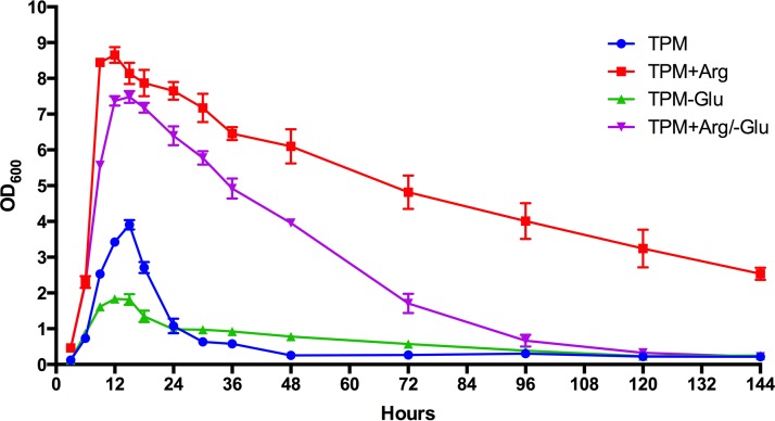
The maximum levels of growth and the lysis patterns of C. botulinum ATCC 3502 were distinctly different in the four primary media tested (Fig. 1). In basal TPM without supplements, maximum growth was approximately 4 optical density (OD) units at 15 h, and then the culture began extensive lysis at 18 h and was nearly completely lysed by 144 h. In TPM without glucose (TPM−Glu), maximum growth was decreased compared to that in basal TPM, with a maximum OD of 1.8, and lysis began at about 15 h and was mainly complete by 144 h. The presence of added arginine in the media markedly increased maximum growth. In TPM+Arg (TPM supplemented with 2% [0.115 M] arginine), the maximum OD attained was approximately 8.8, and lysis commenced at about 15 h but was slow and progressive, and complete lysis was not observed after 144 h. In TPM supplemented with arginine but without glucose (TPM+Arg−Glu), the growth rate was also relatively high and resulted in a maximum OD of approximately 7.5, and lysis was slowed but was largely complete after 144 h. The growth rate during the exponential phase was higher in the two media with added arginine. These results show that arginine strongly stimulated growth, resulting in higher cell densities, but also slowed lysis compared to the results seen with basal TPM and TPM−Glu. Supplementation of arginine and lack of glucose resulted in an increase in pH compared to basal TPM results (Table 1). Arginine supplementation also affected the formation of endospores. In basal TPM and TPM−Glu, the spore counts were approximately 2.3 × 10⁴ and 4.7 × 10⁴ per ml, respectively. In contrast, in media supplemented with arginine, the spore counts were <10 per ml.
FIG 1.
Patterns of growth of C. botulinum strain ATCC 3502 in four different media. Error bars represent standard deviations.
TABLE 1.
Culture pH values of ATCC 3502 in four different media after 24 h or 6 days of incubation
| Medium | pH |
|
|---|---|---|
| Day 1 (24 h) | Day 6 | |
| TPM | 6.0 | 6.3 |
| TPM+Arg | 7.7 | 7.6 |
| TPM−Glu | 6.9 | 7.9 |
| TPM+Arg/−Glu | 8.3 | 8.3 |
BoNT titers in cultures determined by intraperitoneal (i.p.) time-to-death mouse bioassay.
An i.p. time-to-death mouse bioassay was performed on supernatants of the four primary culture media on day 1 and day 6. The four media differed markedly in levels of BoNT formation. The TPM culture had a typical titer of toxicity for these growth conditions of ca. 105 50% lethal doses (LD50)/ml on day 1 and day 6. On day 1, the TPM+Arg culture had a toxicity titer of approximately 102 LD50/ml, and on day 6 it was below 102 LD50/ml (Table 2). The TPM−Glu culture had toxicity titers of about 103 LD50/ml on day 1 and less than 102 LD50/ml on day 6. The TPM+Arg/−Glu culture had a toxicity of about 103 LD50/ml on day 1. On day 6, the toxicity was less than 102 LD50/ml. These results clearly show that arginine and glucose markedly affected BoNT titers in culture. The decrease in BoNT titers in TPM−Glu and TPM+Arg/−Glu between day 1 and day 6 suggests that BoNT is partially inactivated in culture under these conditions.
TABLE 2.
Toxicity (in mouse LD50/ml) of trypsinized culture supernatants from experimental media, determined by mouse intraperitoneal survival time assay
| Medium | Time point (day) | Toxicity (LD50/ml)a |
|---|---|---|
| TPM | 1 | 2.91 × 10⁵ |
| 6 | 1.13 × 10⁵ | |
| TPM+Arg | 1 | 2.42 × 10² |
| 6 | <1 × 10² | |
| TPM−Glu | 1 | 1.37 × 10³ |
| 6 | <1 × 10² | |
| TPM+Arg/−Glu | 1 | 4.25 × 10³ |
| 6 | <1 × 10² |
Values represent the mean of the results from four mice per condition.
Western blot analyses of whole cultures and supernatants of cultures in primary media.
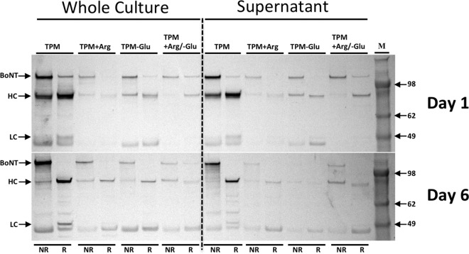
To analyze BoNT formation and proteolytic modification, including HC and LC cleavage, Western blot analyses were performed on samples from the four primary media on days 1 and 6 (Fig. 2). Both the whole culture and the culture supernatant (obtained by centrifugation performed to remove cells) were analyzed to compare total BoNT and extracellular BoNT, respectively. The Western analyses performed using whole culture and supernatant showed a pattern analogous to the titers found in mice.
FIG 2.
Western blot analysis of samples from each medium at the day 1 and day 6 time points, using anti-BoNT/A1 polyclonal antibodies. To allow comparison of total BoNT and extracellular BoNT, the left panel represents samples of the whole culture and the right panel represents samples of the culture supernatant, respectively. Lanes are in pairs. NR indicates a nonreduced sample, and R indicates a reduced sample. HC, BoNT heavy chain; LC, BoNT light chain.
The Western blot analyses showed that the quantity of BoNT was markedly decreased in media supplemented with arginine compared to basal TPM (Fig. 2). The Western blot results for TPM−Glu showed less BoNT than in TPM (Fig. 2). The intensity of the BoNT band from the supernatant of TPM−Glu was decreased from day 1 to day 6. Since only a minor decrease in the BoNT level was seen in the whole culture between day 1 and day 6, the decrease in the BoNT level in the supernatant suggests that extracellular BoNT was partially inactivated in TPM−Glu whereas intracellular BoNT was not. TPM+Arg/−Glu also showed a low level of BoNT production compared to TPM (Fig. 2).
The Western analyses also allowed an estimation of the level of proteolytic activation of BoNT to the toxic dichain form (Fig. 2). At day 1, BoNT was not fully cleaved in all four media, while at day 6 the BoNTs were completely nicked and present as HC and LC with negligible holotoxin observed in the reduced samples.
Immunoprecipitation and SDS-PAGE analysis of BoNT and TC proteins in culture supernatants.
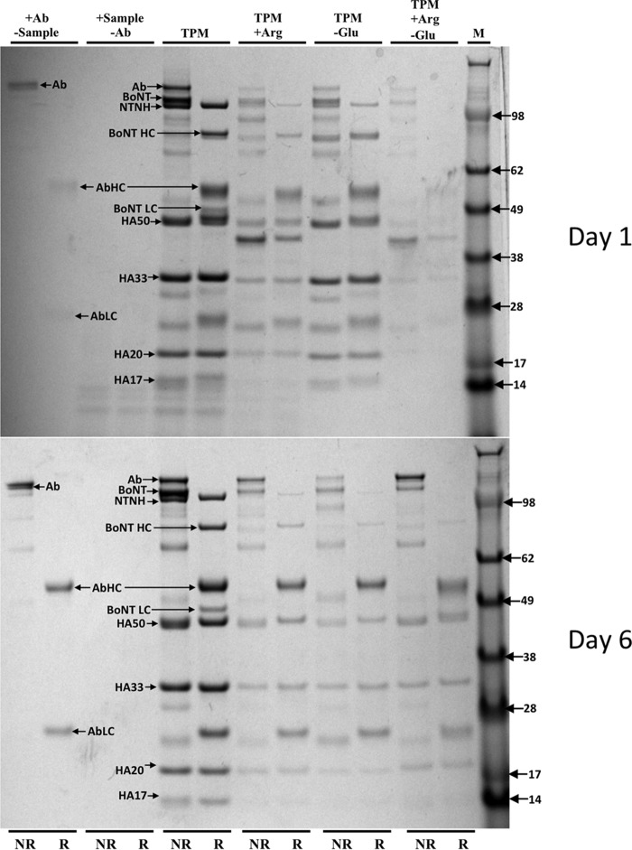
In addition to Western blotting, immunoprecipitation of culture supernatant enabled the analysis of toxin complex (TC) formation. Figure 3 shows results of SDS-PAGE analysis of immunoprecipitation. The analysis was performed using polyclonal antibodies to BoNT and therefore would detect BoNT and bound TC proteins. Figure 4 shows results of SDS-PAGE analysis of an immunoprecipitation performed using a monoclonal antibody to HA50. HA50 is a proteolytic product of HA70 (along with HA20); thus, the immunoprecipitation using the anti-HA50 antibody would detect other complex proteins and BoNT associated with HA50.
FIG 3.
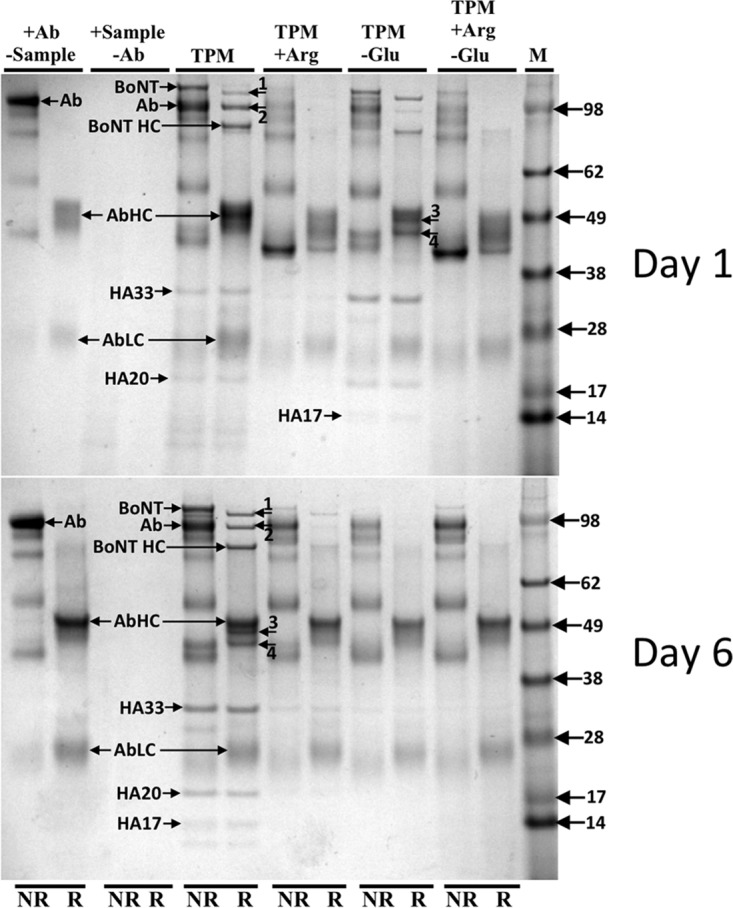
Immunoprecipitation analysis of samples of culture supernatant from each medium at the day 1 and day 6 time points, using anti-BoNT/A1 polyclonal antibodies. Lanes are in pairs. R indicates that the sample was reduced with DTT, and NR indicates a nonreduced sample. Arrow 1, NTNH; arrow 2, truncated NTNH; arrow 3, BoNT light chain; arrow 4, HA50 (breakdown product of HA70 along with HA20). BoNT HC, BoNT heavy chain; Ab, antibody; AbHC, antibody heavy chain; AbLC, antibody light chain.
FIG 4.
Immunoprecipitation analysis of samples of culture supernatant from each medium at the day 1 and day 6 time points, using anti-HA50 antibodies. Lanes are in pairs. R indicates that the sample was reduced with DTT, and NR indicates a nonreduced sample. BoNT LC, BoNT light chain; BoNT HC, BoNT heavy chain; Ab, antibody; AbHC, antibody heavy chain; AbLC, antibody light chain.
In TPM, anti-BoNT and anti-HA50 immunoprecipitations showed prominent bands of BoNT and the TC proteins (NTNH, HA17, HA20, HA33, and HA50) (Fig. 3 and 4). In the BoNT immunoprecipitation of TPM+Arg cultures, BoNT and TC proteins were largely absent on days 1 and 6 (Fig. 3) but were detectable in the HA50 immunoprecipitation (Fig. 4). These data indicate that protein expression of BoNT and TC was repressed. The immunoprecipitation analyses also demonstrated that while BoNT production was strongly repressed in TPM+Arg, the small quantity of BoNT produced still formed a TC.
In the TPM−Glu culture, BoNT and TC proteins were detected in the day 1 culture by both the anti-BoNT/A1 and anti-HA50 antibodies (Fig. 3 and 4). Both the complex proteins and BoNT were present in smaller quantities by day 6, which is consistent with the decrease of BoNT observed in the mouse assay and the Western blot analysis of the culture supernatant. This indicated that the toxin complex was formed and that the TC proteins and BoNT were inactivated. In TPM+Arg/−Glu, very low levels of BoNT and complex proteins were detected in the immunoprecipitation using anti-BoNT/A1 and anti-HA50 antibodies (Fig. 3 and 4).
Western blot analyses of C. botulinum grown in media with arginine metabolites.
To determine whether the repression was specific to arginine, metabolites of arginine, including citrulline, ornithine, and proline, were examined for their effect on expression of BoNT by Western blotting (Fig. 5). The presence of proline and ornithine did not affect expression compared to TPM, while that of citrulline led to repression but not to the degree observed with arginine (Fig. 5).
FIG 5.
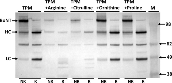
Western blot analysis using BoNT/A1 polyclonal antibodies of samples of ATCC 3502 C. botulinum cultures incubated for 6 days in five different media. Only whole-culture samples are shown. Lanes are in pairs. R indicates that the sample was reduced with DTT, and NR indicates a nonreduced sample. HC, BoNT heavy chain; LC, BoNT light chain.
DISCUSSION
In this study, we have shown that arginine and glucose in a complex medium (TPM) markedly affect C. botulinum type A ATCC 3502 growth, lysis, and spore formation and the expression and stability of BoNT and TC proteins. Earlier studies in our laboratory showed that high levels of arginine (20 g/liter) repressed BoNT formation compared to the results seen with basal levels in C. botulinum Okra B in a chemically defined medium (33). In this study, it was found that BoNT was strongly regulated by arginine in the complex medium TPM. This was unexpected since TPM contains substantial quantities of arginine and other amino acids.
The growth and lysis patterns differed decidedly among the four media. Arginine increased growth rate and approximately doubled maximum growth. The lowest growth rate and lowest maximum growth were observed in TPM−Glu. Under this condition, it is likely that growth was slowed due to energy limitation. The basis for the accelerated growth and relatively high cell densities obtained in media supplemented with arginine does not appear to have been relief from energy limitation since high growth was also obtained in the medium supplemented with arginine but lacking glucose. Arginine could also have been providing rapidly assimilated nitrogen, as well as having other functions in increasing growth and cellular biosynthesis. The increased pH in nonbasal media could be due to catabolism of amino acids with release of ammonia or other basic compounds, as well as to differences in growth and lysis.
To evaluate whether BoNT repression was specific for arginine, related metabolites were examined in TPM. Proline and ornithine did not affect BoNT formation, while citrulline did result in decreased formation of BoNT. Since arginine can be synthesized from citrulline in group I C. botulinum, the decreased BoNT levels seen under this condition could have been due to metabolic changes such as formation of ATP or to conversion of citrulline to arginine. Further work is required to investigate the mechanism of repression by arginine.
Proteolytic (group I) serotype A strains of C. botulinum and related clostridial species such as C. sporogenes have been shown to contain a highly active arginine deiminase (ADI) system but to lack other arginine catabolic systems. The ADI system can provide energy through ATP formation and can also provide nitrogen for cellular biosynthesis. It was postulated in 1989 (33) that regulation of BoNT formation in C. botulinum Okra B was controlled by classical carbon catabolite repression or nitrogen regulation (36). Amino acid catabolism also affects the synthesis of other clostridial toxins, including those produced by C. difficile, C. perfringens, C. septicum, and C. tetani (37, 38). Therefore, amino acid catabolism and associated regulatory mechanisms may share common mechanisms among toxigenic clostridia.
Recently, several mechanisms of BoNT regulation have been shown to occur in C. botulinum. These include control by BotR (39–41), the CodY sensing system (42), two-component regulatory systems (43, 44), and quorum sensing (45). Other mechanisms related to amino acid metabolism and protein translation such as riboswitches could also be involved in regulation. These systems as well as undiscovered regulatory control mechanisms may be involved in nutritional and environmental regulation of BoNT, synthesis, activation, and complex formation in C. botulinum. The molecular mechanisms controlling BoNT formation, stability, lysis, and sporulation represent an intriguing and yet incompletely understood area. Currently, our laboratory is performing genomic and mutational studies to further understand the mechanisms of regulation, with an emphasis on genes in the arginine deiminase pathway, and performing RNA sequencing (RNA-Seq) of cultures grown in basal and high-arginine media. An understanding of these mechanisms will be important in understanding the production of BoNTs in foods and clinical samples, for improvement of BoNT as a pharmaceutical, and to provide insights into the evolutionary functions of BoNT.
MATERIALS AND METHODS
Biosafety and biosecurity.
Our laboratory and personnel are registered with the Federal Select Agent Program for research involving botulinum neurotoxins (BoNTs) and BoNT-producing strains of clostridia. The research program, procedures, documentation, security, and facilities are closely monitored by the University of Wisconsin—Madison Biosecurity Task Force, the University of Wisconsin—Madison Office of Biological Safety, the University of Wisconsin Select Agent Program, and the CDC and the Animal and Plant Health Inspection Service (APHIS) as part of the University of Wisconsin—Madison and CDC Select Agent Program. All personnel routinely receive suitability assessments and completed rigorous and continuing biosafety training, including biosafety level 3 (BSL3) or BSL2 and select agent practices, before participating in laboratory studies involving BoNTs and neurotoxigenic C. botulinum. All animal experiments have been approved by the University of Wisconsin and IACUC.
Strain, media, and growth.
C. botulinum ATCC 3502 was used in this study. This strain was the first to have its genome sequenced (35) and is widely used in studies by several laboratories. It produces BoNT/A1 and a toxin complex (TC) consisting of NTNH, HA17, HA33, and HA70. Initial cultures were grown in TPGY medium, which consists of 5% Trypticase peptone (Difco), 2% yeast extract (Difco), 0.5% Bacto peptone (Difco), 0.4% glucose (Fisher Scientific), and 0.1% cysteine HCl (MP Biomedicals). Cultures were inoculated at a 1:100 dilution from an 18-h TPGY starter culture in an anaerobic chamber and incubated at 35°C. Growth measurements (OD at 600 nm [OD600]) were determined in triplicate in an Eppendorf Biophotometer. For experimental determinations, cultures were grown at 35°C in 10 ml of TPM in Bellco anaerobe tubes fitted with rubber stoppers. TPM is a complex medium that supports high BoNT production in group 1 C. botulinum (22). TPM consists of 2% casein hydrolysate (NZ Case TT; Kerry Ingredients), 1% yeast extract (Difco), and 0.5% glucose (Fisher Scientific). The following four medium conditions were primarily used: (i) TPM (basal medium), (ii) TPM+Arg (TPM supplemented with 2% [0.115 M] arginine [from Sigma-Aldrich]), (iii) TPM−Glu (TPM without glucose), and (iv) TPM+Arg/−Glu (TPM without glucose and with 2% arginine). The following three other conditions were also used for analysis of BoNT synthesis: (i) TPM+citrulline (Sigma-Aldrich) (0.115 M), (ii) TPM+ornithine (Sigma-Aldrich) (0.115 M), and (iii) TPM+proline (Sigma-Aldrich) (0.115 M). For all media, the pH was adjusted to 7.35 and then tubes of media were sparged with nitrogen and sealed before autoclaving to ensure an anaerobic environment.
Spore enumeration.
At the final sampling of 6 days (144 h), 1-ml culture aliquots were collected and heated to 80°C for 20 min to kill vegetative cells. Aliquots were then cooled in ice water, and appropriate dilutions were spread on TYG (3% tryptone, 1.5% yeast extract, 0.1% thioglycolate) 1% agar plates. Colonies were enumerated after 3 days of incubation at 35°C. Tryptone, yeast extract, and agar were from Difco, and thioglycolate was from Sigma-Aldrich.
Immunoprecipitation of toxin complexes (TCs), SDS-PAGE analyses, and Western blotting.
Immunoprecipitation was performed using a Life Technologies Dynabeads protein A kit according to the manufacturer's instructions, with the exception that the recommended wash buffer was replaced by 50 mM sodium citrate at pH 5.5. Immunoprecipitation was performed as previously described (25) with some minor modifications. Briefly, anti-BoNT/A1 polyclonal antibodies produced in our laboratory or an anti-HA50 monoclonal antibody (kindly provided by Larry Stanker) was incubated for 45 min with the protein A-coated Dynabeads at ambient temperature. The antibody-Dynabead complex was incubated for 1 h with 1 ml of culture supernatant (culture aliquots centrifuged for 5 min at 16,000 × g) and then washed 5 times with 50 mM sodium citrate (pH 5.5) to remove nonspecific bound proteins. The antibodies and bound proteins were then eluted from the Dynabeads in 30 μl of elution buffer, analyzed by SDS-PAGE, and subjected to Coomassie staining.
For SDS-PAGE analysis, 10 μl of NuPAGE 4× lithium dodecyl sulfate (LDS) sample buffer was added to 30-μl samples of whole culture, culture supernatant obtained by centrifugation, or eluted immunoprecipitation samples (see below). The samples were split into two 20-μl aliquots and analyzed under nonreduced and reduced conditions (100 mM dithiothreitol [DTT]). Reduction with DTT breaks the disulfide bond joining the heavy chain and the light chain of BoNT, allowing them to separate under SDS-PAGE conditions if the toxin has undergone proteolytic cleavage. Both the reduced and nonreduced sample aliquots were boiled for 5 min. Samples were separated on Life Technologies 4% to 12% Bis-Tris gels in NuPAGE morpholineethanesulfonic acid (MES) SDS running buffer. SeeBlue Plus2 prestained standard (Invitrogen) was used as a molecular weight marker.
For Western blot analyses, 30-μl samples of whole culture and culture supernatants (centrifuged for 5 min at 16,000 × g) were collected at days 1 and 6 and were analyzed by SDS-PAGE under reduced and nonreduced conditions. The separated protein bands were transferred to a polyvinylidene difluoride (PVDF) membrane using a Life Technologies Novex semidry blotter. The membrane was probed with anti-BoNT/A1 polyclonal rabbit antibodies prepared in our laboratory and with a secondary bovine anti-rabbit alkaline phosphatase antibody (Santa Cruz Biotechnology). The membrane was developed using a BCIP/NBT (5-bromo-4-chloro-3-indolylphosphate/Nitro Blue Tetrazolium) kit (Zymed Laboratories).
Mouse bioassay of BoNT.
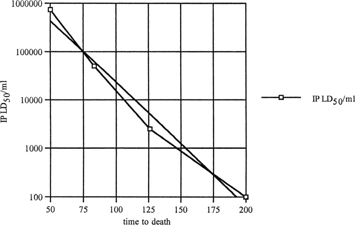
An intraperitoneal (i.p.) time-to-death method was used to estimate the toxicity of the cultures. This method was previously established in a collaboration of E. A. Johnson with Edward J. Schantz in his laboratory (Fig. 6). Though less accurate than using serial dilutions in a quantal mouse bioassay, this method requires far fewer mice to be sacrificed while providing a useful estimate of toxicity. This method is accurate up to approximately 200 min (corresponding to about 102 LD50/ml). When 200 min was exceeded, the LD50 was conservatively recorded as <102 LD50/ml.
FIG 6.
The relationship between time to death and BoNT/A or B toxicity for a 0.5-ml intraperitoneal (IP) injection of a ca. 25-g mouse. The curve fit formula used to calculate toxicity from time to death (in minutes) is as follows: y = 8,334,208.65 × 10−0.025x.
The samples were first trypsinized in order to proteolytically cleave any full-length BoNT. Samples from day 1 and day 6 cultures were centrifuged for 5 min at 16,000 × g, and the supernatants were treated with 5 μg/ml of trypsin (Worthington Biochemical Corporation) and incubated at 37°C for 1 h. Type II-S soybean trypsin inhibitor (Sigma-Aldrich) (20 μg/ml) was added, and samples were incubated at room temperature for 10 min. The trypsinized samples were filtered using a 0.2-μm-pore-size syringe filter (Thermo Scientific), and 0.5 ml of a 1:1 mixture of trypsinized culture supernatant in gelatin phosphate buffer (0.2% gelatin and 30 mM sodium phosphate, pH 6.3) was injected into 4 female ICR mice (Harlan Laboratories). Survival time was recorded and compared to the LD50 standard curve (Fig. 6).
ACKNOWLEDGMENTS
We thank Larry Stanker for his generous gift of monoclonal antibodies.
This work was supported by the National Institute of Allergy and Infectious Diseases (RO1 AI095274) and by funds from the Food Research Institute, University of Wisconsin—Madison.
REFERENCES
- 1.Cherington M. 1998. Clinical spectrum of botulism. Muscle Nerve 21:701–710. doi:. [DOI] [PubMed] [Google Scholar]
- 2.Johnson EA. 2007. Clostridium botulinum, p 401–421. In Doyle MP, Beuchat LR (ed), Food microbiology: fundamentals and frontiers, 3rd ed. ASM Press, Washington, DC. [Google Scholar]
- 3.Arnon SS, Schechter R, Inglesby TV, Henderson DA, Bartlett JG, Ascher MS, Eitzen E, Fine AD, Hauer J, Layton M, Lillibridge S, Osterholm MT, O'Toole T, Parker G, Perl TM, Russell PK, Swerdlow DL, Tonat K, Working Group on Civilian Biodefense . 2001. Botulinum toxin as a biological weapon: medical and public health management. JAMA 285:1059–1070. doi: 10.1001/jama.285.8.1059. [DOI] [PubMed] [Google Scholar]
- 4.Truong DDD, Hallett M, Zachary C (ed). 2009. Manual of botulinum toxin therapy. Cambridge University Press, Cambridge, United Kingdom. [Google Scholar]
- 5.Johnson EA, Montecucco C. 2008. Botulism. Handb Clin Neurol 91:333–368. doi: 10.1016/S0072-9752(07)01511-4. [DOI] [PubMed] [Google Scholar]
- 6.Schiavo G, Matteoli M, Montecucco C. 2000. Neurotoxins affecting neuroexocytosis. Physiol Rev 80:717–766. [DOI] [PubMed] [Google Scholar]
- 7.Restani L, Novelli E, Bottari D, Leone P, Barone I, Galli-Resta L, Strettoi E, Caleo M. 2012. Botulinum neurotoxin A impairs neurotransmission following retrograde transynaptic transport. Traffic 13:1083–1089. doi: 10.1111/j.1600-0854.2012.01369.x. [DOI] [PubMed] [Google Scholar]
- 8.Bomba-Warczak E, Vevea JD, Brittain JM, Figueroa-Bernier A, Tepp WH, Johnson EA, Yeh FL, Chapman ER. 2016. Interneuronal transfer and distal action of tetanus toxin and botulinum neurotoxins A and D in central neurons. Cell Rep 16:1974–1987. doi: 10.1016/j.celrep.2016.06.104. [DOI] [PMC free article] [PubMed] [Google Scholar]
- 9.Hatheway CL. 1995. Botulism: the present status of the disease, p 55–75. In Montecucco C. (ed), Clostridial neurotoxins. Springer, Berlin, Germany. [DOI] [PubMed] [Google Scholar]
- 10.Smith LD, Sugiyama H. 1988. Botulism: the organism, its toxins, the disease, 2nd ed, vol 2 Charles C. Thomas, Springfield, IL. [Google Scholar]
- 11.Johnson EA. 2005. Clostridium botulinum and Clostridium tetani, p 1035–1088. In Borriello SP, Murray PR, Funke G (ed), Topley and Wilson's microbiology and microbial infections, vol 8 Hodder Arnold, London, United Kingdom. [Google Scholar]
- 12.Hill KK, Smith TJ, Helma CH, Ticknor LO, Foley BT, Svensson RT, Brown JL, Johnson EA, Smith LA, Okinaka RT, Jackson PJ, Marks JD. 2007. Genetic diversity among botulinum neurotoxin-producing clostridial strains. J Bacteriol 189:818–832. doi: 10.1128/JB.01180-06. [DOI] [PMC free article] [PubMed] [Google Scholar]
- 13.Jacobson MJ, Lin G, Tepp W, Dupuy J, Stenmark P, Stevens RC, Johnson EA. 2011. Purification, modeling, and analysis of botulinum neurotoxin subtype A5 (BoNT/A5) from Clostridium botulinum strain A661222. Appl Environ Microbiol 77:4217–4222. doi: 10.1128/AEM.00201-11. [DOI] [PMC free article] [PubMed] [Google Scholar]
- 14.Williamson CH, Sahl JW, Smith TJ, Xie G, Foley BT, Smith LA, Fernandez RA, Lindstrom M, Korkeala H, Keim P, Foster J, Hill K. 2016. Comparative genomic analyses reveal broad diversity in botulinum-toxin-producing Clostridia. BMC Genomics 17:180. doi: 10.1186/s12864-016-2502-z. [DOI] [PMC free article] [PubMed] [Google Scholar]
- 15.Holdeman LV, Cato EP, Moore WEC. 1972. Anaerobic bacteriology manual. Anaerobe Laboratory, Virginia Polytechnic Institute and State University, Blacksburg, VA. [Google Scholar]
- 16.Hauschild AHW. 1993. Epidemiology of human foodborne botulism, p 69–104. In Dodds KL. (ed), Clostridium botulinum: ecology and control in foods. Marcel Dekker, New York, NY. [Google Scholar]
- 17.Wainwright RB, Heyward WL, Middaugh JP, Hatheway CL, Harpster AP, Bender TR. 1988. Food-borne botulism in Alaska, 1947–1985: epidemiology and clinical findings. J Infect Dis 157:1158–1162. doi: 10.1093/infdis/157.6.1158. [DOI] [PubMed] [Google Scholar]
- 18.Bandyopadhyay S, Clark AW, DasGupta BR, Sathyamoorthy V. 1987. Role of the heavy and light chains of botulinum neurotoxin in neuromuscular paralysis. J Biol Chem 262:2660–2663. [PubMed] [Google Scholar]
- 19.DasGupta BR, Dekleva ML. 1990. Botulinum neurotoxin type A: sequence of amino acids at the N-terminus and around the nicking site. Biochimie 72:661–664. doi: 10.1016/0300-9084(90)90048-L. [DOI] [PubMed] [Google Scholar]
- 20.Dekleva ML, Dasgupta BR. 1990. Purification and characterization of a protease from Clostridium botulinum type A that nicks single-chain type A botulinum neurotoxin into the di-chain form. J Bacteriol 172:2498–2503. doi: 10.1128/jb.172.5.2498-2503.1990. [DOI] [PMC free article] [PubMed] [Google Scholar]
- 21.Krysinski EP, Sugiyama H. 1981. Nature of intracellular type A botulinum neurotoxin. Appl Environ Microbiol 41:675–678. [DOI] [PMC free article] [PubMed] [Google Scholar]
- 22.Bradshaw M, Dineen SS, Maks ND, Johnson EA. 2004. Regulation of neurotoxin complex expression in Clostridium botulinum strains 62A, Hall A-hyper, and NCTC 2916. Anaerobe 10:321–333. doi: 10.1016/j.anaerobe.2004.07.001. [DOI] [PubMed] [Google Scholar]
- 23.Rodríguez Jovita M, Collins MD, East AK. 1998. Gene organization and sequence determination of the two botulinum neurotoxin gene clusters in Clostridium botulinum type A(B) strain NCTC. 2916. Curr Microbiol 36:226–231. doi: 10.1007/s002849900299. [DOI] [PubMed] [Google Scholar]
- 24.Jacobson MJ, Lin G, Raphael B, Andreadis J, Johnson EA. 2008. Analysis of neurotoxin cluster genes in Clostridium botulinum strains producing botulinum neurotoxin serotype A subtypes. Appl Environ Microbiol 74:2778–2786. doi: 10.1128/AEM.02828-07. [DOI] [PMC free article] [PubMed] [Google Scholar]
- 25.Lin G, Tepp WH, Bradshaw M, Fredrick CM, Johnson EA. 2015. Immunoprecipitation of native botulinum neurotoxin complexes from Clostridium botulinum subtype A strains. Appl Environ Microbiol 81:481–491. doi: 10.1128/AEM.02817-14. [DOI] [PMC free article] [PubMed] [Google Scholar]
- 26.Lin G, Tepp WH, Pier CL, Jacobson MJ, Johnson EA. 2010. Expression of the Clostridium botulinum A2 neurotoxin gene cluster proteins and characterization of the A2 complex. Appl Environ Microbiol 76:40–47. doi: 10.1128/AEM.01882-09. [DOI] [PMC free article] [PubMed] [Google Scholar]
- 27.Gu S, Rumpel S, Zhou J, Strotmeier J, Bigalke H, Perry K, Shoemaker CB, Rummel A, Jin R. 2012. Botulinum neurotoxin is shielded by NTNHA in an interlocked complex. Science 335:977–981. doi: 10.1126/science.1214270. [DOI] [PMC free article] [PubMed] [Google Scholar]
- 28.Fujinaga Y, Inoue K, Watanabe S, Yokota K, Hirai Y, Nagamachi E, Oguma K. 1997. The haemagglutinin of Clostridium botulinum type C progenitor toxin plays an essential role in binding of toxin to the epithelial cells of guinea pig small intestine, leading to the efficient absorption of the toxin. Microbiology 143:3841–3847. doi: 10.1099/00221287-143-12-3841. [DOI] [PubMed] [Google Scholar]
- 29.Sakaguchi G. 1982. Clostridium botulinum toxins. Pharmacol Ther 19:165–194. doi: 10.1016/0163-7258(82)90061-4. [DOI] [PubMed] [Google Scholar]
- 30.Mager J, Kindler SH, Grossowicz N. 1954. Nutritional studies with Clostridium parabotulinum type A. J Gen Microbiol 10:130–141. doi: 10.1099/00221287-10-1-130. [DOI] [PubMed] [Google Scholar]
- 31.Holdeman LV, Smith LD. 1965. Study of the nutritional requirements and toxin production of Clostridium botulinum type F. Can J Microbiol 11:1009–1019. doi: 10.1139/m65-134. [DOI] [PubMed] [Google Scholar]
- 32.Bonventre PF, Kempe LL. 1959. Physiology of toxin production by Clostridium botulinum types A and B. II. Effect of carbohydrate source on growth, autolysis, and toxin production. Appl Microbiol 7:372–374. [DOI] [PMC free article] [PubMed] [Google Scholar]
- 33.Patterson-Curtis SI, Johnson EA. 1989. Regulation of neurotoxin and protease formation in Clostridium botulinum Okra B and Hall A by arginine. Appl Environ Microbiol 55:1544–1548. [DOI] [PMC free article] [PubMed] [Google Scholar]
- 34.Whitmer ME, Johnson EA. 1988. Development of improved defined media for Clostridium botulinum serotypes A, B, and E. Appl Environ Microbiol 54:753. [DOI] [PMC free article] [PubMed] [Google Scholar]
- 35.Sebaihia M, Peck MW, Minton NP, Thomson NR, Holden MT, Mitchell WJ, Carter AT, Bentley SD, Mason DR, Crossman L, Paul CJ, Ivens A, Wells-Bennik MH, Davis IJ, Cerdeno-Tarraga AM, Churcher C, Quail MA, Chillingworth T, Feltwell T, Fraser A, Goodhead I, Hance Z, Jagels K, Larke N, Maddison M, Moule S, Mungall K, Norbertczak H, Rabbinowitsch E, Sanders M, Simmonds M, White B, Whithead S, Parkhill J. 2007. Genome sequence of a proteolytic (group I) Clostridium botulinum strain Hall A and comparative analysis of the clostridial genomes. Genome Res 17:1082–1092. doi: 10.1101/gr.6282807. [DOI] [PMC free article] [PubMed] [Google Scholar]
- 36.Magasanik B. 1976. Classical and postclassical modes of regulation of the synthesis of degradative bacterial enzymes. Prog Nucleic Acid Res Mol Biol 17:99–115. doi: 10.1016/S0079-6603(08)60067-7. [DOI] [PubMed] [Google Scholar]
- 37.Bernheimer AW. 1944. Nutritional requirements and factors affecting the production of toxin of Clostridium septicum. J Exp Med 80:321–331. doi: 10.1084/jem.80.4.321. [DOI] [PMC free article] [PubMed] [Google Scholar]
- 38.Mueller JH, Schoenbach EB, Jezukawicz JJ, Miller PA. 1943. Production of tetanus toxin on peptone-free media. J Clin Invest 22:315–318. doi: 10.1172/JCI101397. [DOI] [PMC free article] [PubMed] [Google Scholar]
- 39.Marvaud JC, Gibert M, Inoue K, Fujinaga Y, Oguma K, Popoff MR. 1998. botR/A is a positive regulator of botulinum neurotoxin and associated non-toxin protein genes in Clostridium botulinum A. Mol Microbiol 29:1009–1018. doi: 10.1046/j.1365-2958.1998.00985.x. [DOI] [PubMed] [Google Scholar]
- 40.Raffestin S, Marvaud JC, Cerrato R, Dupuy B, Popoff MR. 2004. Organization and regulation of the neurotoxin genes in Clostridium botulinum and Clostridium tetani. Anaerobe 10:93–100. doi: 10.1016/j.anaerobe.2004.01.001. [DOI] [PubMed] [Google Scholar]
- 41.Raffestin S, Dupuy B, Marvaud JC, Popoff MR. 2005. BotR/A and TetR are alternative RNA polymerase sigma factors controlling the expression of the neurotoxin and associated protein genes in Clostridium botulinum type A and Clostridium tetani. Mol Microbiol 55:235–249. doi: 10.1111/j.1365-2958.2004.04377.x. [DOI] [PubMed] [Google Scholar]
- 42.Zhang Z, Dahlsten E, Korkeala H, Lindstrom M. 2014. Positive regulation of botulinum neurotoxin gene expression by CodY in Clostridium botulinum ATCC 3502. Appl Environ Microbiol 80:7651–7658. doi: 10.1128/AEM.02838-14. [DOI] [PMC free article] [PubMed] [Google Scholar]
- 43.Connan C, Brueggemann H, Mazuet C, Raffestin S, Cayet N, Popoff MR. 2012. Two-component systems are involved in the regulation of botulinum neurotoxin synthesis in Clostridium botulinum type A strain Hall. PLoS One 7:e41848. doi: 10.1371/journal.pone.0041848. [DOI] [PMC free article] [PubMed] [Google Scholar]
- 44.Zhang Z, Korkeala H, Dahlsten E, Sahala E, Heap JT, Minton NP, Lindstrom M. 2013. Two-component signal transduction system CBO0787/CBO0786 represses transcription from botulinum neurotoxin promoters in Clostridium botulinum ATCC 3502. PLoS Pathog 9:e1003252. doi: 10.1371/journal.ppat.1003252. [DOI] [PMC free article] [PubMed] [Google Scholar]
- 45.Cooksley CM, Davis IJ, Winzer K, Chan WC, Peck MW, Minton NP. 2010. Regulation of neurotoxin production and sporulation by a putative agrBD signaling system in proteolytic Clostridium botulinum. Appl Environ Microbiol 76:4448–4460. doi: 10.1128/AEM.03038-09. [DOI] [PMC free article] [PubMed] [Google Scholar]


