Abstract
Congenital myasthenic syndromes (CMS) are rare and heterogeneous genetic diseases characterized by compromised neuromuscular transmission and clinical features of fatigable weakness; age at onset, presenting symptoms, distribution of weakness, and response to treatment differ depending on the underlying molecular defect. Mutations in one of the multiple genes, encoding proteins expressed at the neuromuscular junction, are currently known to be associated with subtypes of CMS. The most common CMS syndrome identified is associated with mutation in the CHRNE gene, causing principally muscle nicotinic acetylcholine receptor deficiency, that results in reduced receptor density on the postsynaptic membrane.
We describe the clinical, neurophysiological and molecular features of two unrelated CMS Italian families with marked phenotypic variability, carrying the already reported p.T159P mutation in the CHRNE gene. Our report highlights clinical heterogeneity, intrafamily variability in spite of the same genotype and a possible gender effect; it confirms the efficacy and safety of salbutamol in patients who harbor mutations in the epsilon subunit of acetylcholine receptor.
Key words: Congenital myasthenic syndromes, CHRNE gene, phenotype variability
Introduction
Congenital myasthenic syndromes (CMS) comprise heterogeneous genetic diseases characterized by compromised neuromuscular transmission. CMS can be classified as presynaptic, synaptic or postsynaptic, depending on the location of the primary defect within the neuromuscular junction (1, 2). Some patients present signs from birth, or shortly after, especially those with mild presentations, who remain undiagnosed until adolescence. To date, 31 causative genes in SMC have been identified including genes that code for the AChR subunits (CHRNE, CHRNA1, CHRNB1, CHRND and CHRNG), molecules expressed in the neuromuscular junction and, recently, proteins involved in abnormal glycosylation of AChR subunits (1-9). The most common CMS identified is associated with mutations in the CHRNE gene, encoding the epsilon subunit of the acetylcholine receptor (AChR).
We describe the clinical, neurophysiological and molecular features of two unrelated CMS Italian families with marked phenotypic variability, carrying the already reported p.T159P mutation in the CHRNE gene: in one family this mutation is present in homozygous state, whereas in the second family it is compound heterozygous associated with the known p.S235L mutation (10, 11).
Case reports
Family 1
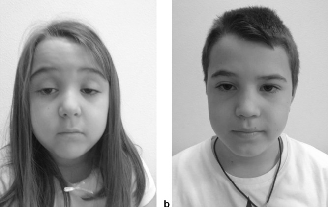
Patient 1 is a female, 4 years old, second child of third cousins healthy parents. Since first months she presented bilateral ptosis, difficulties in sucking and dysphagia, leading to ab ingestis pulmonitis at 8 months of age. Psychomotor development was normal, but mild weakness, unsteady gait and fatigability since early infancy, and slight fluctuations of symptoms with worsening during the evening, were referred. Neurological examination at 4 years of age, showed bilateral ptosis (Fig. 1a), facial muscles weakness, nasal voice, generalized hypotonia, muscle weakness more marked at lower limbs, positive Gower's sign, anserine ambulation, and running inability. AChR antibodies were absent. Electromyography revealed mild myopathic alterations; single fibre test was not performed because of patient's poor compliance. Muscle biopsy revealed aspecific myopathic features. At follow up, 6 months later, clinical evolution was stable. Parents noticed substantial improvement during treatment with salbutamol for a trivial respiratory disease; a post synaptic CMS was suspected. Oral treatment with salbutamol was started: a marked improvement of ptosis, weakness and activities of daily living was reported, without side effects.
Figure 1.
Patient 1 and patient 2 from family 1. The female presents marked bilateral ptosis (a) while the older brother shows slight bilateral ptosis (b).
Patient 2, now 10 years old, is the older brother of Patient 1. After his sister's hospitalization, he underwent neurological examination showing only slight bilateral ptosis (Fig.1b), and very mild lower limb girdle weakness (MRC: 4+).
All 12 exons of the CHRNE gene were sequenced following the already reported protocol (12). The analysis in family 1 revealed a previously identified c.475A>C mutation in exon 6 (p.T159P), in homozygous form (10). Genetic analysis in her older brother (Patient 2) revealed the same homozygous p.T159P mutation. The healthy parents carry one mutant allele each (Fig. 2).
Figure 2.
Families 1 and 2: genomic DNA of propositi (arrows) and family members. Closed symbols indicate affected individuals carrying two mutant alleles. Half shaded symbols represent asymptomatic carriers harbouring a single mutant allele. (a) Pedigree of the family 1: the p.T159P mutation is present in homozygous form in the sons, while in heterozygous form in carrier parents. (b) Pedigree of the family 2: the children present compound heterozygous mutations p.T159P and p.S235L, whereas the mother and the father are carrier of p.T159P and p.S235L, respectively.
Family 2
Patient 3 is a girl, now 20 years old, second child of healthy non consanguineous parents. Since first months of life she presented with bilateral ptosis and axial weakness. Subsequently ophthalmoparesis, diurnal fluctuations of ptosis, facial weakness, fatigability, difficulties in running and climbing stairs were reported. At age 4 years a diagnosis of CMS was reached. Clinical conditions remained stable during adolescence; electromyographic study revealed mild myopathic changes in upper and lower limbs and repetitive nerve stimulation (RNS) of facial nerve showed a pathological decremental response. At last observation, 20 years of age, she presented with marked bilateral ptosis, almost complete ophthalmoparesis, axial weakness, positive Gower's sign, and running inability.
Patient 4 is the younger brother of Patient 3, now 13 years old. He similarly showed since birth presence of ptosis, ophthalmoparesis and mild axial weakness, that remained stable during subsequent years. The electrophysiological findings were similar to those observed in his sister. Treatment with Pyridostigmine was uneffective.
Direct sequencing of the CHRNE gene in both siblings revealed the known p.T159P mutation associated with a second already reported mutation c.704C>T (p.S235L) in exon 7, both mutations were present in heterozygous form (10, 11). The mother was the carrier of p.T159P mutation, and the father of the p.S235L mutation (Fig. 2).
Table 1 summarizes the clinical aspects of the 4 patients of 2 families described in this study.
Table 1.
Clinical characteristics of the patients.
| Patient | Gender | Onset age/ symptoms |
Evolution | Cinical findings at diagnosis | Treatment/ response |
|
|---|---|---|---|---|---|---|
| Family 1 |
1 | Female | First months/ bilateral ptosis, difficulties in sucking and dysphagia |
Worsened | Bilateral ptosis, facial muscles weakness, nasal voice, generalized hypotonia, limb girdle weakness more marked at lower limbs, positive Gower's sign, anserine ambulation |
Salbutamol/effective |
| 2 | Male | Early infancy/ mild ptosis |
Stable | Mild bilateral ptosis and mild lower limb girdle weakness |
No treatment | |
| Family 2 |
3 | Female | First months/ ptosis |
Worsened in infancy/ stable in adolescence |
Bilateral ptosis, ophthalmoparesis, axial weakness, positive Gower's sign |
Pyridostigmine/ uneffective |
| 4 | Male | First months/ ptosis |
Worsened in infancy/ stable in adolescence |
Bilateral ptosis, ophthalmoparesis and mild axial weakness |
Pyridostigmine/ uneffective |
|
Genetic analysis
In patients all 12 exons and the adjacent splice donor and acceptor sequences of the CHRNE gene were sequenced, using genomic DNA isolated from blood, following the already reported protocol (5), while in their healthy parents the only mutated exons were analysed. The PCR products were purified by EuroSAP (Euroclone) and sequenced by bidirectional sequencing using the BigDye Terminator v3.1 Cycle Sequencing Kit (Thermo Fisher Scientific), on an 3130xl Genetic Analyzer (Thermo Fisher Scientific). The obtained sequences were analysed with SeqScape v.3.0 software (Thermo Fisher Scientific) and compared with reference wild-type sequence (GenBank CHRNE accession numbers: NM_000080.3).
Informed consent
Written informed consent for genetic analysis and for photos from children of Family 1 was obtained from probands' relatives and their familial members.
Discussion
All CMS patients share same clinical features, but age at onset, presenting symptoms, distribution of weakness, and response to treatment differ depending on the molecular mechanism that results from the genetic defect (1, 2).
We report four Italian CMS patients harboring CHRNE mutations and showing marked clinical variability, ranging from isolated mild ptosis to marked ptosis associated with ophthalmoparesis, facial and lower limbgirdle weakness (Patients 3 and 4) and intrafamily phenotypic variability in both families.
Genotype is different in the two families. In Family 1 the known p.T159P mutation is present in homozygous state whereas in Family 2 the p.T159P mutation is associated with the known p.S235L mutation (10, 11).
The p.T159P mutation is localized on the long cytoplasmatic N-terminal portion of the epsilon protein, which contains several loop regions which are critical for receptor function (6). Expression study showed that this mutation causes principally AChR deficiency (10, 14). The p.T159P mutation was previously identified in one CMS proband in compound heterozygous whit a second one (p.A411P) (10).
The p.S235L mutation is localized at the end of the membrane-spanning M1 domain of the epsilon protein, which joins covalently the four a-helical segments M1- M4 to the extracellular domain, hence this mutation may change this structural link (13). The p.S235L mutation was previously found in one Portuguese CMS patient associated with a second p.70insG mutation, presenting the clinical signs of ptosis, ophthalmoparesis, dysphagia, proximal weakness, and electrophysiological studies revealed a RNS decrement (11). Also in our patients the p.S235L mutation in compound heterozygous state seems to aggravate the phenotype.
In siblings of Family 1, harbouring p.T159P mutation in homozygous state, a marked clinical variability is evident. Marked phenotypic variability has been already described in two siblings with CMS due to mutations in MUSK gene: the sister was reported to be much more severely affected than the brother and a gender-effect was hypothesized since menstrual periods and fever worsened her symptoms (15). Although Patient 1 was in a prepuberal age, our report confirmed the hypothesis of a gender effect in the phenotypic expression. Our report underlines intrafamily clinical variability in spite of the same genotype and a possible gender effect; confirms the efficacy and safety of salbutamol in patients who harbour mutations in the epsilon of AchR (16).
Acknowledgements
We are very grateful to the parents for providing consent for this study.
References
- 1.Cruz PM, Palace J, Beeson D. Congenital myasthenic syndromes and the neuromuscular junction. Curr Opin Neurol. 2014;27:566–575. doi: 10.1097/WCO.0000000000000134. [DOI] [PubMed] [Google Scholar]
- 2.Engel AG, Shen XM, Selcen D, Sine SM. Congenital myasthenic syndromes: pathogenesis, diagnosis, and treatment. Lancet Neurol. 2015;14:420–434. doi: 10.1016/S1474-4422(14)70201-7. [DOI] [PMC free article] [PubMed] [Google Scholar]
- 3.Chang T, Cossins J, Beeson D. A rare c.183_187dupCTCAC mutation of the acetylcholine receptor CHRNE gene in a South Asian female with congenital myasthenic syndrome: a case report. BMC Neurol. 2016;16:195–195. doi: 10.1186/s12883-016-0716-y. [DOI] [PMC free article] [PubMed] [Google Scholar]
- 4.Bauché S, O'Regan S, Azuma Y, et al. Impaired presynaptic highaffinity choline transporter causes a congenital myasthenic syndrome with episodic apnea. Am J Hum Genet. 2016;99:753–761. doi: 10.1016/j.ajhg.2016.06.033. [DOI] [PMC free article] [PubMed] [Google Scholar]
- 5.O'Grady GL, Verschuuren C, Yuen M, et al. Variants in SLC18A3, vesicular acetylcholine transporter, cause congenital myasthenic syndrome. Neurology. 2016;87:1442–1448. doi: 10.1212/WNL.0000000000003179. [DOI] [PMC free article] [PubMed] [Google Scholar]
- 6.O'Connor E, Töpf A, Müller JS, et al. Identification of mutations in the MYO9A gene in patients with congenital myasthenic syndrome. Brain. 2016;139(Pt 8):2143–2153. doi: 10.1093/brain/aww130. [DOI] [PMC free article] [PubMed] [Google Scholar]
- 7.Shen XM, Scola RH, Lorenzoni PJ, et al. Novel synaptobrevin-1 mutation causes fatal congenital myasthenic syndrome. Ann Clin Transl Neurol. 2017;4:130–138. doi: 10.1002/acn3.387. [DOI] [PMC free article] [PubMed] [Google Scholar]
- 8.Lam CW, Wong KS, Leung HW, Law CY. Limb girdle myasthenia with digenic RAPSN and a novel disease gene AK9 mutations. Eur J Hum Genet. 2017;25:192–199. doi: 10.1038/ejhg.2016.162. [DOI] [PMC free article] [PubMed] [Google Scholar]
- 9.Salpietro V, Lin W, Vedove AD, et al. Homozygous mutations in VAMP1 cause a presynaptic congenital myasthenic syndrome. Ann Neurol. 2017;81:597–603. doi: 10.1002/ana.24905. [DOI] [PMC free article] [PubMed] [Google Scholar]
- 10.Wang HL, Ohno K, Milone M, et al. Fundamental gating mechanism of nicotinic receptor channel revealed by mutation causing a congenital myasthenic syndrome. J Gen Physiol. 2000;116:449–462. doi: 10.1085/jgp.116.3.449. [DOI] [PMC free article] [PubMed] [Google Scholar]
- 11.Mihaylova V, Scola RH, Gervini B, et al. Molecular characterisation of congenital myasthenic syndromes in Southern Brazil. J Neurol Neurosurg Psychiatry. 2010;81:973–977. doi: 10.1136/jnnp.2009.177816. [DOI] [PubMed] [Google Scholar]
- 12.Brugnoni R, Maggi L, Canioni E, et al. Identification of previously unreported mutations in CHRNA1, CHRNE and RAPSN genes in three unrelated Italian patients with congenital myasthenic syndrome. J Neurol. 2010;257:1119–1123. doi: 10.1007/s00415-010-5472-0. [DOI] [PubMed] [Google Scholar]
- 13.Unwin N. Refined structure of the nicotinic acetylcholine receptor at 4A resolution. J Mol Biol. 2005;346:967–989. doi: 10.1016/j.jmb.2004.12.031. [DOI] [PubMed] [Google Scholar]
- 14.Ohno K, Wang HL, Milone M, et al. Congenital myasthenic syndrome caused by decreased agonist binding affinity due to a mutation in the acetylcholine receptor epsilon subunit. Neuron. 1996;17:157–170. doi: 10.1016/s0896-6273(00)80289-5. [DOI] [PubMed] [Google Scholar]
- 15.Maggi L, Brugnoni R, Scaioli V, et al. Marked phenotypic variability in two siblings with congenital myasthenic syndrome due to mutations in MUSK. J Neurol. 2013 doi: 10.1007/s00415-013-7118-5. [DOI] [PMC free article] [PubMed] [Google Scholar]
- 16.Sadeh M, Shen XM, Engel AG. Beneficial effect of albuterol in congenital myasthenic syndrome with epsilon-subunit mutations. Muscle Nerve. 2011;44:289–291. doi: 10.1002/mus.22153. [DOI] [PMC free article] [PubMed] [Google Scholar]


