Abstract
In our previous study, we demonstrated the propagation of mouse-passaged scrapie isolates with long incubation periods (L-type) derived from natural Japanese sheep scrapie cases in murine hypothalamic GT1-7 cells, along with disease-associated prion protein (PrPSc) accumulation. We here analyzed the susceptibility of GT1-7 cells to scrapie prions by exposure to infected mouse brains at different passages, following interspecies transmission. Wild-type mice challenged with a natural sheep scrapie case (Kanagawa) exhibited heterogeneity of transmitted scrapie prions in early passages, and this mixed population converged upon one with a short incubation period (S-type) following subsequent passages. However, when GT1-7 cells were challenged with these heterologous samples, L-type prions became dominant. This study demonstrated that the susceptibility of GT1-7 cells to L-type prions was at least 105 times higher than that to S-type prions and that L-type prion-specific biological characteristics remained unchanged after serial passages in GT1-7 cells. This suggests that a GT1-7 cell culture model would be more useful for the economical and stable amplification of L-type prions at the laboratory level. Furthermore, this cell culture model might be used to selectively propagate L-type scrapie prions from a mixed prion population.
Introduction
Scrapie is a transmissible spongiform encephalopathy (TSE) of sheep and goats. TSE, also known as prion disease, is a family of fatal neurodegenerative disorders that affect both humans and animals [1]. The diversity of scrapie prions in sheep has been well investigated [2–6]. Currently, it has believed that sheep scrapie consists of more than 20 strains with different biological phenotypes, including different incubation periods; lesion profiles; biochemical properties of the disease-associated prion protein (PrPSc), a misfolded form of the cellular prion protein (PrPC); and neuroanatomical PrPSc distribution patterns in inbred mice [7–9]. Thus far, there have been no reports of scrapie prions being transmitted to humans. However, a panel of scrapie prions can be transmitted to several lines of transgenic mice overexpressing human PrPC [10]. More recently, scrapie prions were successfully transmitted to primates [11]. Thus, it is important to distinguish and analyze the biological and pathological differences among scrapie prions to determine whether any exhibit zoonotic potential.
We previously reported that two different mouse-passaged scrapie prion types were isolated from a single natural scrapie case (Kanagawa) of sheep by interspecies transmission to mice [4]. These isolates were designated as short-type (S-type) and long-type (L-type) based on their incubation periods and pathologies [4, 5]. Further, we reported that murine hypothalamic GT1-7 cells produced de novo PrPSc in response to L-type prions but not to S-type prions [5]. In this study, we demonstrated through mouse bioassays that the biological properties of L-type prions remained unchanged even after serial passages in GT1-7 cells. Our data suggest that GT1-7 cells can be used to selectively propagate L-type scrapie prions from a mixed prion population during the early transmission of sheep scrapie prions to mouse.
Materials and methods
Mouse-passaged sheep scrapie prions in this study
Three mouse-passaged field scrapie prion isolates (Tsukuba-2, Ka/O, and Ka/W) [3, 4] were used in this study. ICR/CD-1 mice infected with L-type prion isolates (Tsukuba-2 and Ka/O) exhibit clinical symptoms of polydipsia and polyuria with a long incubation period (approximately 280 days) and restricted PrPSc distribution in the brain stem [3, 5]. In contrast, mice infected with the S-type prion isolate (Ka/W) exhibit a short incubation period (approximately 150 days) and marked vacuolation and PrPSc distribution throughout the entire brain area [4].
Mouse inoculation
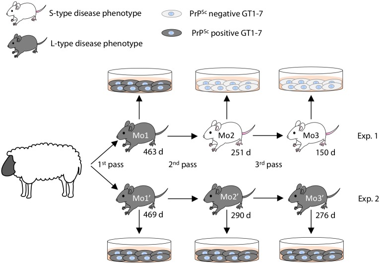
Animal experiments were carried out in strict accordance with the regulations outlined in the Guide for the Care and Use of Laboratory Animals of the National Institute of Animal Health (NIAH) and Guidelines for Proper Conduct of Animal Experiments, 2006 by the Science Council of Japan. All animal experiments were performed with the approval of the Animal Ethical Committee and the Animal Care and Use Committee of the NIAH (approval ID: 11–008 and 15–005). For the primary passage, the brain stem of a scrapie affected sheep (Kanagawa) was intracerebrally inoculated into mice (n = 16) as described in our previous report [4]. Two out of the 16 mice were chosen for the secondary passage (referred as Mo1 of Exp. 1 and Mo1′ of Exp. 2 in Fig 1, respectively). Likewise, two mouse brains were selected for the tertiary (Mo2 and Mo2′) and the quaternary (Mo3 and Mo3′) passages. The same brain homogenates were also exposed to GT1-7 cells. All intracerebral inoculations were performed under sevoflurane anesthesia. Three-week-old outbred ICR mice (SLC, Shizuoka, Japan) were inoculated intracerebrally with 20 μL of 10% mouse brain homogenates (2 mg brain equivalent) or GT1-7 cell homogenates (2 × 10⁵ cells). These homogenates were serially diluted before use. Mice were checked daily for the presence of clinical signs, such as emaciation, rough fur, hunched posture, and polyuria. All challenged mice were sacrificed under sevoflurane anesthesia when they presented with clinical disease. Survival period was determined as the time from inoculation to the clinical endpoint or sudden death, at which point brains were collected for analysis. The left hemispheres were immediately stored at −80°C for bioassay and western blot (WB) analysis, and the right hemispheres were fixed with 10% neutral-buffered formalin (pH 7.4) containing 10% methanol for histopathology and immunohistochemistry. Prion phenotypes were assessed in diseased mice according to survival period and/or neuroanatomical PrPSc distribution patterns [5].
Fig 1. Passage history of scrapie sources used in this study.
For the serial passage of scrapie prions in mice, the brain of a single mouse was chosen from the previous passage. The selected mice were named Mo1, Mo2, and Mo3 in Exp. 1 and Mo1′, Mo2′, and Mo3′ in Exp. 2. Mice that clinically and/or pathologically developed L-type disease are shown in grey, while those that developed S-type disease are shown in white. Survival days are indicated underneath each mouse. PrPSc-positive and -negative GT1-7 cells are shown in grey and white, respectively.
Exposure of GT1-7 cells to brain homogenates from scrapie-infected mice
Subcultured GT1-7 cells were exposed to brain homogenates of diseased mice as described previously [5]. Briefly, cells were grown on 6-well plates at 105 cells/well. After 2 days of incubation, the medium was replaced with 1 mL medium containing 20 μL of 10% (w/v) brain homogenate as mentioned in the above section. GT1-7 cells exposed to each brain homogenate are referred as GT/Mo1, GT/Mo2, GT/Mo3, GT/Mo1′, GT/Mo2′, and GT/Mo3′. For tissue cell culture endpoint titration assay, cells were exposed to 1 mL medium containing 20 μL of 10-fold serial dilutions of brain homogenate. Another 1 mL of plain medium was added to each well, and cells were incubated for an additional day. Then, the medium was removed, and the cells were washed three times with serum-free medium and seeded into new 6-well plates for the first passage (P1). For subsequent passages, cells were seeded in a new well at a 1:4 dilution every 4 or 5 days, and the remaining cells were cultured in T25 flasks for evaluation of PrPSc by WB. Each passage and assay was performed twice until the 10th passage (P10).
Extraction of PrPSc from brains and cells
Brain homogenates (20%) were mixed with an equal volume of detergent buffer containing 4% (w/v) Zwittergent 3–14 (Merck Japan, Tokyo, Japan), 1% (w/v) Sarkosyl (Sigma-Aldrich Japan, Tokyo, Japan), 100 mM NaCl, and 50 mM Tris-HCl (pH 7.6) and were incubated for 30 min with collagenase (Wako, Osaka, Japan; final concentration of 500 μg/mL) at 37°C. To detect PrPSc in GT1-7 cells, confluent cells were washed with phosphate-buffered saline (PBS) and then lysed with lysis buffer containing 50 mM Tris-HCl (pH 7.6), 150 mM NaCl, 0.5% (w/v) Triton X-100, 0.5% (w/v) sodium deoxycholate, and 2 mM EDTA. After 2 min of centrifugation at 6,500 ×g at 4°C, the supernatant was collected. Then, samples prepared from brains and cells were incubated for 30 min with proteinase K (PK) (Roche Diagnosis Japan, Tokyo, Japan; final concentration of 40 μg/mL) at 37°C. PK digestion was terminated with 2 mM 4-(2-aminoethyl) benzenesulfonyl fluoride hydrochloride (Pefabloc; Roche Diagnostics Japan). Samples were mixed with a 2-butanol/methanol mixture (5:1), and PrPSc was precipitated by centrifugation at 20,000 ×g for 10 min at 20°C. Pellets were resuspended in Laemmli sample buffer and subjected to WB.
Immunoprecipitation
Before immunoprecipitation, concentrations of infected brain homogenates were adjusted to contain approximately the same amount of PK-resistant PrPSc according to the intensities of the bands obtained by WB. Infected brain homogenates were mixed with brain homogenates prepared from PrP knockout mice to normalize the amount of total protein in each sample. Then, normalized brain homogenates were lysed with an equal volume of buffer containing 0.01% (w/v) Triton X-100, 0.01% (w/v) sodium deoxycholate, 100 mM NaCl, and 10 mM Tris-HCl (pH 7.6) for a final concentration of 5% (w/v). After vortexing, lysed brain homogenates were centrifuged at 500 ×g for 15 min at 4°C, and the supernatants were collected. Then, supernatants were diluted to a final concentration of 0.5% (w/v) with buffer containing 3% (w/v) Tween 20 and 3% (w/v) Triton X-100 in PBS. Protein G Dynabeads (Life Technologies Japan, Tokyo, Japan) were blocked with 4% (w/v) blocking solution (BlockAce; DS Pharma Biomedical, Osaka, Japan). Diluted supernatants (250 μL) were incubated with PrPSc-specific monoclonal antibody (mAb) 3H6 [12, 13] at a concentration of 1 μg/mL in 500-μL reaction volumes for 2.5 h with rotation at room temperature. Protein G Dynabeads (40 μL) were added and incubated for 2 h on the rotor at room temperature. Beads were washed five times with PBS containing 2% (w/v) Tween 20 and 2% (w/v) Triton X-100. Then, beads were resuspended in Laemmli sample buffer and subjected to WB.
Western blotting
Samples were electrophoresed on NuPAGE Novex 12% Bis-Tris gels with NuPAGE MOPS-SDS running buffer in accordance with the manufacturer’s instructions (Life Technologies, Carlsbad, CA, USA). Proteins were transferred onto an Immobilon-P membrane (Millipore, Billerica, MA, USA). The blotted membrane was incubated with anti-PrP mAb T2-HRP [14] at 4°C overnight. After washing twice with PBS containing 0.05% (v/v) Tween 20 (PBS-T), signals were developed with a chemiluminescent substrate (SuperSignal; Thermo Fisher Scientific K.K., Yokohama, Japan). For semi-quantitation, blots were imaged using a Fluorchem system (Alpha Innotech, San Leandro, CA, USA) and analyzed using image reader software (AlphaEaseFC; Alpha Innotech) according to the manufacturer’s instructions.
Histopathology and immunohistochemistry
Formalin-fixed brains were immersed in 98% formic acid to reduce infectivity and then embedded in paraffin wax. Serial sections were stained with hematoxylin and eosin for evaluation of neuropathological changes. After epitope retrieval, PrPSc immunocytochemistry was performed using the mAbs T1 [14] or SAF84 (Bertin Pharma, Montigny le Bretonneux, France) [15]. Immunoreactions were developed using the anti-mouse universal immunoperoxidase polymer [Nichirei Histofine Simple Stain MAX-PO (M); Nichirei, Tokyo, Japan] as the secondary antibody and were visualized with 3,3′-diaminobenzedine tetrachloride as the chromogen.
Results
Susceptibility of GT1-7 cells to scrapie prions during interspecies transmission
As shown in Fig 1, GT1-7 cells produced de novo PrPSc in response to exposure to primary passaged scrapie (Kanagawa)-infected mouse brains in two sets of transmission experiments (Fig 1, Exp. 1 and Exp. 2). However, GT1-7 cells did not produce de novo PrPSc following exposure to Mo2 and Mo3 brains in Exp. 1 (Fig 1, Exp. 1 and S1 Fig). In contrast, de novo PrPSc was detected in GT1-7 cells exposed to Mo2′ and Mo3′ brains in Exp. 2 (Fig 1, Exp. 2 and S1 Fig). To resolve the discrepancy between the two experiments, accumulated prions in the brains and GT1-7 cells were analyzed.
Heterogeneity of prions in mice infected with sheep Kanagawa scrapie isolate
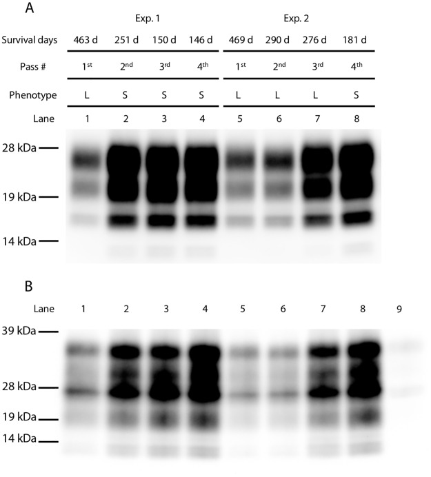
As shown in Exp. 2 of Fig 1 and Table 1, mice challenged with the brain homogenates of the sheep scrapie Kanagawa case exhibited L-type disease from the primary to 3rd passages. Because of the species barrier, survival periods in the primary passaged mice were long; therefore, disease phenotype was determined by the neuroanatomical PrPSc distribution patterns of their brains. In contrast to Exp. 2, the disease phenotype changed from L-type to S-type in Exp. 1 at the 2nd passage. As shown in Fig 2A, mice that developed S-type disease accumulated PK-resistant PrPSc more rapidly than mice that developed the L-type disease. Weak PrPSc signals bound to mAb 3H6 were detected in the primary passaged mouse brains in both experiments. The intensity of the PrPSc signal bound to mAb 3H6 was clearly elevated in the brains of mice that developed S-type disease at the 2nd and subsequent passages (Fig 2B, lanes 2–4). In contrast, PrPSc signals bound to mAb 3H6 in the brains of mice that developed L-type disease remained weak at the 2nd passage (Fig 2B, lane 6), but the signal intensities gradually increased at the 3rd passage (Fig 2B, lane 7). Lane 8 in Fig 2 shows a mouse whose disease phenotype changed from L-type to S-type at the 4th passage, and its survival period was shortened to 181 days. Both PK-resistant and mAb 3H6-specific PrPSc signal intensities of this mouse were clearly higher than those detected in the previous passage in Exp. 2. These results may indicate that heterogeneous prions were present in a single mouse challenged with the brain homogenates of scrapie-affected sheep Kanagawa.
Table 1. Infection of mice with L- and S-type prions with or without passaging in GT1-7 cells.
| Inoculum 1 | PrPSc in inoculum 2 | Subsequent passages in mice | |||
|---|---|---|---|---|---|
| n/n0 3 | Survival days 4 | Prion phenotype | |||
| Exp. 1 6 | Mo1 brain (463 days, L-type) 5 | + | 8/9 | 196 ± 31.6 | S-type |
| 1/9 | 289 | L-type | |||
| GT1-7 exposed to Mo1 brain | + | 9/9 | 278 ± 5.7 | L-type | |
| Mo2 brain (251 days, S-type) | + | 10/10 | 148 ± 8.9 | S-type | |
| GT1-7 exposed to Mo2 | – | 8/9 | 288 ± 36.8 | S-type | |
| 1/9 | 375 | L-type | |||
| Mo3 brain (150 days, S-type) | + | 5/5 | 143 ± 4.3 | S-type | |
| GT1-7 exposed to Mo3 brain | – | 9/9 | 246 ±17.6 | S-type | |
| Exp. 2 | Mo1’ brain (469 days, L-type) | + | 5/10 | 220 ± 7.2 | S-type |
| 5/10 | 290 ± 6.4 | L-type | |||
| GT1-7 exposed to Mo1’ brain | + | 9/9 | 285 ± 15.4 | L-type | |
| Mo2’ brain (295 days, L-type) | + | 1/11 | 195 | S-type | |
| 10/11 | 273 ±13.8 | L-type | |||
| GT1-7 exposed to Mo2’ brain | + | 9/9 | 277±14.7 | L-type | |
| Mo3’ brain (276 days, L-type) | + | 5/5 | 191 ± 7.1 | S-type | |
| GT1-7 exposed to Mo3’ brain | + | 10/10 | 280 ± 13.1 | L-type | |
1 Inoculum (brain or GT1-7 cells) was intracerebrally injected into ICR mice.
2 PrPSc accumulation was confirmed by western blotting: +, PrPSc positive;–, PrPSc negative.
3 Number of diseased mice out of total number of challenged mice.
4 Survival periods were determined as the time from inoculation to the clinical endpoint or death (mean survival days ± standard deviation).
5 Survival periods and prion phenotype of the individual mouse used for the inoculation are indicated.
6 Experiment numbers are the same as in Fig 1.
Fig 2. Monitoring PrPSc accumulation in diseased mice throughout passaging.
(A) Representative western blot of PrPSc in mouse brains at different passages. Experiment number, survival days, passage number, disease phenotype, and lane number are indicated on the top of the panel. Each lane included 0.2 mg brain equivalents. (B) Immunoprecipitation assay with mAb 3H6. Lane number is indicated on the top of the panel. In lane 9, uninfected brain homogenate was probed with mAb 3H6. In all other lanes, PrPSc was detected with mAb T2-HRP. Molecular markers are shown on the left side of each panel.
Disease phenotypes of mice challenged with PrPSc-positive and PrPSc-negative GT1-7 cells
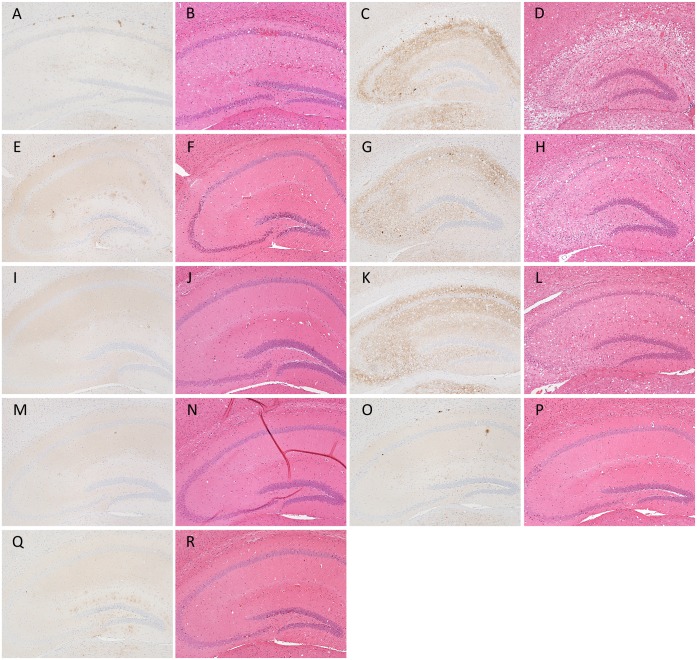
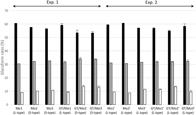
To determine whether the biological properties of L- and S-type prions were maintained after serial passages in GT1-7 cell culture, brain homogenates collected from the mice that developed L-type or S-type disease at each passage number were exposed to cells and subcultured until passage #10 (P10), and then these cells were intracerebrally injected into ICR mice. WB analysis demonstrated that GT1-7 cells exposed to the brain homogenates from mice that developed L-type disease harbored PrPSc, while the cells exposed to the brain homogenates from mice that developed S-type disease did not (S1 Fig). All mice challenged with PrPSc-positive GT1-7 cells developed L-type disease with a 100% attack rate (Table 1 and Fig 3) and had similar mean survival periods (277–285 days) when 10% brain homogenates were inoculated (273–290 days) as shown in Table 1. Of the mice challenged with PrPSc-negative GT1-7 cells, only one mouse developed L-type disease (2nd passage of Exp. 1 in Table 1 and Fig 3I and 3J), and the others developed S-type disease with longer incubation periods (2nd and 3rd passages of Exp. 1 in Table 1 and Fig 3G and 3H) as compared to those of mice inoculated with 10% brain homogenates of S-type-diseased mice (Ka/W column of mouse bioassay in Table 2). A clear difference in the glycoprofiles of PrPSc was not observed between mice challenged with mouse-passaged and cell-passaged prions (Fig 4). These results indicate that the biological features of L-type prions can be sustained in PrPSc-positive GT1-7 cells after serial passages. However, the PrPSc glycoprofiles of L-type prion-infected GT1-7 cells were clearly different from those of the brains (Fig 4 and S2 Fig), indicating the involvement of not only prion strains (isolates) but also host cellular factors in the PrPSc glycoform ratio.
Fig 3. Representative images of PrPSc deposition types associated with each prion phenotype in ICR mice.
Sections of the hippocampus were subjected to immunostaining of PrPSc and H&E staining. Typical PrPSc distribution patterns and histopathology of the hippocampus affected with L-type (A and B) and S-type (C and D) prions are shown. Sections of the hippocampi of mice inoculated with cell homogenates prepared from GT/Mo1 (E and F), GT/Mo2 (G, H, I and J), GT/Mo3 (K and L), GT/Mo1 ′ (M and N), GT/Mo2′ (O and P), and GT/Mo3′ (Q and R) were also subjected to immunostaining of PrPSc and H&E staining. Immunohistochemical detection of PrPSc was performed using the mAb SAF84.
Table 2. Comparison of the susceptibility of GT1-7 cells and ICR mice to L- and S-type prions.
| Assay | GT1-7 at P10 1 | ICR mice 2 | |||
|---|---|---|---|---|---|
| PrPSc accumulation | Survival periods (n/n0) 3 | ||||
| Isolate (Phenotype 4) | Ka/O (L-type) | Ka/W (S-type) | Tsukuba-2 (L-type) | Ka/W (S-type) | |
| Log10 dilution of inoculum 7 | -1 | + + 5 | – – | 288 ± 5.0 (4/4) | 147 ± 3.8 (5/5) |
| -2 | + + | N.D 6 | 326 ± 32.8 (5/5) | 155 ± 3.1 (5/5) | |
| -3 | + + | N.D | 355 ± 22.1 (3/3) | 168 ± 3.4 (5/5) | |
| -4 | + + | N.D | 461 ± 21.5 (4/4) | 177 ± 2.9 (5/5) | |
| -5 | + – | N.D | 496 (1/5) | 194 ± 4.8 (5/5) | |
| -6 | – – | N.D | >496 (0/5) | 246 ± 13.8 (5/5) | |
| -7 | N.D | N.D | >496 (0/5) | 203, 234, 235 (3/5) | |
| -8 | N.D | N.D | >496 (0/5) | >496 (0/5) | |
| Infectious units 8 | 106.7 TICD50 | N.D | 106.4 LD50 | 108.8 LD50 | |
1 PrPSc accumulation in GT1-7 cells was investigated at 10th passage (P10).
2 Mouse endpoint titration assay was conducted.
3 Survival periods are shown as mean survival days ± standard deviation. Number of diseased mice out of total number of inoculated mice is shown as n/n0.
4 Prion phenotypes are indicated.
5 PrPSc accumulation was confirmed by western blotting: +, PrPSc positive;–, PrPSc negative. Results of duplicate wells are indicated.
6 No data.
7 Mouse brain homogenate was diluted from 10−1 to 10−8. 10−1 indicates 10% brain homogenate.
8 Infectious units (TCID50 or LD50 per gram of brain equivalents) were calculated by Behrens-Karber’s formula.
Fig 4. Glycoform profiles of PrPSc in mice infected with GT1-7 cell culture-derived S-type and L-type prions.
PrPSc glycoform percentages of original mouse brains used for the infection of GT1-7 cells (Mo1, Mo2, Mo3, Mo1′, Mo2′, and Mo3′) were compared to those of mouse brains inoculated with GT1-7 cell homogenates (GT/Mo1, GT/Mo2, GT/Mo3, GT/Mo1′, GT/Mo2′, and GT/Mo3′). Results of GT1-7 cell homogenate-inoculated mice are shown as the mean ± standard deviation (n = 4). The PrPSc glycoprofile of GT/Mo2-inoculated mice in this graph shows the mean glycoform ratio of the mice that developed S-type disease. The inoculum (brain or GT1-7 cells) was intracerebrally injected into ICR mice. Experiment numbers are the same as in Fig 1.bar graph shows di-glycosylated (black columns), mono-glycosylated (gray columns), and unglycosylated (white columns) forms of PrPSc. L- and S-type at the bottom of each lane indicate the disease phenotypes. PrPSc was detected with mAb T2-HRP.
Endpoint titration assays of L-type and S-type scrapie prions in GT1-7 cells and ICR mice
To compare the susceptibility to L-type and S-type prions between conventional mice and GT1-7 cells, serial dilutions of L- or S-type prion-infected mouse brain homogenates were used to challenge to ICR mice and GT1-7 cells, respectively (Table 2). Because of the heterogeneity in mouse-passaged L-type scrapie isolates, the Tsukuba-2 mouse-passaged scrapie isolate, which was purified by limiting dilution, was used as a substitute for Ka/O isolates. GT1-7 cells exposed to a 10−5 dilution of brain homogenate from a mouse that developed L-type disease produced de novo PrPSc in 1 out of the 2 wells; however, they did not produce de novo PrPSc in response to a 10−6 dilution (Table 2 and S2 Fig). Prion titers of the tested brain were estimated at 106.7 TCID50/g of brain equivalents (eq). A bioassay using ICR mice challenged with the same dilutions demonstrated that 1 out of the 5 mice clinically and pathologically developed L-type disease in response to the injection of a 10−5 dilution, indicating that the tested brain contained 106.3 LD50/g of brain eq (Table 2). In contrast, 3 out of the 5 mice challenged with a 10−7 dilution of S-type prion-infected brain homogenate developed S-type disease, indicating that this brain contained 108.8 LD50/g of brain eq of S-type prions. However, no PrPSc was detected from GT1-7 cells exposed to the same dilutions or even from those exposed to a 10−1 dilution (Table 2).
Discussion
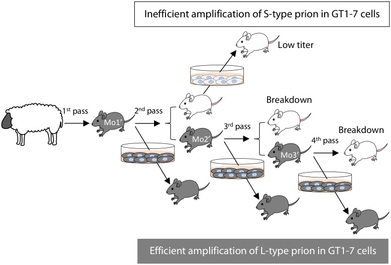
At the primary passage, extremely long incubation periods are commonly observed during interspecies transmission. This results from the species barrier. It is well known that incubation periods and neuropathological features are stabilized after a few passages in the same mouse strains [16]. In contrast, it has been reported that the scrapie disease phenotype, including incubation periods, clinical signs, and neuropathology, can suddenly change, even after serial passages in the same mouse strain [17, 18]. This phenomenon is called “breakdown”, and it was observed at the 4th passage of L-type prions in this study (Table 1 and Fig 5). Interestingly, we found that the PrPSc-specific mAb 3H6 preferred to be bound to the PrPSc that accumulated in mice that developed S-type disease according to immunoprecipitation assays. The amount of PrPSc bound to mAb 3H6 in a mouse brain selected from the 3rd passage of L-type prions (lane 7 in Fig 2B) tended to be higher than that of the brains selected from previous passages (lanes 5 and 6 in Fig 2B), and the mice inoculated with this brain exhibited breakdown at the 4th passage. These data strongly suggest that the proportions of L- and S-type prions in the brain are altered during serial transmission in mice. In addition, mouse endpoint titration assays for both S- and L-type prions enable us to speculate that S-type prions replicated faster than L-type prions in mice, as mice that received 102.8 infectious units of S-type prions developed the disease earlier than those that received 106.4 infectious units of L-type prions (Table 2). This indicates the possibility that S-type prions easily become dominant throughout subsequent passages in mice when both prions co-exist in a single mouse brain. Experimental mice are routinely used to amplify scrapie prions. We propose that close attention must be paid in order to maintain L-type prions in laboratory mice. In contrast to mice, GT1-7 cells efficiently amplify PrPSc and sustain high prion infectivity in response to L-type prions but not to S-type prions. Moreover, the biological properties of L-type prions remain unchanged after serial passages in GT1-7 cells. Taken together, this cell culture model could be convenient for the stable amplification of L-type prions.
Fig 5. Illustration of breakdown during the transmission of L-type prions in mice and effective amplification in GT1-7 cells.
Grey mice and cells indicate the presence of L-type prions, while white mice and cells indicate S-type prions. Mice exhibiting the L-type disease still harbor S-type scrapie prions, resulting in the appearance of mice with the S-type disease at every passage from P2 to P4. GT1-7 cells accumulate PrPSc in response to L-type prions and can be used to re-propagate mice with L-type prions by injecting mice with cell homogenates. In contrast, GT1-7 cells challenged with S-type prions do not accumulate PrPSc, but they do harbor very low levels of S-type prion infectivity.
It requires over 280 days to obtain the bioassay results regarding L-type prions even when mice are challenged with 10% brain homogenates. However, using PrPSc accumulation as an index, GT1-7 cell culture assay could produce results in approximately 60 to 70 days. In addition, the sensitivity of tissue cell culture assay to L-type prions was comparable to that of mouse bioassay. In contrast, the amplification of S-type prions in GT1-7 cells was extremely inefficient (Table 2). An unexpected finding of this study was that the mice challenged with PrPSc-negative GT1-7 cells developed S-type disease (2nd and 3rd passages in Exp. 1 of Table 1). These cells were subcultured more than 10 times before injection into mice. It is likely that the original brain homogenate exposed to GT1-7 cells was diluted to a level below the detection limit of the mouse bioassay; that is, we cannot exclude the possibility that very low levels of S-type prions continue to replicate even in PrPSc-positive GT1-7 cells. Further study will be needed to resolve this issue. Nevertheless, we here demonstrated that the susceptibility of GT1-7 cells to L-type prions was at least 105 times higher than that to S-type prions. We conclude that GT1-7 cells would be useful for the selective propagation of L-type prions. Recently, the relationship between the seeding activity of PrPSc and prion infectivity has been revealed through the use of protein misfolding cyclic amplification (PMCA) and real-time quaking-induced conversion (RT-QuIC) [19–22]. Therefore, combining the tissue cell culture assays developed here with PMCA and/or RT-QuIC may provide new opportunities for the purification of L-type prions from a mixed prion population.
It remains unclear why L-type prions are efficiently amplified in GT1-7 cells, unlike in the mouse brain. One possibility is that conformational differences between L- and S-type prions directly affect their effective replication in GT1-7 cells. Indeed, our immunoprecipitation assays may indicate that L-type prions have structurally different PrPSc conformations from S-type prions. The other possibility is that host-encoded cofactors, which may be lacking in GT1-7 cells, are required for the efficient replication of S-type prions. Previous reports done by others also support the selective susceptibility of neuronal populations in animals and in cell culture models to specific prion strains or isolates [23–27]. Our data may suggest the possibility that prion strain-specific cofactors are differentially expressed among various cells. Cofactors for prion replication, such as phospholipids and polyanions, have been mainly identified in in vitro PrPSc amplification studies [28–30]. Further study is required to clarify the cofactors that are clearly involved in the effective replication of specific prions in in vivo models.
Supporting information
Representative western blot of GT1-7 cells exposed to diseased brains derived from different mouse passages (1st to 3rd). Experiment number, passage number, protein loaded (μg), and lane number are indicated at the top of each lane. GT1-7 cells were collected at P10, and PrPSc was detected with mAb T2-HRP. Mr indicates the protein marker. Molecular markers are shown on the left. Three laboratory scrapie prion strains (22L, ME7, and Chandler) were used as positive and negative infection controls. As previously reported [5], GT1-7 cells were susceptible to 22L and Chandler but not to ME7.
(TIF)
Glycoform ratios of GT1-7 cells exposed to Mo3′ brain homogenates were calculated at passage #8 (P8) and #10 (P10). PrPSc was detected with mAb T2-HRP. The bar graph shows di-glycosylated (black columns), mono-glycosylated (gray columns), and unglycosylated (white columns) forms of PrPSc. Values are expressed as the mean ± standard deviation (n = 3).
(TIF)
Representative western blot of GT1-7 cells exposed to serial dilutions of brain homogenates from mice with the L-type disease phenotype at P10. Isolate name and prion phenotype of the inoculum are indicated at the top. The log10 dilution factor of the brain homogenate and the amount of protein loaded (μg) are also indicated at the top of each lane. PrPSc was detected with mAb T2-HRP. Molecular markers are shown on the left.
(TIF)
Acknowledgments
We are grateful to Naoko Tabeta, Ritsuko Miwa, Naomi Furuya, Junko Yamada, and the other animal laboratory staff of NIAH for technical and animal care assistance.
Data Availability
All relevant data are within the paper and its Supporting Information files.
Funding Statement
K. Miyazawa received the Grants-in-Aid from the Ministry of Health, Labor, and Welfare of Japan. K. Miyazawa, K. Masujin, HO, YM and TY received the basic research fund of National Agriculture and Food Research Organization (NARO).
References
- 1.Ryou C. Prions and prion diseases: fundamentals and mechanistic details. J Microbiol Biotechnol. 2007;17(7):1059–70. Epub 2007/12/07. . [PubMed] [Google Scholar]
- 2.Horiuchi M, Nemoto T, Ishiguro N, Furuoka H, Mohri S, Shinagawa M. Biological and biochemical characterization of sheep scrapie in Japan. Journal of clinical microbiology. 2002;40(9):3421–6. doi: 10.1128/JCM.40.9.3421-3426.2002 [DOI] [PMC free article] [PubMed] [Google Scholar]
- 3.Hirogari Y, Kubo M, Kimura KM, Haritani M, Yokoyama T. Two different scrapie prions isolated in Japanese sheep flocks. Microbiology and immunology. 2003;47(11):871–6. . [DOI] [PubMed] [Google Scholar]
- 4.Masujin K, Shu Y, Okada H, Matsuura Y, Iwamaru Y, Imamura M, et al. Isolation of two distinct prion strains from a scrapie-affected sheep. Archives of virology. 2009;154(12):1929–32. Epub 2009/10/31. doi: 10.1007/s00705-009-0534-2 . [DOI] [PMC free article] [PubMed] [Google Scholar]
- 5.Miyazawa K, Okada H, Iwamaru Y, Masujin K, Yokoyama T. Susceptibility of GT1-7 cells to mouse-passaged field scrapie isolates with a long incubation. Prion. 2014;8(4):306–13. doi: 10.4161/pri.32232 . [DOI] [PMC free article] [PubMed] [Google Scholar]
- 6.Carp RI, Callahan SM. Variation in the characteristics of 10 mouse-passaged scrapie lines derived from five scrapie-positive sheep. The Journal of general virology. 1991;72(Pt 2):293–8. doi: 10.1099/0022-1317-72-2-293 [DOI] [PubMed] [Google Scholar]
- 7.Bruce ME. TSE strain variation. British medical bulletin. 2003;66:99–108. . [DOI] [PubMed] [Google Scholar]
- 8.Fraser H, Dickinson AG. The sequential development of the brain lesion of scrapie in three strains of mice. Journal of comparative pathology. 1968;78(3):301–11. . [DOI] [PubMed] [Google Scholar]
- 9.Thackray AM, Hopkins L, Lockey R, Spiropoulos J, Bujdoso R. Emergence of multiple prion strains from single isolates of ovine scrapie. The Journal of general virology. 2011;92(Pt 6):1482–91. doi: 10.1099/vir.0.028886-0 . [DOI] [PubMed] [Google Scholar]
- 10.Cassard H, Torres JM, Lacroux C, Douet JY, Benestad SL, Lantier F, et al. Evidence for zoonotic potential of ovine scrapie prions. Nature communications. 2014;5:5821 doi: 10.1038/ncomms6821 . [DOI] [PubMed] [Google Scholar]
- 11.Comoy EE, Mikol J, Luccantoni-Freire S, Correia E, Lescoutra-Etchegaray N, Durand V, et al. Transmission of scrapie prions to primate after an extended silent incubation period. Scientific reports. 2015;5:11573 doi: 10.1038/srep11573 . [DOI] [PMC free article] [PubMed] [Google Scholar]
- 12.Ushiki-Kaku Y, Endo R, Iwamaru Y, Shimizu Y, Imamura M, Masujin K, et al. Tracing conformational transition of abnormal prion proteins during interspecies transmission by using novel antibodies. The Journal of biological chemistry. 2010;285(16):11931–6. Epub 2010/02/24. doi: 10.1074/jbc.M109.058859 . [DOI] [PMC free article] [PubMed] [Google Scholar]
- 13.Ushiki-Kaku Y, Shimizu Y, Tabeta N, Iwamaru Y, Ogawa-Goto K, Hattori S, et al. Heterogeneity of abnormal prion protein (PrP(Sc)) in murine scrapie prions determined by PrP(Sc)-specific monoclonal antibodies. The Journal of veterinary medical science / the Japanese Society of Veterinary Science. 2014;76(2):285–8. doi: 10.1292/jvms.13-0409 [DOI] [PMC free article] [PubMed] [Google Scholar]
- 14.Shimizu Y, Kaku-Ushiki Y, Iwamaru Y, Muramoto T, Kitamoto T, Yokoyama T, et al. A novel anti-prion protein monoclonal antibody and its single-chain fragment variable derivative with ability to inhibit abnormal prion protein accumulation in cultured cells. Microbiology and immunology. 2010;54(2):112–21. doi: 10.1111/j.1348-0421.2009.00190.x . [DOI] [PubMed] [Google Scholar]
- 15.Okada H, Sato Y, Sata T, Sakurai M, Endo J, Yokoyama T, et al. Antigen retrieval using sodium hydroxide for prion immunohistochemistry in bovine spongiform encephalopathy and scrapie. Journal of comparative pathology. 2011;144(4):251–6. doi: 10.1016/j.jcpa.2010.10.001 . [DOI] [PubMed] [Google Scholar]
- 16.Beringue V, Vilotte JL, Laude H. Prion agent diversity and species barrier. Veterinary research. 2008;39(4):47 Epub 2008/06/04. doi: 10.1051/vetres:2008024 . [DOI] [PubMed] [Google Scholar]
- 17.Bruce ME, Dickinson AG. Biological evidence that scrapie agent has an independent genome. The Journal of general virology. 1987;68(Pt 1):79–89. doi: 10.1099/0022-1317-68-1-79 [DOI] [PubMed] [Google Scholar]
- 18.Beck KE, Sallis RE, Lockey R, Vickery CM, Beringue V, Laude H, et al. Use of murine bioassay to resolve ovine transmissible spongiform encephalopathy cases showing a bovine spongiform encephalopathy molecular profile. Brain pathology. 2012;22(3):265–79. Epub 2011/09/17. doi: 10.1111/j.1750-3639.2011.00526.x . [DOI] [PMC free article] [PubMed] [Google Scholar]
- 19.Matsuura Y, Ishikawa Y, Bo X, Murayama Y, Yokoyama T, Somerville RA, et al. Quantitative analysis of wet-heat inactivation in bovine spongiform encephalopathy. Biochemical and biophysical research communications. 2013;432(1):86–91. doi: 10.1016/j.bbrc.2013.01.081 . [DOI] [PubMed] [Google Scholar]
- 20.Yoshioka M, Matsuura Y, Okada H, Shimozaki N, Yamamura T, Murayama Y, et al. Rapid assessment of bovine spongiform encephalopathy prion inactivation by heat treatment in yellow grease produced in the industrial manufacturing process of meat and bone meals. BMC veterinary research. 2013;9(1):134 doi: 10.1186/1746-6148-9-134 . [DOI] [PMC free article] [PubMed] [Google Scholar]
- 21.Wilham JM, Orru CD, Bessen RA, Atarashi R, Sano K, Race B, et al. Rapid end-point quantitation of prion seeding activity with sensitivity comparable to bioassays. PLoS pathogens. 2010;6(12):e1001217 Epub 2010/12/15. doi: 10.1371/journal.ppat.1001217 . [DOI] [PMC free article] [PubMed] [Google Scholar]
- 22.Takatsuki H, Satoh K, Sano K, Fuse T, Nakagaki T, Mori T, et al. Rapid and Quantitative Assay of Amyloid-Seeding Activity in Human Brains Affected with Prion Diseases. PloS one. 2015;10(6):e0126930 doi: 10.1371/journal.pone.0126930 . [DOI] [PMC free article] [PubMed] [Google Scholar]
- 23.Yokoyama T, Masujin K, Schmerr MJ, Shu Y, Okada H, Iwamaru Y, et al. Intraspecies prion transmission results in selection of sheep scrapie strains. PloS one. 2010;5(11):e15450 doi: 10.1371/journal.pone.0015450 . [DOI] [PMC free article] [PubMed] [Google Scholar]
- 24.Beck KE, Vickery CM, Lockey R, Holder T, Thorne L, Terry LA, et al. The interpretation of disease phenotypes to identify TSE strains following murine bioassay: characterisation of classical scrapie. Veterinary research. 2012;43(1):77 Epub 2012/11/03. doi: 10.1186/1297-9716-43-77 . [DOI] [PMC free article] [PubMed] [Google Scholar]
- 25.Oelschlegel AM, Geissen M, Lenk M, Riebe R, Angermann M, Schaetzl H, et al. A bovine cell line that can be infected by natural sheep scrapie prions. PloS one. 2015;10(1):e0117154 doi: 10.1371/journal.pone.0117154 . [DOI] [PMC free article] [PubMed] [Google Scholar]
- 26.Grassmann A, Wolf H, Hofmann J, Graham J, Vorberg I. Cellular aspects of prion replication in vitro. Viruses. 2013;5(1):374–405. doi: 10.3390/v5010374 . [DOI] [PMC free article] [PubMed] [Google Scholar]
- 27.Bruce ME. Scrapie strain variation and mutation. British medical bulletin. 1993;49(4):822–38. . [DOI] [PubMed] [Google Scholar]
- 28.Zurawel AA, Walsh DJ, Fortier SM, Chidawanyika T, Sengupta S, Zilm K, et al. Prion nucleation site unmasked by transient interaction with phospholipid cofactor. Biochemistry. 2014;53(1):68–76. doi: 10.1021/bi4014825 . [DOI] [PMC free article] [PubMed] [Google Scholar]
- 29.Supattapone S. Synthesis of high titer infectious prions with cofactor molecules. The Journal of biological chemistry. 2014;289(29):19850–4. doi: 10.1074/jbc.R113.511329 . [DOI] [PMC free article] [PubMed] [Google Scholar]
- 30.Ma J. The role of cofactors in prion propagation and infectivity. PLoS pathogens. 2012;8(4):e1002589 Epub 2012/04/19. doi: 10.1371/journal.ppat.1002589 . [DOI] [PMC free article] [PubMed] [Google Scholar]
Associated Data
This section collects any data citations, data availability statements, or supplementary materials included in this article.
Supplementary Materials
Representative western blot of GT1-7 cells exposed to diseased brains derived from different mouse passages (1st to 3rd). Experiment number, passage number, protein loaded (μg), and lane number are indicated at the top of each lane. GT1-7 cells were collected at P10, and PrPSc was detected with mAb T2-HRP. Mr indicates the protein marker. Molecular markers are shown on the left. Three laboratory scrapie prion strains (22L, ME7, and Chandler) were used as positive and negative infection controls. As previously reported [5], GT1-7 cells were susceptible to 22L and Chandler but not to ME7.
(TIF)
Glycoform ratios of GT1-7 cells exposed to Mo3′ brain homogenates were calculated at passage #8 (P8) and #10 (P10). PrPSc was detected with mAb T2-HRP. The bar graph shows di-glycosylated (black columns), mono-glycosylated (gray columns), and unglycosylated (white columns) forms of PrPSc. Values are expressed as the mean ± standard deviation (n = 3).
(TIF)
Representative western blot of GT1-7 cells exposed to serial dilutions of brain homogenates from mice with the L-type disease phenotype at P10. Isolate name and prion phenotype of the inoculum are indicated at the top. The log10 dilution factor of the brain homogenate and the amount of protein loaded (μg) are also indicated at the top of each lane. PrPSc was detected with mAb T2-HRP. Molecular markers are shown on the left.
(TIF)
Data Availability Statement
All relevant data are within the paper and its Supporting Information files.


