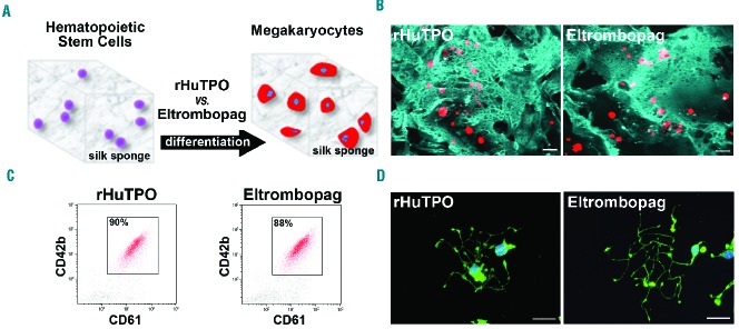
Figure 3.
Eltrombopag stimulates ex vivo megakaryocyte differentiation within the 3D silk bone marrow model. (A) Aqueous silk solution was mixed with salt particles and dried at room temperature overnight. After leaching out the salt, the resulting porous silk sponge was trimmed and sterilized. CD34+ hematopoietic stem cells from human cord blood were then seeded into the sponges and cultured for 13 days in the presence of 10 ng/mL recombinant human thrombopoietin (rHuTPO) or 200 ng/mL eltrombopag. (B) Confocal microscopy analysis of CD61+ megakaryocytes after 13 days of differentiation within the silk sponges under the different tested conditions (red=CD61; blue=silk; scale bar=100 μm). (C) Flow cytometry analysis of samples from the silk-sponges demonstrated an almost comparable percentage of CD61+CD42b+ cells between rHuTPO and eltrombopag. (D) Representative β1-tubulin staining of proplatelet formation by megakaryocytes collected from the silk sponge scaffold and seeded on fibrinogen. Both rHuTPO and eltrombopag supported normal proplatelet extension (green=β1-tubulin; blue=nuclei; scale bar=20 μm).

