Abstract
Acquisition of α2-6 sialoside receptor specificity by α2-3 specific highly-pathogenic avian influenza viruses (H5N1) is thought to be a prerequisite for efficient transmission in humans. By in vitro selection for binding α2-6 sialosides, we identified four variant viruses with amino acid substitutions in the hemagglutinin (S227N, D187G, E190G, and Q196R) that revealed modestly increased α2-6 and minimally decreased α2-3 binding by glycan array analysis. However, a mutant virus combining Q196R with mutations from previous pandemic viruses (Q226L and G228S) revealed predominantly α2-6 binding. Unlike the wild type H5N1, this mutant virus was transmitted by direct contact in the ferret model although not by airborne respiratory droplets. However, a reassortant virus with the mutant hemagglutinin, a human N2 neuraminidase and internal genes from an H5N1 virus was partially transmitted via respiratory droplets. The complex changes required for airborne transmissibility in ferrets suggest that extensive evolution is needed for H5N1 transmissibility in humans.
Keywords: Highly pathogenic avian influenza virus, Hemagglutinin, Host cell receptor, Virus attachment, H5N1, Sialoglycan, Host range, Pandemic virus emergence
Introduction
Circulation of H5N1 highly pathogenic avian influenza virus in Asia, Europe and Africa has resulted in more than 550 human infections since 2003, with a case fatality ratio exceeding 58% (WHO-CSR, 2011). This number of human cases is just a minute fraction of the large number of people likely exposed to H5N1 during thousands of H5N1 outbreaks reported in poultry (Kilpatrick, 2006; OIE, 2007; Wang et al., 2008), indicating that virus transmission from birds to humans is very inefficient. Likewise, further virus transmission from person-to-person is rare (Kandun et al., 2006; Wang et al., 2008). However, the few cases of suspected human-to-human spread are a threat to public health because of the possibility that modest functional changes in the virus may confer transmissibility in the human population causing a new influenza pandemic (Olsen et al., 2005; Yamada et al., 2006).
Efficient infection and transmission of avian influenza viruses between birds depend in part on the high affinity of the viral hemagglutinin (HA) for α2-3 sialoglycan receptors which are typically found on cells of the avian intestinal and respiratory tracts (Ito et al., 2000; Vines et al., 1998). In contrast, human influenza A viruses bind with high affinity to α2-6-sialoglycans and poorly if at all to α2-3-sialoglycans (Carroll and Paulson, 1985; Chen et al., 2011; Gambaryan et al., 1995, 2005b; Rogers and D’Souza, 1989; Rogers and Paulson, 1983; Stevens et al., 2006a). The fact that α2-6 specificity was exhibited by the earliest pandemic influenza viruses with HA genes of avian origin in 1957 (H2) and 1968 (H3) has suggested that an emerging H5N1 virus with preference for α2-6-sialoglycan receptors could also become highly infectious and contagious for humans (Chutinimitkul et al., 2010; Knossow et al., 2002; Maines et al., 2006, 2011; Matrosovich et al., 2000; Salomon et al., 2006). Several H5N1 clinical isolates from humans have acquired incremental α2-6-sialoglycan binding activity without concomitant loss of α2-3-sialoglycan binding (Yamada et al., 2006). However, lack of transmission to household members or healthcare personnel indicates that additional functional changes are a prerequisite for high infectivity and transmissibility in humans.
A significant challenge for pandemic preparedness is to develop a predictive understanding of the structural and functional changes in the avian H5 HA that could impart high transmissibility in humans, drawing from the H1, H2 or H3 HA subtypes from the three pandemics of the past century. Transmission studies in the ferret model revealed lack of respiratory droplet H5N1 transmission, even when reassorted with human influenza virus “internal genes”, suggesting inadequate HA and NA functions (Maines et al., 2006). Studies with the 1918 H1N1 pandemic virus showed that its transmissibility in a ferret model correlated with preferential binding of α2-6-sialoglycan by HA and simultaneous loss of α2-3-sialoglycan binding (Tumpey et al., 2007b). In the present study, we exploited glycomic and genetic approaches to identify variant H5N1 viruses with predominant binding to α2-6-sialoglycans. We used α2-6-sialoglycan-coated magnetic beads to drive in vitro evolution of a contemporary H5N1 virus in consecutive passages to identify spontaneous variants that increased α2-6 specificity. We suggest that in vitro-selected mutant viruses will help identify HA structural and functional changes associated with human-like receptor recognition that will be valuable for understanding the changes needed to affect receptor specificity changes in influenza, and for monitoring the evolution of H5N1 field viruses toward a potentially pandemic form.
Results
In vitro selection of H5N1 receptor variants
The current model for emergence of pandemic influenza viruses from avian sources postulates a switch in sialic acid receptor preference from α2-3 sialoglycan to α2-6 sialoglycan structures (Knossow et al., 2002; Rogers et al., 1985; Tumpey et al., 2007b). Mutations in the HA gene of recent H5N1 isolates collected from humans indicate that receptor specificity can evolve in this direction, either as a consequence of selection in the human host, or possibly in response to the abundance of α2-6 sialoglycan receptors in the intestine of terrestrial poultry, such as chicken and quail (Gambaryan et al., 2005a; Guo et al., 2007; Yamada et al., 2006). To examine the functional evolution of H5 HA receptor specificity in the laboratory, we implemented an in vitro receptor-binding virus enrichment approach that recapitulates in vivo selection. Synthetic 6′-sialyl (N-acetyl-lactosamine) (6′ SLN) was used as the affinity ligand mimicking the human receptor to capture spontaneous viral receptor variants on the surface of magnetic beads. Starting with a pool of 108 EID50 of A/Vietnam/1203/2004 (VN04 virus), we performed four consecutive rounds of in vitro binding and elution followed by isolation of 150 individual virus clones by plaque purification and characterization by sequence analysis.
Eleven of 150 (7.3%) virus plaques sequenced had one non-synonymous mutation in the HA1 region of the HA. These 11 variant virus plaques corresponded to 8 unique substitutions in the HA (Table 1). Substitutions at positions 187, 190, 196 and 227 (H3 numbering) were located in the receptor binding site (RBS) or its close proximity, whereas position 157 is located at the tip of the molecule. In contrast, positions 39, and 295 are located more distantly from this site toward the stem, whereas position 255 is predicted to be buried at the trimer interface. Some of the mutations near the receptor binding pocket had been previously documented in natural isolates of H5N1 viruses (Yamada et al., 2006). Interestingly, the Q196R mutation is found exclusively in three human isolates (A/Iraq/659/2006, A/Iraq/756/2006, and A/Vietnam/3028II/2004-clone3; GenBank accessions EU146876 and EU146878.1) and one avian isolate (A/chicken/Reshoty/02/2006; accession CY047483). The S227N mutation has also been reported primarily in human isolates (with the single exception of A/wild duck/Liaoning/8/2006; HM172084) and was noted to mediate increased α2-6-sialoglycan receptor binding (Shinya et al., 2005; Yamada et al., 2006). The higher frequency of H5N1 viruses with these changes among human cases is consistent with the positive selection of variants with higher avidity for α2-6 sialosides during replication in the human airway. The D187G and E190G mutations were not present in any other H5N1 isolates. However, previously reported mutations E190D and N186K at or near these positions markedly reduced α2-3 sialoside binding, consistent with a functional role for the E190D mutation (Stevens et al., 2006b; Yamada et al., 2006). Taken together, these results support the relevance of the in vitro evolution approach to examine the development of H5 mutations that increase avidity for the human-like α2-6 sialoglycan receptors.
Table 1.
In vitro selection of avian influenza variants with α2-6 sialoglycan beads.
| Mutation in HAa | H5 numbering | Frequency |
|---|---|---|
| Wildtype VN04 | – | 139 |
| A39T | 29 | 1 |
| K157Q | 153 | 1 |
| D187G | 183 | 1 |
| E190G | 186 | 1 |
| Q196R | 192 | 2 |
| S227N | 223 | 1 |
| E255K | 251 | 3 |
| H295Q | 292 | 1 |
Mutation is denoted as the amino acid in the wildtype virus (in single letter amino acid notation) followed by the position in the mature HA and the amino acid found in the variant virus. Positions are shown in the H3 numbering system, equivalent to A29, K153, D183, E186, Q192, S223, E251, and H292 in the H5 protein.
Differential interaction of H5N1 virus variants with sialoglycans
To analyze the receptor-binding phenotype of viruses selected by in vitro evolution we employed two binding assays with different characteristics; a sialoglycan-ELISA and a sialoglycan microarray. The sialoglycan ELISA measures multivalent binding of polymeric molecules containing isomers of sialyl-lactosamine (3′SLN-PAA as a model avian-type receptor and 6′SLN-PAA as human-type receptor) to virions adsorbed onto a polystyrene multiwell plate (Gambaryan and Matrosovich, 1992). The sialoglycan microarray detects the multivalent binding of virions to a diverse spectrum of naturally occurring sialylated oligosaccharides covalently-linked to a glass surface (Blixt et al., 2004; Stevens et al., 2006a).
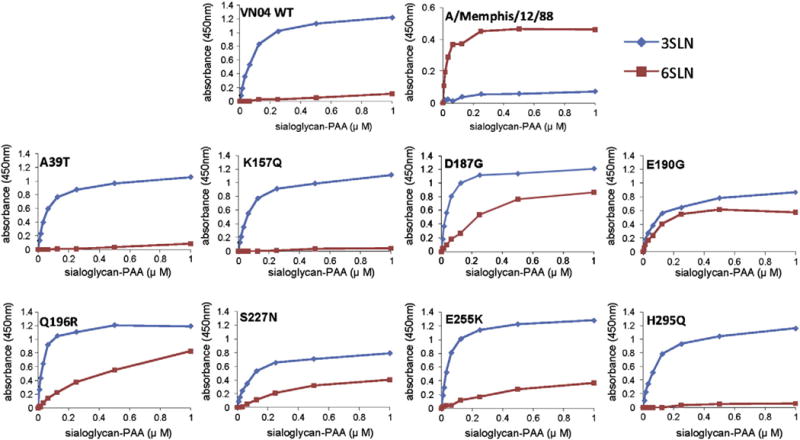
In the ELISA based assay, the parental wild-type (wt) VN04 virus showed an absolute preference for α2-3-sialoglycan (3′SLN) binding, opposite to the α2-6 preference of a seasonal H3N2 human virus, A/Memphis/12/1988 (Fig. 1). Five of the 8 variant VN04 viruses; D187G, E190G, Q196R, S227N, and E255K, had increased binding to 6′ sialyl-lactosamine relative to WT parental virus (Fig. 1). However, the binding to 6′SLN was observed at much higher sialoglycan concentrations compared to A/Memphis/12/1988 virus, indicating that the interactions were weak relative to the 3′SLN binding. Variants S227N, D187G, E190G, and Q196R had more significant gains in 6′ SLN binding than variant E255K. Since the 6′SLN-PAA used in this assay is the same sialoside conjugate used in the in vitro selection procedure, it is notable that this assay readily detects subtle increases in avidity to α2-6 sialosides.
Fig. 1.

Solid phase receptor binding assay of H3N2 and H5N1 variant viruses compared to wildtype. Binding of sialylglycopolymers 3′SLN-PAA (blue line) and 6′SLN-PAA (red lines) by influenza H3N2 and H5N1 viruses performed as described in Materials and methods section. The x axis denotes the concentration of biotinylated sialoglycan (μM) added to the wells, the y axis indicates the resulting absorbance reading. Data shown are representative from three independent binding experiments.
Except for the E255K mutant, the mutations showing increased binding to α2-6 sialosides are located within two distinct structural elements of the receptor binding site of HA; amino acid residues 187, 190, 196 are located in the 190-helix, whereas residue 227 occupies a central position in the 220-loop (Wiley and Skehel, 1987). These elements are critical determinants of interactions with galactose-2 and distal monosaccharides toward the reducing end of sialoglycan receptors (Ha et al., 2001; Stevens et al., 2006b). Therefore, structural changes of variant HAs are fully consistent with the observed receptor function differences. Mutations at positions 39, 157, and 295 did not affect sialoside binding specificity as compared to the parental virus, consistent with their more distant location relative to the receptor binding site.
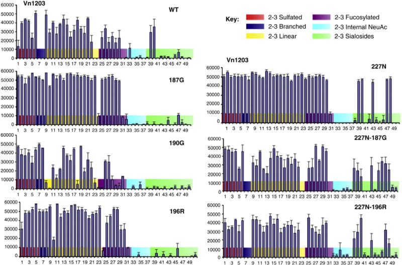
To achieve a better understating of the repertoire of glycans bound by variant viruses, we used a glycan microarray that contains 50 sialoglycans covalently attached to the glass surface (Blixt et al., 2004; Stevens et al., 2006a, 2006b). The in vitro selected variants with increased 6′ SLN sialoglycan binding in the ELISA assay were analyzed by glycan microarray. To rule out the contribution of mutations in other virion surface proteins, these variants were re-derived by reverse genetics (rg) and phenotypically re-confirmed by ELISA (data not shown). The glycan array showed that the wt VN04-rg virus has strong preference for α2-3-linked sialoglycans (Fig. 2), and weak binding to α2-6-linked sialoglycans, with significant binding to only two α2-6 sialylated biantennary glycans (Fig. 2, glycan #39–40). Compared to the wt VN04 virus, the three 190-helix variant viruses showed minimal increased binding to α2-6 sialoglycans, resembling the profile of wt VN04 (Fig. 2), whereas the S227N variant showed a substantial increase in binding to several additional α2-6-sialoglycans, including a long α2-6-sialylated di-N-acetyllactosamine (Fig. 2, glycan #47) and fucosylated α2-6 tri-N-acetyllactosamine (Fig. 2, glycan #48). Both of these sequences contain the 6′SLN sequence used in the in vitro selection experiments. However, increased binding affinity toward the shorter α2-6-linked sialyl-lactosamine (6′SLN) was not detected using the glycan microarray (glycans #44–45).
Fig. 2.

Glycan microarray analysis of H5N1 variant viruses and including dual mutations from previous pandemic viruses. Different types of sialoglycans on the array (x-axis) are indicated by colors; the structure of each numbered glycan is provided in Table S1. Vertical bars denote mean binding signal (fluorescence intensity) and T bar extensions indicate the standard error. Glycan microarray analysis of H5N1 variant viruses and including dual mutations from previous pandemic viruses. Different types of sialoglycans on the array (x-axis) are indicated by colors; the structure of each numbered glycan is provided in Table S1. Vertical bars denote mean binding signal (fluorescence intensity) and T bar extensions indicate the standard error.
The specificity analysis using both the ELISA and glycan microarray assays demonstrated that the in vitro selection protocol yielded variants with weak but increased avidity for binding α2-6 sialoglycans, with minimal changes in binding to α2-3-sialoglycans. The ELISA assay appeared to be more sensitive than microarray for picking up these changes, which we attribute to a lower binding detection threshold (Figs. 1–2). In contrast, the glycan microarray is valuable for simultaneously assessing the impact of the change in specificity with respect to the diverse α2-3 and α2-6 sialoglycan structures found in nature. Thus, both assays are valuable.
Receptor binding by viruses with dual 190-helix and 220-loop mutations
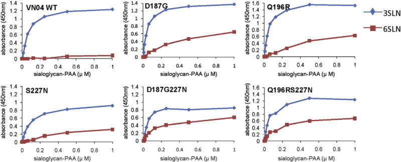
In the human pandemics of the previous century, at least two mutations in the receptor binding pocket have been implicated in changing receptor specificity from preferential recognition of α2-3-sialoglycans to recognition of α2-6 sialoglycans (Matrosovich et al., 2000; Rogers et al., 1983; Tumpey et al., 2007a). For recent human H5N1 isolates, two or more changes have been implicated in the increase α2-6-linked sialoside recognition and binding to human airway tissues (Chutinimitkul et al., 2010; Yamada et al., 2006). Based on the sporadic detection of HA S227N substitutions in human H5N1 isolates, we reasoned that any of the 3 in vitro-selected mutations in the 190 helix region of HA could be potentially co-selected (e.g. S227N-D187G), yielding dual mutations that would synergize to increase the specificity for α2-6 sialoglycans. To test this, double mutants pairing S227N with one of the three 190-helix mutations were generated by reverse genetics to analyze their receptor binding properties by sialoglycan ELISA and arrays. The sialoglycan-ELISA assay revealed a slightly additive effect of the pairwise mutations on sialoglycan binding; 227N-187G, and 227N-196R showed a slightly increased α2-6-linked sialyl-lactosamine (6′SLN) binding at lower glycan concentrations as compared to viruses with a single amino acid change (Fig. 3).
Fig. 3.

Solid phase receptor binding assay of H5N1 variant with dual mutations close to the receptor binding site. Binding of sialylglycopolymers 3′SLN-PAA (blue line) and 6′SLN-PAA (red lines) by influenza H5N1 variants was described in Materials and methods section. The x and y axis are as in Fig. 1. Data shown are representative from three independent binding experiments.
This increase in avidity was also evident in the glycan array analysis of 227N-187G, and 227N-196R double mutant viruses, which demonstrated binding to additional α2-6-sialoglycans, including sulfated α2-6-linked sialyllactosamine (Fig. 2, glycan #38) and α2-6 sialylated N, N′-diacetyllactosediamine (LacDiNac, #47); both glycans have been detected in human or animal tissue (Fig. 2) and are reported to be recognized by human influenza viruses (Crottet et al., 1996; Degroote et al., 2003; Stevens et al., 2010). During propagation of the VN04 227N-190G mutant in eggs or cell culture, additional mutations in HA1 were detected as subpopulations, suggesting that additional mutations are required for efficient growth of this double mutant in these laboratory hosts. Therefore, the receptor binding properties of this double mutant virus were not further characterized.
Taken together, these results suggested that a combination of mutations arising independently within the 190-helix and 220-loop of H5N1 HA molecule may synergize to enhance binding of α2-6 sialoglycan receptors. A combination of 187G, 196R and 227N mutations selected in this in vitro study revealed increased binding to a limited number α2-6 sialoglycans while retaining a strong preference for α2-3 sialoglycans.
Introduction of α2-6 specificity determinants of H2 and H3 pandemic viruses into H5N1 receptor variants
Previous studies showed that the mutations conferring α2-6 specificity in H2 and H3 human pandemic viruses (i.e. Q226L/G228S) produced only an incremental switch in α2-6 specificity when introduced into the H5 HA framework (Stevens et al., 2006a, 2008). The corresponding mutant H5N1 viruses retained a relatively high avidity for α2-3-receptors (Stevens et al., 2008), which is not found in human H1, H2 and H3 isolates. Additional amino acid changes present in circulating strains of H5N1 close to RBS or different mutations in the 220 loop were found to further polarize the receptor binding of 226L/228S mutant H5N1 viruses toward α2-6 glycans. In particular, incorporating an Arginine at position 193 and lack of glycosylation at Asparagine 158 found in the widely circulating clade 2.2 viruses showed the most prominent “receptor switch” (Stevens et al., 2008). Since the 196R variant identified by in vitro selection has also been identified among recent human isolates from clade 2.2 viruses, we evaluated the three in vitro selected 190-loop mutations 187G, 190G, and 196R for their effect on receptor specificity of viruses with an H5 clade 2.2 framework containing the 220-loop receptor switch mutations; A/egret/Egypt/1162/2006-226L/228S (EG06-226L/228S).
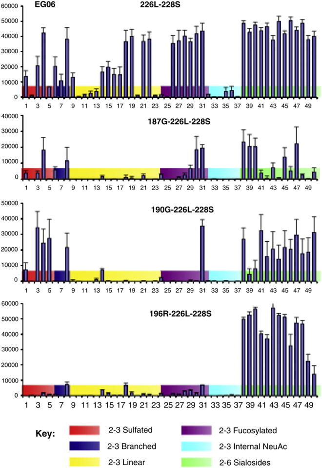
Glycan array analysis showed that introducing any one of the three 190 loop mutations into the EG06-226L226S framework significantly increased the switch to α2-6 specificity (Fig. 4). In particular, the EG06-187G/226L/228S or EG06-190G/226L/228S viruses showed a notable decreased affinity for α2-3 sialoglycans. Remarkably, the EG06-196R/226L/228S virus exhibited little α2-3 sialoglycan binding and strong binding to α2-6 sialoglycans (Fig. 4). Thus, unlike the natural human H5N1 isolates that exhibit weak binding to α2-6 sialosides and strong binding to α2-3 sialosides, this mutant exhibits a nearly complete switch in specificity, characteristic of the pandemic human influenza viruses of 1957 and 1968.
Fig. 4.

Glycan microarray analysis of clade 2 H5N1 viruses with dual mutations close to the receptor binding site. The color conventions and glycan key for the charts are described in Fig. 2.
In vivo studies in the ferret model
To establish the in vivo consequences of receptor specificity changes identified in vitro, these mutant H5N1 viruses were analyzed in a ferret model. Virus was administered to ferrets by intranasal inoculation and the following day infected animals were co-housed with naive ferrets to test for transmission by direct contact and adjacent to cages with additional uninfected ferrets to test for transmission via airborne respiratory droplets. After intranasal inoculation into ferrets, the wild type rg EG06 virus reached a peak titer ~104 pfu/ml in nasal washes at 3 days post infection (data not shown). No infection of contact ferrets was detected by virus shedding or seroconversion (Table 2), as reported in earlier studies with H5N1 viruses (Maines et al., 2006). In contrast, when the ferrets were infected with EG06-196R/226L/228S virus, viral shedding was detected in the nasal secretions of direct contact ferrets, although no infection was evident in ferrets exposed to respiratory droplets (Table 2). The results suggest that a measurable increase in H5N1 virus transmission via direct contact in ferrets can be imparted by modification of HA molecules approximating a human virus receptor specificity.
Table 2.
Presence of virus in nasal secretions and seroconversion of inoculated ferrets and their contacts.
| Virus | Inoculated ferrets
|
Direct contact ferrets
|
Aerosol contact ferrets
|
|||
|---|---|---|---|---|---|---|
| Virus detection | Sero-conversiona | Virus detection | Sero-conversion | Virus detection | Sero-conversion | |
| EG06 WT | 2/2b | 2/2 | 0/2 | 0/2 | 0/2 | 0/2 |
| EG06-196R/226L/228S | 2/2 | 2/2 | 2/2 | 2/2 | 0/2 | 0/2 |
| c EG06-196R/226L/228S-H3N2 NA | 2/2 | 2/2 | 2/2 | 2/2 | 1/2 | 1/2 |
| c EG06-196R/226L/228S-H1N1 NA | 2/2 | 2/2 | 2/2 | 2/2 | 0/2 | 0/2 |
Hemagglutination-inhibition antibody titer of the convalescent serum sample is ≥4-fold greater than pre-inoculation sample.
Number of ferrets testing positive/total ferrets tested.
Denotes internal genes from A/Vietnam/1203/2004 (H5N1).
Efficient human-to-human transmission of influenza virus re-quires not only proper receptor binding, but also a balanced neuraminidase activity and other viral replication characteristics determined by the replicative machinery encoded by the internal genes. To investigate their role in transmission, the EG06 H5 HA mutant was placed into a VN04 virus background by reverse genetics. The N1 neuraminidase of H5N1 is predicted to maintain α2-3 sialoside specificity as a result of its long co-evolution with H5 HA molecules that bind α2-3 sialoglycans in avian species (Raab and Tvaroska, 2010). A reverse genetics virus with HA of EG06-196R/226L/228S, the N2 neuraminidase from a human seasonal H3N2 virus, A/Brisbane/10/2007, and 6 internal genes from a clade 1 H5N1 virus, VN04, known to replicate efficiently in ferrets (Maines et al., 2011), was generated and evaluated in the ferret transmission model. As shown in Table 2, both direct contact ferrets that were housed in the same cage with inoculated ferrets became infected, as evidenced by virus shedding and seroconversion results. In addition, viral shedding was also detected in one of two ferrets housed in adjacent cages and exposed only via respiratory droplets. An analogous reverse genetics virus with the N1 NA from a human seasonal H1N1 virus, A/Brisbane/59/2007 resulted in efficient direct contact but not respiratory droplet transmission (Table 2).
The results of these investigations indicate that combinations of mutations surrounding the receptor binding pocket of the H5 HA are able to switch receptor specificity to one that is strongly α2-6 specific, characteristic of human influenza viruses, and that this receptor specificity change is sufficient to increase transmissibility between ferrets. However, lack of efficient transmission by a virus that contained a human virus neuraminidase demonstrates that a switch to α2-6 specificity may be required, but is not sufficient for emergence of a pandemic virus from H5N1. Rather, efficient transmission of H5N1 or a reassortant virus containing H5N1 genes will require optimization of both the HA, NA and internal genes.
Discussion
Highly pathogenic avian influenza viruses (H5N1) have become enzootic in avian populations in many countries, e.g. Vietnam, Indonesia, and Egypt, where they continue to evolve rapidly (Melidou, 2009; Sims, 2007). Despite very high case-fatality proportions in some countries, human cases remain sporadic and isolated, suggesting that efficient human infection and transmissibility depend on additional functional changes in the virus. Poor binding to α2-6-linked receptors is believed to be a major barrier preventing efficient human infection and transmissibility. However, extensive circulation of H5N1 viruses in terrestrial poultry and sporadic infections in mammals have raised the concern of evolution toward α2-6-linked human-like receptor specificity as observed in the H9N2 viruses circulating in poultry and pigs (Song et al., 2009; Takano et al., 2009; Wan and Perez, 2007; Wan et al., 2008). A powerful approach to study this process was reported by Rogers et al. (1985), who used in vitro methodologies to select a virus with human receptor specificity from an avian precursor, mimicking in vivo adaptation to humans. In this study, we applied a similar protocol to understand the potential of H5N1 viruses to increase the chances of infecting and transmitting in humans. We opted for a 6′ SLN glycan as receptor analog for affinity selection because wt H5N1 viruses exhibited excellent binding to 3′SLN but not 6′SLN with the critical α2-6 linkage (Stevens et al., 2006a).
This in vitro evolution study led to the identification of four amino acid substitutions located in the 190-helix and 220-loop of the HA receptor binding site, two of which (196R and 227N) have been found almost exclusively in H5N1 viruses isolated from human cases, e.g. in A/Vietnam/3028II/2004 clone 3 (Yamada et al., 2006). A recent study (Watanabe et al., 2011) showed that a substitution from Q to H at position 192 (i.e. Q196H in the corresponding H3 numbering used in this study) in the HA of clade 2.2.1 H5N1 viruses circulating in Egypt also revealed increased binding to α2,6 sialoglycans. These substitutions mediated minimal changes in α2-3-sialoglycan binding and limited increases in α2-6-sialoglycan binding. Notably, most receptor binding changes were detected by the ELISA assay and not in glycan microarrays, demonstrating that the ELISA based assay may be more sensitive at detecting slight changes in avidity to α2-6-sialoglycans. These studies confirmed previous observations regarding the detection of receptor variants by the sialoglycan-ELISA method (Yamada et al., 2006). However, as shown previously, these subtle receptor binding changes were not sufficient for efficient infection and transmission of H5N1 viruses bearing 196R and 227N mutations in ferrets, an animal model believed to exhibit similar requirements to humans for influenza receptor specificity (Yen et al., 2007). Reverse genetics generated viruses bearing pairwise combinations of independently selected 187G and 196R with 227N mutations revealed incremental increases in binding to α2-6 sialoglycans, but the viruses retained a strong preference to α2-3 sialoglycans. These modest changes of receptor specificity are in stark contrast with the very dramatic switch observed for the avian viruses responsible for the pandemics of 1957 and 1968 (Matrosovich et al., 2000; Pappas et al., 2010; Rogers and Paulson, 1983), demonstrating that single or even double mutations in the H5 framework are not sufficient to effect the switch to the ‘human type’ receptor specificity.
Having identified several mutations by in vitro evolution that might contribute to a switch in receptor specificity, we assessed their potential impact on the specificity of currently circulating clade 2 H5N1 viruses. The clade 2.2 H5N1 viruses have become enzooting in poultry in many Asian countries and Egypt, accounting for a majority of documented human infections in recent years (WHO-CSR, 2011). An additional motivation for choosing a clade 2.2 virus to evaluate the Q226L/G228S mutations was based on the identification of the Q196R in the HA of human isolates of this clade. In a previous investigation, multiple clades of H5N1 viruses bearing the critical H2 and H3 mutations imparting human-like receptor binding (Q226L/G228S) demonstrated a decreased but still significant affinity for α2-3 sialoglycans (Stevens et al., 2008). Interestingly, when these 220-loop mutations were combined with a 190-helix mutation (Q196R) yielding the EG06-196R/226L/228S triple mutant virus (clade 2.2) we observed a major loss of α2-3 sialoglycan binding, while retaining strong binding to α2-6 sialoglycans, a pattern similar to that observed in the 1957 and 1968 pandemic viruses (Matrosovich et al., 2000; Pappas et al., 2010; Rogers and Paulson, 1983).
Previous studies indicated that H5N1 viruses with altered receptor specificity may have reduce fitness and compromised replication and/or transmission efficiency in vivo (Maines et al., 2011). The replication characteristics of the EG06-196R/226L/228S triple mutant virus in a ferret model resulted in increased transmission efficiency to direct contact animals housed in the same cage. However, a low level of droplet transmission to ferrets in adjacent cages was only detected when the EG06-196R/226L/228S HA was inserted into the backbone of the highly virulent A/Vietnam/1203/04 virus carrying the NA of a human H3N2 virus (A/Brisbane/10/2007). These data reinforce the complex functional requirements for in vivo generation of respiratory droplets that can infect other animals. Although still poorly understood, such requirements may include functional compatibility between HA and NA resulting in efficient virus attachment to cells of the upper respiratory tract and subsequent release of progeny virions (Wagner et al., 2002).
Related studies reported by Maines et al. analyzed reassortant viruses that contained the internal genes of an H5N1 virus that was not transmissible in ferrets and the HA and NA genes from a human H3N2 virus that was transmissible (Maines et al., 2006). Despite the efficient replication of the reassortant viruses in the upper airway of ferrets, these viruses exhibited poor transmission by direct contact, and failed to transmit via respiratory droplets despite having a human virus HA and NA (Maines et al., 2006). The lack of increased transmission with the addition of the human HA and NA could be due to many factors. However, it is worth noting that we selected the internal genes of the VN04 H5N1 virus for our reassortant viruses because of its efficient replication in inoculated ferrets, yet it exhibited inefficient transmission by direct contact and no transmission by respiratory droplets to contact ferrets. In contrast, there was reduced virulence in inoculated ferrets with the EG06 strain (Maines et al., 2011). Thus, the selection of internal genes that support efficient replication of the virus may be important for revealing phenotypic changes affecting early events in the replication cycle, such as those mediated by the HA and NA.
In conclusion, this in vitro evolution study led to identification of viruses with amino acid changes that impart increased α2-6 sialoside receptor binding. One of these mutations introduced in a clade 2.2 H5N1 virus contributed toward increased respiratory droplet transmission between ferrets only if combined with mutations observed in previous pandemic viruses, as well as a human N2 NA and the internal genes from a clade 1 H5N1 virus. The complex genetic changes required by a clade 2.2 H5N1 virus to reach a low level of transmissibility in ferrets would indicate that considerable functional evolution is still required for acquisition of transmissibility in humans.
Materials and methods
Viruses and cells
Highly pathogenic H5N1 viruses, A/Vietnam/1203/2004 (clade 1) and A/egret/Egypt/1162/2006 (clade 2.2) herein VN04 and EG06 respectively, were obtained from the WHO Global Influenza Surveillance Network (WHO/FAO/OIE, 2007). The viruses were propagated in the allantoic sac of 10–11 day embryonated chicken eggs for 24 h at 37 °C and aliquoted stocks were stored at −80 °C. Viral infectivity was determined by plaque assay on MDCK cells or by endpoint dilution in chicken eggs as described (WHO, 2002). Madin–Darby canine kidney (MDCK) cells (cat # CCL-34) and Human Embryonic Kidney 293 (HEK293) cells (cat # CRL-1573) were obtained from the American Type Culture Collection and propagated in DMEM with 10% fetal bovine serum. All experiments using H5 subtype highly pathogenic avian influenza viruses were conducted under biosafety level 3 containment (http://www.cdc.gov/OD/ohs/biosfty/bmbl5/bmbl5toc.htm) including enhancements required by the U.S. Department of Agriculture and Select Agent Program (Richmond and Nesby-O’Dell, 2002), available at: http://www.access.gpo.gov/nara/cfr/waisidx_06/9cfr121_06.html.
Receptor specificity variant virus selection
Neu5Acα2-6Galβ1-4GlcNAcβ-SpNH-polyacrylamide-30kD (6′SLN-PAA) was prepared as previously described (Collins et al., 2006). The 6′SLN-PAA-coated matrix was prepared by incubating streptavidin magnetic beads (Dynabeads M280) with 6′SLN-PAA-Biotin and subsequently removing the unbound glycans by buffer exchange, as described previously (Collins et al., 2006). A VN04 virus pool containing 108 EID50 was incubated with 107 α2-6-sialoglycan magnetic beads in a 0.45 ml volume for 10 min at 25 °C in DMEM/0.4% BSA and rinsed extensively to remove unbound virus. Virus attached to beads was eluted by incubation with Clostridium perfringens sialidase (1 U/ml) for 3 h at 37 °C followed by magnetic separation. Released virus was amplified by propagation in MDCK cells as described, except that the adsorption step was done at 4 °C for 15–20 min followed by culture medium exchange. The binding, elution, amplification cycle was repeated 4 times to allow for sufficient expansion of variants. Isolation of clonal virus populations entailed identification of well-isolated plaques formed on MDCK monolayers under agarose as described previously (Tobita et al., 1975). A total of 150 well-separated plaques were individually selected from the material eluted after the fourth cycle of binding and elution. Viral stocks from each clone were propagated in MDCK cells.
RT-PCR, nucleotide sequencing
Total RNA was extracted from tissue culture supernatant or allantoic fluid using the QiAmp Viral RNA minikit (Qiagen) and reverse transcribed to cDNA using a proprietary one-step reaction system (RT-PCR kit Qiagen) and primers. PCR amplification of the coding region of the influenza HA gene was performed as previously described using universal gene-specific primer sets (primer sequences available on request). The resulting DNA amplicons served as templates for automated sequencing on an Applied Biosystems 3100 automated DNA sequencer using cycle sequencing dye terminator chemistry (ABI). Mixed base calls (RNA quasispecies) were evaluated by comparing chromatographs for both strands of the amplicon cDNA.
Virus receptor specificity analysis by sialoglycan ELISA
The procedure was adapted from Gambaryan et al. (Gambaryan and Matrosovich, 1992). Briefly, polystyrene microplates were coated with fetuin (10 μg/ml) for 16 h. After four rinses with washing buffer (0.01% Tween 80 in 0.2× PBS), plates were incubated with influenza virus (32 HA units/50 μl in PBS). After incubation at 4 °C overnight, the plates were rinsed four times with washing buffer. Wells were filled with biotinylated 3′-SLN-PAA or 6′-SLN-PAA (Consortium for Functional Glycomics, http://www.functionalglycomics.org) in washing buffer, supplemented with 1 μM zamanivir (GSK, Middlesex UK) and 0.02% bovine serum albumin and incubated for 2.5 h on ice. Assay plates were again rinsed four times with washing buffer, and incubated with streptavidin-horseradish peroxidase (HRP)-conjugate at 4 °C for 1 h. Assay plates were rinsed five times with washing buffer and incubated with TMB/hydrogen peroxide substrate (BD Pharmingen) for 20 min at room temperature; absorbance was determined at 450 nm after stopping the reaction with 0.1 N HCl. Assays were repeated three times.
Glycan specificity of virions by microarray analysis
Analysis of the receptor specificity of influenza virus using glycan microarrays was done essentially as described previously (Blixt et al., 2004; Stevens et al., 2006a). Custom microarrays for influenza research were produced for the CDC on NHS activated glass slides (Schott Nexterion, Mainz, Germany) using a glycan library provided by the Consortium for Functional Glycomics (Table S1 for list of glycan structures). Virus preparations were diluted to 1 ml into phosphate buffered saline containing 3% (w/v) bovine serum albumin (PBS-BSA) to HA titers of 128. Virus suspensions were applied to slides and subsequently incubated in a closed container with gentle agitation for 1 h. Unbound virus was washed off by dipping slides sequentially in PBS. The slides were then overlaid with corresponding primary antibodies (sheep anti-VN04) diluted in PBS-BSA, followed by incubation with biotinylated secondary antibody for 30 min. Slides were washed briefly with PBS as above followed by application of the avidin-Alexa fluro 635 conjugates. All steps were performed at 4 °C. After drying the slides in a steam of nitrogen they were immediately scanned (ProScanArray HT slide scanner, Perkin Elmer or Genepix Molecular Device) followed by image analysis with ImaGene 6.1 software (Biodiscovery, Inc., El Segundo, CA).
Derivation of viruses by reverse genetics
Reassortant viruses were generated from plasmids by a reverse genetics approach (Fodor et al., 1999; Hoffmann and Webster, 2000; Neumann and Kawaoka, 1999). To generate viruses with amino acid changes in HA, mutations were introduced using an overlap extension PCR approach (Urban et al., 1997). All new plasmids were sequenced to verify the absence of unwanted mutations. Viruses derived by plasmid transfection of HK293 cells were propagated in MDCK or eggs. The HA1 of resulting virus stocks were sequenced to detect the emergence of revertants during amplification.
Infections in the ferret model
Male ferrets, 6–12 months of age (Triple F Farms, Sayre, PA) serologically negative by hemagglutination inhibition (HI) assay for currently circulating human seasonal influenza viruses (Matsuoka et al., 2009) were used to assess viral transmissibility in vivo. Two animals were inoculated intranasally with 106 PFU of virus diluted in 0.5 ml PBS and housed in individual cages with perforated side-walls as described previously (Maines et al., 2006). The following day (1 day post-inoculation), one additional “direct contact” ferret was housed in the same cage with each inoculated ferret. Two “respiratory droplet contact” ferrets were placed in adjacent cages (one per cage) with a perforated side-wall leaving a 5 cm gap between cages. Nasal washes were collected as previously described (Zitzow et al., 2002) from all ferrets on alternating days for 11 days and the concentration of viable virus in these samples was determined by endpoint titration in MDCK cells. Pre- and post-exposure ferret sera collected 14 days after virus inoculation were analyzed in HI assay to assess seroconversion. The HI assay was performed with 0.5% turkey red blood cells as previously described (WHO, 2002). Animal studies were conducted under a protocol approved by the CDC Animal Care and Use Committee.
Supplementary Material
Acknowledgments
Glycan microarrays were produced for the Centers for Disease Control using the glycan library of CFG developed under funding by the National Institute of General Medical Sciences, Grant GM62116. We thank Dr. Kenneth Earhart (NAMRU-3, Cairo), the Ministry of Public Health of Egypt and Dr. Le Quynh Mai, National Institute of Hygiene and Epidemiology, Hanoi, Vietnam, for providing H5N1 viruses. We thank Jacqueline Katz for providing A/Memphis/12/1988 (H3N2) virus seed and M. Jaber Hossain for help and advice on animal studies. We wish to acknowledge the Consortium for Functional Glycomics Grant GM62116 for synthetic glycans used in binding assays. We also thank the Centers for Disease Control Animal Resources Branch for exceptional animal care and Anna Crie for assistence in manuscript preparation. The findings and conclusions in this report are those of the authors and do not necessarily represent the views of the Centers for Disease Control and Prevention or the Agency for Toxic Substances and Disease Registry.
Footnotes
Supplementary materials related to this article can be found online at doi:10.1016/j.virol.2011.10.006.
References
- Blixt O, Head S, Mondala T, Scanlan C, Huflejt ME, Alvarez R, Bryan MC, Fazio F, Calarese D, Stevens J, Razi N, Stevens DJ, Skehel JJ, van Die I, Burton DR, Wilson IA, Cummings R, Bovin N, Wong CH, Paulson JC. Printed covalent glycan array for ligand profiling of diverse glycan binding proteins. Proc Natl Acad Sci U S A. 2004;101:17033–17038. doi: 10.1073/pnas.0407902101. [DOI] [PMC free article] [PubMed] [Google Scholar]
- Carroll SM, Paulson JC. Differential infection of receptor-modified host cells by receptor-specific influenza viruses. Virus Res. 1985;3:165–179. doi: 10.1016/0168-1702(85)90006-1. [DOI] [PubMed] [Google Scholar]
- Chen LM, Rivailler P, Hossain J, Carney P, Balish A, Perry I, Davis CT, Garten R, Shu B, Xu X, Klimov A, Paulson JC, Cox NJ, Swenson S, Stevens J, Vincent A, Gramer M, Donis RO. Receptor specificity of subtype H1 influenza A viruses isolated from swine and humans in the United States. Virology. 2011;412:401–410. doi: 10.1016/j.virol.2011.01.015. [DOI] [PMC free article] [PubMed] [Google Scholar]
- Chutinimitkul S, van Riel D, Munster VJ, van den Brand JM, Rimmelzwaan GF, Kuiken T, Osterhaus AD, Fouchier RA, de Wit E. In vitro assessment of attachment pattern and replication efficiency of H5N1 influenza A viruses with altered receptor specificity. J Virol. 2010;84:11802–11813. doi: 10.1128/JVI.02737-09. [DOI] [PMC free article] [PubMed] [Google Scholar]
- Collins BEBE, Blixt OO, Han SS, Duong BB, Li HH, Nathan JKJK, Bovin NN, Paulson JCJC. High-affinity ligand probes of CD22 overcome the threshold set by cis ligands to allow for binding, endocytosis, and killing of B cells. J Immunol. 2006;177:2994–3003. doi: 10.4049/jimmunol.177.5.2994. [DOI] [PubMed] [Google Scholar]
- Crottet P, Kim YJ, Varki A. Subsets of sialylated, sulfated mucins of diverse origins are recognized by L-selectin. Lack of evidence for unique oligosaccharide sequences mediating binding. Glycobiology. 1996;6:191–208. doi: 10.1093/glycob/6.2.191. [DOI] [PubMed] [Google Scholar]
- Degroote S, Maes E, Humbert P, Delmotte P, Lamblin G, Roussel P. Sulfated oligosaccharides isolated from the respiratory mucins of a secretor patient suffering from chronic bronchitis. Biochimie. 2003;85:369–379. doi: 10.1016/s0300-9084(03)00022-1. [DOI] [PubMed] [Google Scholar]
- Fodor E, Devenish L, Engelhardt OG, Palese P, Brownlee GG, Garcia-Sastre A. Rescue of influenza A virus from recombinant DNA. J Virol. 1999;73:9679–9682. doi: 10.1128/jvi.73.11.9679-9682.1999. [DOI] [PMC free article] [PubMed] [Google Scholar]
- Gambaryan AS, Matrosovich MN. A solid-phase enzyme-linked assay for influenza virus receptor-binding activity. J Virol Methods. 1992;39:111–123. doi: 10.1016/0166-0934(92)90130-6. [DOI] [PubMed] [Google Scholar]
- Gambaryan AS, Piskarev VE, Yamskov IA, Sakharov AM, Tuzikov AB, Bovin NV, Nifant’ev NE, Matrosovich MN. Human influenza virus recognition of sialyloligosaccharides. FEBS Lett. 1995;366:57–60. doi: 10.1016/0014-5793(95)00488-u. [DOI] [PubMed] [Google Scholar]
- Gambaryan A, Tuzikov A, Pazynina G, Bovin N, Balish A, Klimov A. Evolution of the receptor binding phenotype of influenza A (H5) viruses. Virology. 2005a;344:432–438. doi: 10.1016/j.virol.2005.08.035. [DOI] [PubMed] [Google Scholar]
- Gambaryan A, Yamnikova S, Lvov D, Tuzikov A, Chinarev A, Pazynina G, Webster R, Matrosovich M, Bovin N. Receptor specificity of influenza viruses from birds and mammals: new data on involvement of the inner fragments of the carbohydrate chain. Virology. 2005b;334:276–283. doi: 10.1016/j.virol.2005.02.003. [DOI] [PubMed] [Google Scholar]
- Guo CT, Takahashi N, Yagi H, Kato K, Takahashi T, Yi SQ, Chen Y, Ito T, Otsuki K, Kida H, Kawaoka Y, Hidari KI, Miyamoto D, Suzuki T, Suzuki Y. The quail and chicken intestine have sialyl-galactose sugar chains responsible for the binding of influenza A viruses to human type receptors. Glycobiology. 2007;17:713–724. doi: 10.1093/glycob/cwm038. [DOI] [PubMed] [Google Scholar]
- Ha Y, Stevens DJ, Skehel JJ, Wiley DC. X-ray structures of H5 avian and H9 swine influenza virus hemagglutinins bound to avian and human receptor analogs. Proc Natl Acad Sci U S A. 2001;98:11181–11186. doi: 10.1073/pnas.201401198. [DOI] [PMC free article] [PubMed] [Google Scholar]
- Hoffmann E, Webster RG. Unidirectional RNA polymerase I-polymerase II transcription system for the generation of influenza A virus from eight plasmids. J Gen Virol. 2000;81:2843–2847. doi: 10.1099/0022-1317-81-12-2843. [DOI] [PubMed] [Google Scholar]
- Ito T, Suzuki Y, Suzuki T, Takada A, Horimoto T, Wells K, Kida H, Otsuki K, Kiso M, Ishida H, Kawaoka Y. Recognition of N-glycolylneuraminic acid linked to galactose by the alpha2,3 linkage is associated with intestinal replication of influenza A virus in ducks. J Virol. 2000;74:9300–9305. doi: 10.1128/jvi.74.19.9300-9305.2000. [DOI] [PMC free article] [PubMed] [Google Scholar]
- Kandun ININ, Wibisono HH, Sedyaningsih ERER, Yusharmen WW, Hadisoedarsuno WW, Purba HH, Santoso CC, Septiawati EE, Tresnaningsih BB, Heriyanto DD, Yuwono SS, Harun SS, Soeroso SS, Giriputra PJPJ, Blair AA, Jeremijenko HH, Kosasih SDSD, Putnam GG, Samaan MM, Silitonga KKH, Chan LLLLM, Poon WW, Lim AA, Klimov SS, Lindstrom YY, Guan RR, Donis JJ, Katz NN, Cox MM, Peiris TMTM, Uyeki TMTM. Three Indonesian clusters of H5N1 virus infection in 2005. N Engl J Med. 2006;355:2186–2194. doi: 10.1056/NEJMoa060930. [DOI] [PubMed] [Google Scholar]
- Kilpatrick AM. Predicting the global spread of H5N1 avian influenza. Proc Natl Acad Sci U S A. 2006;103:19368–19373. doi: 10.1073/pnas.0609227103. [DOI] [PMC free article] [PubMed] [Google Scholar]
- Knossow M, Gaudier M, Douglas A, Barrere B, Bizebard T, Barbey C, Gigant B, Skehel JJ. Mechanism of neutralization of influenza virus infectivity by antibodies. Virology. 2002;302:294–298. doi: 10.1006/viro.2002.1625. [DOI] [PubMed] [Google Scholar]
- Maines TR, Chen LM, Matsuoka Y, Chen H, Rowe T, Ortin J, Falcon A, Nguyen TH, Mai le Q, Sedyaningsih ER, Harun S, Tumpey TM, Donis RO, Cox NJ, Subbarao K, Katz JM. Lack of transmission of H5N1 avian-human reassortant influenza viruses in a ferret model. Proc Natl Acad Sci U S A. 2006;103:12121–12126. doi: 10.1073/pnas.0605134103. [DOI] [PMC free article] [PubMed] [Google Scholar]
- Maines TR, Chen LM, Van Hoeven N, Tumpey TM, Blixt O, Belser JA, Gustin KM, Pearce MB, Pappas C, Stevens J, Cox NJ, Paulson JC, Raman R, Sasisekharan R, Katz JM, Donis RO. Effect of receptor binding domain mutations on receptor binding and transmissibility of avian influenza H5N1 viruses. Virology. 2011;413:139–147. doi: 10.1016/j.virol.2011.02.015. [DOI] [PMC free article] [PubMed] [Google Scholar]
- Matrosovich M, Tuzikov A, Bovin N, Gambaryan A, Klimov A, Castrucci MR, Donatelli I, Kawaoka Y. Early alterations of the receptor-binding properties of H1, H2, and H3 avian influenza virus hemagglutinins after their introduction into mammals. J Virol. 2000;74:8502–8512. doi: 10.1128/jvi.74.18.8502-8512.2000. [DOI] [PMC free article] [PubMed] [Google Scholar]
- Matsuoka Y, Lamirande EW, Subbarao K. The ferret model for influenza. Curr Protoc Microbiol. 2009 doi: 10.1002/9780471729259.mc15g02s13. Chapter 15, Unit 15G 12. [DOI] [PubMed] [Google Scholar]
- Melidou A. Avian influenza A(H5N1) — current situation. Euro Surveill. 2009;14 doi: 10.2807/ese.14.18.19199-en. [DOI] [PubMed] [Google Scholar]
- Neumann G, Kawaoka Y. Genetic engineering of influenza and other negative-strand RNA viruses containing segmented genomes. Adv Virus Res. 1999;53:265–300. doi: 10.1016/s0065-3527(08)60352-8. [DOI] [PubMed] [Google Scholar]
- OIE. Update on avian influenza in animals (Type H5) 2007 http://www.oie.int/downld/AVIAN%20INFLUENZA/A_AI-Asia.htm2007.
- Olsen SJSJ, Ungchusak KK, Sovann LL, Uyeki TMTM, Dowell SFSF, Cox NJNJ, Aldis WW, Chunsuttiwat SS. Family clustering of avian influenza A (H5N1) Emerg Infect Dis. 2005;11:1799–1801. doi: 10.3201/eid1111.050646. [DOI] [PMC free article] [PubMed] [Google Scholar]
- Pappas C, Viswanathan K, Chandrasekaran A, Raman R, Katz JM, Sasisekharan R, Tumpey TM. Receptor specificity and transmission of H2N2 subtype viruses isolated from the pandemic of 1957. PLoS One. 2010;5:e11158. doi: 10.1371/journal.pone.0011158. [DOI] [PMC free article] [PubMed] [Google Scholar]
- Raab M, Tvaroska I. The binding properties of the H5N1 influenza virus neuraminidase as inferred from molecular modeling. J Mol Model. 2010;17:1445–1456. doi: 10.1007/s00894-010-0852-z. [DOI] [PubMed] [Google Scholar]
- Richmond JY, Nesby-O’Dell SL. Laboratory security and emergency response guidance for laboratories working with select agents Centers for Disease Control and Prevention. MMWR Recomm Rep. 2002;51:1–6. [PubMed] [Google Scholar]
- Rogers GN, D’Souza BL. Receptor binding properties of human and animal H1 influenza virus isolates. Virology. 1989;173:317–322. doi: 10.1016/0042-6822(89)90249-3. [DOI] [PubMed] [Google Scholar]
- Rogers GN, Paulson JC. Receptor determinants of human and animal influenza virus isolates: differences in receptor specificity of the H3 hemagglutinin based on species of origin. Virology. 1983;127:361–373. doi: 10.1016/0042-6822(83)90150-2. [DOI] [PubMed] [Google Scholar]
- Rogers GN, Paulson JC, Daniels RS, Skehel JJ, Wilson IA, Wiley DC. Single amino acid substitutions in influenza haemagglutinin change receptor binding specificity. Nature. 1983;304:76–78. doi: 10.1038/304076a0. [DOI] [PubMed] [Google Scholar]
- Rogers GN, Daniels RS, Skehel JJ, Wiley DC, Wang XF, Higa HH, Paulson JC. Host-mediated selection of influenza virus receptor variants. Sialic acid-alpha 2,6Gal-specific clones of A/duck/Ukraine/1/63 revert to sialic acid-alpha 2,3Gal-specific wild type in ovo. J Biol Chem. 1985;260:7362–7367. [PubMed] [Google Scholar]
- Salomon R, Franks J, Govorkova EA, Ilyushina NA, Yen HL, Hulse-Post DJ, Humberd J, Trichet M, Rehg JE, Webby RJ, Webster RG, Hoffmann E. The polymerase complex genes contribute to the high virulence of the human H5N1 influenza virus isolate A/Vietnam/1203/04. J Exp Med. 2006;203:689–697. doi: 10.1084/jem.20051938. [DOI] [PMC free article] [PubMed] [Google Scholar]
- Shinya K, Hatta M, Yamada S, Takada A, Watanabe S, Halfmann P, Horimoto T, Neumann G, Kim JH, Lim W, Guan Y, Peiris M, Kiso M, Suzuki T, Suzuki Y, Kawaoka Y. Characterization of a human H5N1 influenza A virus isolated in 2003. J Virol. 2005;79:9926–9932. doi: 10.1128/JVI.79.15.9926-9932.2005. [DOI] [PMC free article] [PubMed] [Google Scholar]
- Sims LD. Lessons learned from Asian H5N1 outbreak control. Avian Dis. 2007;51:174–181. doi: 10.1637/7637-042806R.1. [DOI] [PubMed] [Google Scholar]
- Song H, Wan H, Araya Y, Perez DR. Partial direct contact transmission in ferrets of a mallard H7N3 influenza virus with typical avian-like receptor specificity. Virol J. 2009;6:126. doi: 10.1186/1743-422X-6-126. [DOI] [PMC free article] [PubMed] [Google Scholar]
- Stevens J, Blixt O, Glaser L, Taubenberger JK, Palese P, Paulson JC, Wilson IA. Glycan microarray analysis of the hemagglutinins from modern and pandemic influenza viruses reveals different receptor specificities. J Mol Biol. 2006a;355:1143–1155. doi: 10.1016/j.jmb.2005.11.002. [DOI] [PubMed] [Google Scholar]
- Stevens J, Blixt O, Tumpey TM, Taubenberger JK, Paulson JC, Wilson IA. Structure and receptor specificity of the hemagglutinin from an H5N1 influenza virus. Science. 2006b;312:404–410. doi: 10.1126/science.1124513. [DOI] [PubMed] [Google Scholar]
- Stevens J, Blixt O, Chen LM, Donis RO, Paulson JC, Wilson IA. Recent avian H5N1 viruses exhibit increased propensity for acquiring human receptor specificity. J Mol Biol. 2008;381:1382–1394. doi: 10.1016/j.jmb.2008.04.016. [DOI] [PMC free article] [PubMed] [Google Scholar]
- Stevens J, Chen LM, Carney PJ, Garten R, Foust A, Le J, Pokorny BA, Manojkumar R, Silverman J, Devis R, Rhea K, Xu X, Bucher DJ, Paulson J, Cox NJ, Klimov A, Donis RO. Receptor specificity of influenza A H3N2 viruses isolated in mammalian cells and embryonated chicken eggs. J Virol. 2010;84:8287–8299. doi: 10.1128/JVI.00058-10. [DOI] [PMC free article] [PubMed] [Google Scholar]
- Takano R, Nidom CA, Kiso M, Muramoto Y, Yamada S, Shinya K, Sakai-Tagawa Y, Kawaoka Y. A comparison of the pathogenicity of avian and swine H5N1 influenza viruses in Indonesia. Arch Virol. 2009;154:677–681. doi: 10.1007/s00705-009-0353-5. [DOI] [PMC free article] [PubMed] [Google Scholar]
- Tobita K, Sugiura A, Enomote C, Furuyama M. Plaque assay and primary isolation of influenza A viruses in an established line of canine kidney cells (MDCK) in the presence of trypsin. Med Microbiol Immunol (Berl) 1975;162:9–14. doi: 10.1007/BF02123572. [DOI] [PubMed] [Google Scholar]
- Tumpey TM, Maines TR, Van Hoeven N, Glaser L, Solorzano A, Pappas C, Cox NJ, Swayne DE, Palese P, Katz JM, Garcia-Sastre A. A two-amino acid change in the hemagglutinin of the 1918 influenza virus abolishes transmission. Science. 2007a;315:655–659. doi: 10.1126/science.1136212. [DOI] [PubMed] [Google Scholar]
- Tumpey TMTM, Maines TRTR, Van Hoeven NN, Glaser LL, Solórzano AA, Pappas CC, Cox NJNJ, Swayne DEDE, Palese PP, Katz JMJM, García-Sastre AA. A two-amino acid change in the hemagglutinin of the 1918 influenza virus abolishes transmission. Science. 2007b;315:655–659. doi: 10.1126/science.1136212. [DOI] [PubMed] [Google Scholar]
- Urban A, Neukirchen S, Jaeger KE. A rapid and efficient method for site-directed mutagenesis using one-step overlap extension PCR. Nucleic Acids Res. 1997;25:2227–2228. doi: 10.1093/nar/25.11.2227. [DOI] [PMC free article] [PubMed] [Google Scholar]
- Vines A, Wells K, Matrosovich M, Castrucci MR, Ito T, Kawaoka Y. The role of influenza A virus hemagglutinin residues 226 and 228 in receptor specificity and host range restriction. J Virol. 1998;72:7626–7631. doi: 10.1128/jvi.72.9.7626-7631.1998. [DOI] [PMC free article] [PubMed] [Google Scholar]
- Wagner R, Matrosovich M, Klenk HD. Functional balance between haemagglutinin and neuraminidase in influenza virus infections. Rev Med Virol. 2002;12:159–166. doi: 10.1002/rmv.352. [DOI] [PubMed] [Google Scholar]
- Wan H, Perez DR. Amino acid 226 in the hemagglutinin of H9N2 influenza viruses determines cell tropism and replication in human airway epithelial cells. J Virol. 2007;81:5181–5191. doi: 10.1128/JVI.02827-06. [DOI] [PMC free article] [PubMed] [Google Scholar]
- Wan H, Sorrell EM, Song H, Hossain MJ, Ramirez-Nieto G, Monne I, Stevens J, Cattoli G, Capua I, Chen LM, Donis RO, Busch J, Paulson JC, Brockwell C, Webby R, Blanco J, Al-Natour MQ, Perez DR. Replication and transmission of H9N2 influenza viruses in ferrets: evaluation of pandemic potential. PLoS One. 2008;3:e2923. doi: 10.1371/journal.pone.0002923. [DOI] [PMC free article] [PubMed] [Google Scholar]
- Wang H, Feng Z, Shu Y, Yu H, Zhou L, Zu R, Huai Y, Dong J, Bao C, Wen L, Wang H, Yang P, Zhao W, Dong L, Zhou M, Liao Q, Yang H, Wang M, Lu X, Shi Z, Wang W, Gu L, Zhu F, Li Q, Yin W, Yang W, Li D, Uyeki TM, Wang Y. Probable limited person-to-person transmission of highly pathogenic avian influenza A (H5N1) virus in China. Lancet. 2008;371:1427–1434. doi: 10.1016/S0140-6736(08)60493-6. [DOI] [PubMed] [Google Scholar]
- Watanabe Y, Ibrahim MS, Ellakany HF, Kawashita N, Mizuike R, Hiramatsu H, Sriwilaijaroen N, Takagi T, Suzuki Y, Ikuta K. Acquisition of human-type receptor binding specificity by new H5N1 influenza virus sublineages during their emergence in birds in Egypt. PLoS Pathog. 2011;7:e1002068. doi: 10.1371/journal.ppat.1002068. [DOI] [PMC free article] [PubMed] [Google Scholar]
- WHO. WHO manual on animal influenza diagnosis and surveillance. 2002 http://www.wpro.who.int/NR/rdonlyres/EFD2B9A7-2265-4AD0-BC98-97937B4FA83C/0/manualonanimalaidiagnosisandsurveillance.pdf2002.
- WHO/FAO/OIE. Towards a unified nomenclature system for the highly pathogenic H5N1 avian influenza viruses. 2007 http://www.who.int/csr/disease/avian_influenza/guidelines/nomenclature/en/2007.
- WHO-CSR. Cumulative number of confirmed human cases of avian influenza A/(H5N1) reported to WHO (13 May 2011) 2011 http://www.who.int/csr/disease/avian_influenza/country/cases_table_2011_05_13/en/index.html2011.
- Wiley DC, Skehel JJ. The structure and function of the hemagglutinin membrane glycoprotein of influenza virus. Annu Rev Biochem. 1987;56:365–394. doi: 10.1146/annurev.bi.56.070187.002053. [DOI] [PubMed] [Google Scholar]
- Yamada S, Suzuki Y, Suzuki T, Le MQ, Nidom CA, Sakai-Tagawa Y, Muramoto Y, Ito M, Kiso M, Horimoto T, Shinya K, Sawada T, Kiso M, Usui T, Murata T, Lin Y, Hay A, Haire LF, Stevens DJ, Russell RJ, Gamblin SJ, Skehel JJ, Kawaoka Y. Haemagglutinin mutations responsible for the binding of H5N1 influenza A viruses to human-type receptors. Nature. 2006;444:378–382. doi: 10.1038/nature05264. [DOI] [PubMed] [Google Scholar]
- Yen HL, Lipatov AS, Ilyushina NA, Govorkova EA, Franks J, Yilmaz N, Douglas A, Hay A, Krauss S, Rehg JE, Hoffmann E, Webster RG. Inefficient transmission of H5N1 influenza viruses in a ferret contact model. J Virol. 2007;81:6890–6898. doi: 10.1128/JVI.00170-07. [DOI] [PMC free article] [PubMed] [Google Scholar]
- Zitzow LA, Rowe T, Morken T, Shieh WJ, Zaki S, Katz JM. Pathogenesis of avian influenza A (H5N1) viruses in ferrets. J Virol. 2002;76:4420–4429. doi: 10.1128/JVI.76.9.4420-4429.2002. [DOI] [PMC free article] [PubMed] [Google Scholar]
Associated Data
This section collects any data citations, data availability statements, or supplementary materials included in this article.


