Abstract
The depth sensitivity functions for AC amplitude, phase (PH) and DC intensity signals have been obtained in the frequency domain (where the source amplitude is modulated at radio-frequencies) by making use of analytical solutions of the photon diffusion equation in an infinite slab geometry. Furthermore, solutions for the relative contrast of AC, PH and DC signals when a totally absorbing plane is placed at a fixed depth of the slab have also been obtained. The solutions have been validated by comparisons with gold standard Monte Carlo simulations. The obtained results show that the AC signal, for modulation frequencies < 200 MHz, has a depth sensitivity with similar characteristics to that of the continuous-wave (CW) domain (source modulation frequency of zero). Thus, the depth probed by such a signal can be estimated by using the formula of penetration depth for the CW domain (Sci. Rep. 6, 27057 (2016)). However, the PH signal has a different behavior compared to the CW domain, showing a larger depth sensitivity at shallow depths and a less steep relative contrast as a function of depth. These results mark a clear difference in term of depth sensitivity between AC and PH signals, and highlight the complexity of the estimation of the actual depth probed in tissue spectroscopy.
OCIS codes: (170.3660) Light propagation in tissues; (170.5280) Photon migration; (170.5270) Photon density waves; (170.3880) Medical and biological imaging; (170.3890) Medical optics instrumentation; (170.6510) Spectroscopy, tissue diagnostics
1. Introduction
Depth information is crucial in biomedical optics applications since it can help to identify the part of tissue probed by the detected light. The penetration depth of photons migrating in random media has been largely investigated in the time-domain (TD) and in the continuous wave (CW) domain by several research groups [1–10]. Recently, the photon penetration depth for a slab, in TD and CW domain, has been studied by means of a comprehensive statistical approach. Analytical formulae based on the diffusion equation (DE) for the mean maximum depth and the mean average depth reached by the detected photons have been obtained [11].
Another common domain in biomedical diffuse optics is the so-called “frequency-domain (FD)” where the light source is amplitude modulated at radio-frequencies (tens of megahertz), and, often, in a sinusoidal fashion. The injection of light in this manner generates “diffuse photon density waves” (DPDWs) [12–19] with wavelengths of several centimeters, characterized by an amplitude and a phase. In the TD and CW domain the measurable quantities are proportional to the number of detected photons and thus there is a direct intuitive link between trajectories and observable quantities. However, in the FD we do not have anymore this intuitive relationship between observable quantities, e.g. the phase, and the number of detected photons. This fact marks a clear difference between FD and TD/CW, where results from TD/CW cannot be simply generalized to FD.
So far, the penetration depth in the FD has been investigated only by few studies [20–23]. This problem has been mainly approached by placing optical absorbing heterogeneities (theoretically and experimentally) at different positions inside the medium. By exploiting this strategy, Sevick et al. [20] studied the phase and the amplitude of the (modulated) re-emerging light as a function of the depth of an absorbing object. Houston et al. [21] found that an enhanced depth penetration is obtained with FD fluorescence measurements as compared to CW. Bevilacqua et al. [22], by means of Monte Carlo (MC) simulations and formulas derived with the DE, found that the difference in tissue sampling between CW domain and FD methods is small. Nissilä et al. [23] compared a TD imaging system with a FD imaging system, obtaining higher contrast with the FD system. Last but not least, we must not forget seminal work using the concept of DPDW [18, 19]. In these contributions, parameters such as the diffusion or decay length have been used to estimate penetration depth. However, explicit mathematical expressions have never been derived allowing to describe the exact behavior of the amplitude or the phase of the FD signals as a function of depth and medium geometry. Summarizing, the important point emerging from these few examples is that the results cannot be explained by simply looking at the penetration depth of the photons. It also appears that there is a need of a more general theory that describes the depth sensitivity of the FD observable signals. Such a theory should provide in all generality the dependence of the observable quantities on the depth reached by the detected photons.
Theoretically, the FD and the TD models are expected to be equivalent via a Fourier transform. But, the reality of this mathematical equivalence has in principle a general validity only for the signals reconstruction, and the equivalence for the penetration depth cannot be given for granted. In fact, it is easy to understand the meaning of “a photon reaches a given depth” (and thus carries information from there), while the sentence becomes immediately obscure if one says “the phase (FD observable) reaches a given depth”. Actually, the phase does not “reach a depth”, but its value depends in a complex manner on the fact that some photons have reached this depth. Thus, in the context of the FD theory it is preferable to speak about depth sensitivity of the phase or the amplitude, instead of penetration depth. This terminology will be adopted in the present work to remember that there exists a non-trivial and complex relationship between the FD observable signals and the related photon path distribution. Further, we introduce also the relative contrast, defined as the change in signal produced by insertion of a totally absorption plane at a depth z. This quantity represents a cumulative indication that the signal is affected by the medium set at depths > z (for an exact definition of the depth sensitivity see Sec. 2).
Let us now define the FD observable signals that we will consider in this work. In the FD, we have three measurable quantities [18]: 1) the average intensity (DC intensity), totally equivalent to the CW signal; 2) the amplitude of the intensity oscillations (AC) providing the amplitude of the modulated transmitted light; 3) the phase (PH) of the intensity wave, related to the time-delay experienced by the intensity-wave. In this work, we study the intrinsic characteristics in term of depth sensitivity of these three measurable quantities. We perform our investigation by means of the photon DE in a slab, studying how DC, AC and PH values depend on the maximum depth reached by the detected photons. We stress that a special attention will be given to AC and PH signals. The DC signal, equivalent to the CW signal and for which a statistical general theory on the penetration depth is already available, is used as a reference special case to help the reading/interpretation of the AC and PH results. We also note that from now onward the acronyms DC and CW are used with the same meaning [11].
2. Theory and methods
In this section, we present a general view on the depth sensitivity of the FD observable signals: DC, AC and PH.
2.1. General view
The FD signal involves a time (t) dependent sinusoidally modulated light source at a frequency ν. The light source term, , can be written in complex notation as (see e.g. [18, 19])
| (1) |
where ω = 2πν is the angular modulation frequency and r = (x, y, z) the spatial coordinates. The symbol ∼ appearing over the functions along this manuscript is to remember the complex notation and dependence on the variable ω. For the sake of clarity, we have chosen the convention to explicitly write function variables only when necessary. The terms qDC0(r) and qAC0(r) are the DC and AC parts of the source strength. The time dependent DE for a modulated light source is
| (2) |
where cn is the speed of light in the medium with a refractive index n; μa is the absorption coefficient and; is the diffusion coefficient with the reduced scattering coefficient . The complete solution of Eq. (2) for the fluence rate, , can be written as the sum of DC and AC solutions, i.e.,
| (3) |
The AC solution of the DE with the source term given by Eq. (1) must be oscillating at the same angular frequency ω as the source. Thus, must have the form:
| (4) |
Note that may also depend on ω. Consequently, the reflectance can be written as [24]
| (5) |
The FD reflectance, , is given by a CW term, RDC (r), that in practice is the CW solution, plus a time dependent sinusoidal term, . In fact, the average reflectance RT (r) over a period (T) of the oscillating source, and representing the CW solution, is
| (6) |
where T = 1/ν. Therefore, for the slab the penetration depth for RT (r) simply coincides with the CW penetration depth calculated in [11] (see Eq. (24) in [11]). This fact implicitly means that the trajectories followed by detected photons when the FD signal, RT (r), is measured, obey to the same common rules of photon migration as those considered in the TD and CW domain. However, as explained above, this interpretation cannot be applied for the AC and PH signals.
2.2. Depth sensitivity of AC and PH signals
For any experimental investigator it is important to know how the measurable quantities are sensitive to the depth reached by the detected photons. In what follow we derive the relation governing the depth sensitivity of the AC and PH measurements and the photon maximum depth.
By substituting Eqs. (1), (3) and (4) in Eq. (2) and comparing the oscillating and stationary terms, we obtain the DE for the AC component of the fluence:
| (7) |
Equation (7) is a DE in the CW domain because it does not have a time-dependent term. Note that Eq. (7) is the equation classically used also in DPDW [12, 13], that in the present contribution is specifically solved for AC and PH and for an infinite-slab geometry, in order to obtain the depth sensitivity and the relative contrast. Thus, the general solution for can be obtained by using the classical CW solution of the DE, provided that the coefficient μa is replaced with the term μa − iω/cn. We also note that for obtaining the CW fluence, ΦDC (r), we must solve Eq. (2) without the time derivative. Then, from ΦDC (r) the reflectance RDC (r) can be obtained as usual [11]. Thus, the reflectance, , of the AC component from a diffusive slab of thickness s0 and source-detector distance ρ can be derived as
| (8) |
(remember that the symbol ∼ means implicit ω dependence). The coordinate perpendicular to the photon injection plane is z and this coordinate will be also used to denote the depth. The quantities RDC (s0, ρ, μa), and are the observable signals experimentally measured in the FD, and usually denoted as DC, AC and PH signals. In the present theoretical context, and are expressed as
| (9) |
| (10) |
Let now define the depth sensitivity for the AC and PH signals as sAC (z, ρ, ω) and sPH (z, ρ, ω), respectively, using the concept of maximum depth. Accordingly, the variable z in the functions sAC (z, ρ, ω) and sPH (z, ρ, ω) will be used with this meaning and in the contest of the depth sensitivity functions z will denote the maximum depth reached by detected photons inside the medium. The measured DC, AC and PH signals are generated by all the photons hitting the detector and their maximum depths can vary in the whole range of the medium’s depth. Now, for a slab of thickness s0, the idea is to study the contribution to the AC and PH signals generated only by the photons that have reached a given maximum depth 0 < z < s0 before the detection, and to see how this partial contribution changes when considering photons that have reached z+δz, i.e., for an infinitesimal increment δz of the maximum depth z. In this sense, the difference between the AC and PH fractions of signal for z and z+δz, per a δz displacement, defines the sensitivity to the maximum depth. In order to compare depth sensitivities for slabs of different thickness s0, the values are normalized by the conventional measured AC or PH values. Note also that inside the homogeneous slab there is no change in the refractive index n. Thus, it is necessary to define a slab model with no Fresnel reflection at depth z (for a more in-depth discussion on this point see [11]). This model is distinguished from the classical one (i.e., with Fresnel reflection at s0) by using the symbol “′”. We can thus define, sAC (z, ρ, ω) and sPH (z, ρ, ω) for the slab of thickness s0, in the range 0 ≤ z ≤ s0, as
| (11) |
and
| (12) |
These sensitivity functions describe how and , measured at ρ, depend on photons with a maximum penetration depth z. Note that Eqs. (11) and (12) can have negative values (see following sections).
Since among the FD signals there is also the DC (RDC (r), see Eq. (6)), we also write the depth sensitivity, sDC (z, ρ), in the CW domain, as
| (13) |
In this special case, sDC (z, ρ) is always positive and Eq. (13) can be also viewed as a probability density function. In fact, sDC (z, ρ) is related to the CW domain already studied in [11]. Thus sDC (z, ρ) not only represents the depth sensitivity of the DC observable, but also directly represents the probability density for a photon to reach a maximum depth z in a slab of thickness s0. This interpretation cannot be applied to sAC (z, ρ, ω) (Eq. (11)) and sPH (z, ρ, ω) (Eq. (12)) because the possible negative values do not permit to define them as probability density functions.
We note that in the above DE solutions for the depth sensitivity it is used an isotropic source of unitary strength placed at a depth inside that slab. This source is used to model an external pencil beam of unitary strength impinging onto the slab. Thus, the slab thickness has to be larger than zs for obvious physical reasons.
2.3. Relative contrast of a totally absorbing plane in a slab
A totally absorbing plane placed inside a slab at depth z is a useful and effective way to fix the maximum depth, z, of detected photons and thus is also a test on the penetration depth of the detected photons. By reporting the relative contrast obtained with the insertion and removal of the totally absorbing plane for different values of the maximum depth z, it is possible to provide a complete characterization of the penetration depth of any signal detected from the slab. The relative contrast for a slab of thickness s0 where at z is inserted the totally absorbing plane can be calculated both for the AC and PH signals making use of Eqs. (11) and (12) as
| (14) |
| (15) |
The above formulas are the simplest test that can be performed on the penetration depth of FD signals: the dependence of the contrast from the maximum depth z can be used to identify at which depth the detected signal is more sensitive to variations induced by the totally absorbing plane. An analogous formula can be also written for the DC contrast, CDC (z, ρ), as
| (16) |
The relative contrast of a totally absorbing plane can also provide a test of minimum detectability on the measured FD signals generated by photons that have a certain maximum depth z. In the context of this work we have considered an ideal noise free situation. When the value of the contrast is lower than the threshold of few per cent we can expect to have low probability that such effects can be detected through the measured signals. This approach has been utilized in other related contexts such as in [17]. This is an indirect way to evaluate the penetration depth of the detected signal in the sense that the information from a certain maximum depth z of the medium is detectable whenever the absolute value of the contrast from such depth is higher than a threshold value. In fact, the AC and PH contrast, due to the characteristics of sAC (z′, ρ, ω) and sPH (z′, ρ, ω), can also assume negative value.
Since the PH from a slab can assume very low values it may be posed the question whether the choice to represent the contrast for the PH by a relative contrast function is appropriate. Indeed, when the background value for the PH, , is lower than its variations, it may better to use the absolute contrast instead of the relative contrast. But in the context of this work, in order to guarantee a homogeneous representation of the results, we keep the above definitions for the relative contrast. This is also allowed by the fact that, for the thick slabs and large source-detector separations here considered (typical in biomedical optics applications), we never have background values of the phase close to zero.
2.4. Monte Carlo simulations
We have validated the above proposed approach for the depth sensitivity by means of gold standard MC simulations [25, 26]. Firstly, we have implemented a MC code in the FD so that every detected photon is counted with its weight factor Wi multiplied by the exponential term exp(−iωti), with ti time of flight of the photon. Then, the FD reflectance is calculated by the modified shortcut method [27] as , with Nd total number of detected photons, N total number of emitted photons and A area of the detector. The modulus of returns the AC component of the FD reflectance, while its phase gives the desired PH of the FD reflectance. From the calculation of it is then possible to evaluate all the quantities calculated with Eqs. (11)- -(16).
3. Results
The aim of this section is to show that the theoretical approach for the depth sensitivity proposed in Secs. 2.2–2.3 is consistent with the results of MC simulations whenever diffusion conditions hold. At the same time, we compare the depth sensitivity of AC, PH and DC/CW signals.
As explained in Sec. 2, the DE calculations of Eqs. (11)- -(16) are implemented for a slab using the classical CW DE solutions, RDC (z, ρ, μa) and , for this geometry [11, 24, 28].
Fig. 1 shows a comparison of Eqs. (11) and (12) with the results of MC simulations for a slab with s0 = 100 mm, , μa = 0.005 mm−1, ρ = 20 mm, ν = 100 MHz (data plotted in black), n = 1.4 and the refractive index of the external medium equal to 1, respectively. In the panel of sAC (z, ρ, ω) it is also shown the data obtained for ν = 0 (data plotted in red) that reproduce exactly the CW sensitivity sDC (z, ρ). It can be noted only a slight difference between sAC and sDC. The same refractive indexes are used throughout all the comparisons presented in this section. The agreement for sAC (z, ρ, ω) and sPH (z, ρ, ω) between DE and MC is excellent for z > 5 mm. For smaller z values, we notice a lower agreement for sPH (z, ρ, ω). An explanation of this result is given by the fact that when the thickness of a diffusive slab is very small the accuracy of the DE solutions for reflectance is lower than the typical accuracy of DE solutions. However, a detailed analysis on the phenomenon is out of the scope of this work.
Fig. 1.
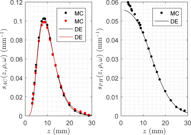
Comparison between DE and MC results for the depth sensitivity of AC and PH signals, sAC (z, ρ, ω) and sPH (z, ρ, ω), plotted versus the maximum depth z for a slab 100 mm thick, with ρ =20 mm, , μa =0.005 mm−1, ν =100 MHz (data plotted in black) and ν =0 (data plotted in red), and refractive indexes of slab and external, 1.4 and 1, respectively. We stress that the data in red (ν =0) represent the DC sensitivity sDC (z, ρ).
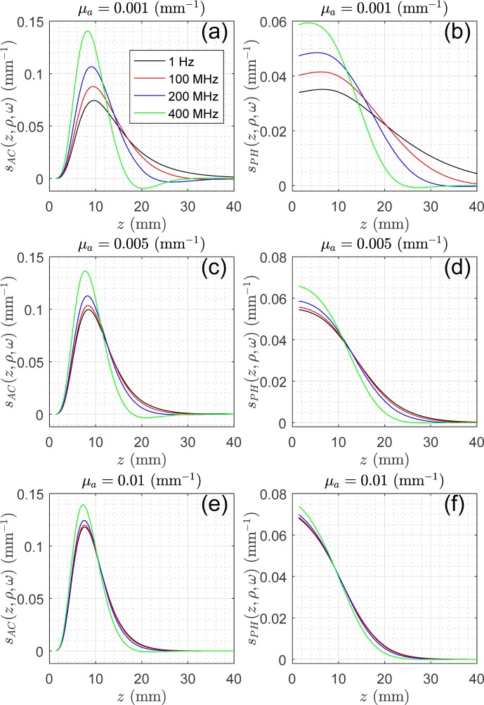
In Fig. 2 the depth sensitivity for AC, and PH signals obtained with the DE is plotted for μa = {0.001, 0.005, 0.01} mm−1. This covers a wide range of absorption values typical of some biological tissues. Figure 2 pertains to a slab of s0 = 100 mm, with , ρ = 20 mm and ν = {10−6, 100, 200, 400} MHz. The results show that for the larger depths the AC and PH signals have larger sensitivity for the lower modulation frequencies in agreement with Bevilacqua et al. [22]. The values of sAC for ν =1 Hz are practically coincident with the DC sensitivity sDC. For larger absorption values the curves at different frequencies tend to be closer and for a very high absorption almost match. For μa = 0.1 mm−1 (limit values for biological tissues in the near infrared window) the curves at the different frequencies are indistinguishable (data not shown). This agrees with the expectation that for very high absorption, given the very short path-lengths of the detected photons, only the shallow depths are probed. This fact reduces the differences between the signals at different frequencies.
Fig. 2.
Depth sensitivity obtained with the DE for AC and PH signals, sAC and sPH, versus the maximum depth z for a slab 100 mm thick, ρ =20 mm, , μa =0.001, 0.005 and 0.01 mm−1, ν = {10−6, 100, 200, 400} MHz and refractive indexes of slab and external, 1.4 and 1, respectively.
In Fig. 3 sAC (z, ρ, ω) and sPH (z, ρ, ω) are compared with the DC depth sensitivity, sDC (z, ρ), for s0 = 100 mm, , μa =0.005 mm−1, ρ =20 mm and ν = {100, 200, 400} MHz. The comparison reveals that the AC depth sensitivity has similar characteristics to the CW depth sensitivity except when frequencies > 200 MHz are considered. Results for frequencies > 400 MHz (data not shown) show that larger is the frequency, the lower is the sensitivity of the signals at larger depth. Concerning the PH depth sensitivity, we note that sPH (z, ρ, ω) shows significant differences as compared to sDC (z, ρ). At low z values, sPH (z, ρ, ω) has the highest values showing that the phase is very sensitive to variations occurring at low values of the depth differently from sDC (z, ρ). For this reason, the comparison between PH and CW requires a more complex interpretation. To this purpose, the next figures describing the relative contrast of a totally absorbing plane in a slab may offer an easier and concise reading of the results.
Fig. 3.
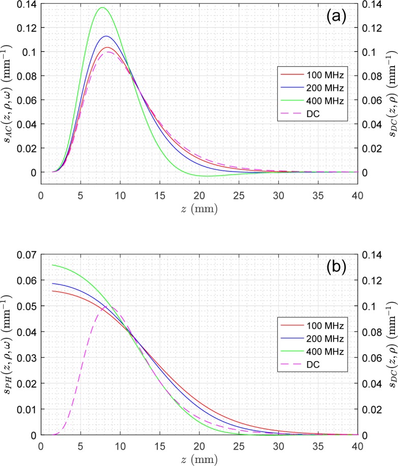
Comparison of the depth sensitivity functions sAC (z, ρ, ω), sPH (z, ρ, ω), and sDC (z, ρ) obtained with the DE. The results pertain to a slab 100 mm thick, ρ =20 mm, , μa =0.005 mm−1, ν = {100, 200, 400} MHz and refractive indexes of slab and external, 1.4 and 1, respectively.
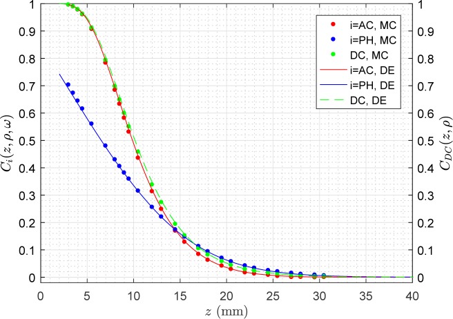
In Fig. 4 it is plotted a comparison of the relative contrast of a totally absorbing plane in a slab calculated with Eqs. (14) - - (16) and with the results of MC simulations for a slab 100 mm thick, with , μa =0.005 mm−1, ρ =20 mm, ν =100 MHz. The agreement between analytical solutions and MC results reveals the correctness of the approach here adopted. The relative contrast offers an excellent agreement also for lower values of z. These results confirm the very similar behavior of AC and CW signals. The values of the AC contrast are for any z lower than the values for the DC signal. The behavior of the PH is different from those of DC and AC signals, showing lower values at shallow depths and a bit higher values for large depths.
Fig. 4.
It pertains to the same optical properties and geometry as Fig. 1 but for the relative contrast of a totally absorbing plane placed at z inside the slab. The DE and the MC results for AC, PH and DC have been plotted versus the maximum depth z.
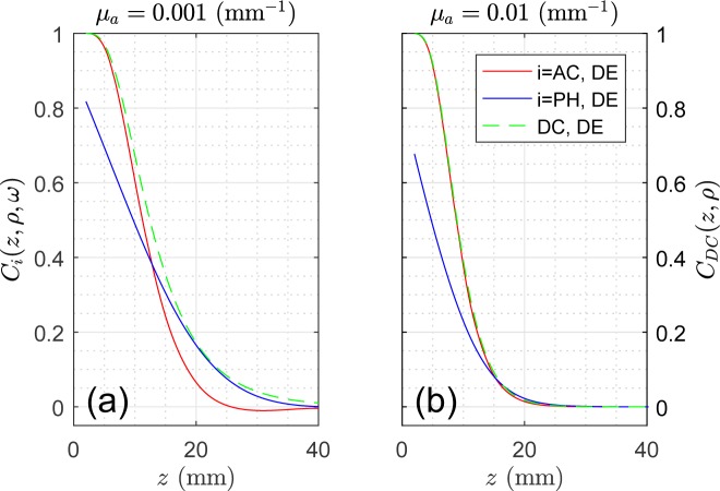
In Fig. 5 the relative contrast of a totally absorbing plane in a slab for AC, PH and DC signals is further investigated by plotting the DE data for μa =0.001 mm−1 and μa =0.01 mm−1. For μa =0.01 mm−1, typical of many biological tissues, AC and DC become almost indistinguishable, while for the PH lower values are still noticeable at shallow depths and slightly larger values (the differences with the DC are hardly noticeable) for large depths. For μa =0.001 mm−1 we note the larger difference between AC and DC with the DC showing always an higher contrast. We have not reported the DE results for very short depths, since the diffusion approximation used to obtain the DE model does not hold in such physical conditions.
Fig. 5.
Relative DE contrast of a totally absorbing plane in a slab: comparison of CAC (z, ρ, ω), CPH (z, ρ, ω), and CDC (z, ρ) versus the z position of the plane. The results pertain to a slab 100 mm thick, ρ =20 mm, , μa = {0.001, 0.01} mm−1, ν =100 MHz and refractive indexes of slab and external, 1.4 and 1, respectively.
In Fig. 6, pertaining to the case of Fig. 4, the relative contrast of a totally absorbing plane in a slab for AC and PH is also plotted for some values of ν = {10−6, 100, 200, 400} MHz. The values of CAC for ν =1 Hz are practically coincident with the DC contrast CDC. Figure 6 shows that the increase of ν determines a shrinkage of the range of depths where the variations of the contrast fall and this range tends to be more confined to the swallow depths. This fact can be expected from the behavior of the depth sensitivity functions previously analyzed.
Fig. 6.
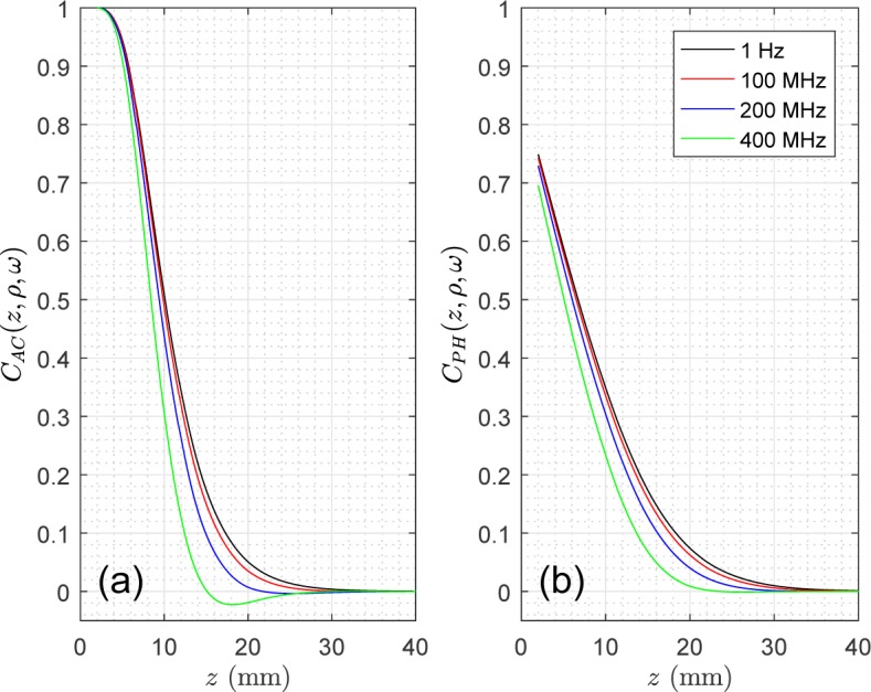
Relative DE contrast of a totally absorbing plane in a slab for AC and PH versus the position of the plane z, for ν = {10−6, 100, 200, 400} MHz. The results pertain to a slab 100 mm thick, ρ =20 mm, , μa =0.005 mm−1, and refractive indexes of slab and external, 1.4 and 1, respectively.
Similar results to those shown in this section can be obtained for other ρ (results not shown), if diffusion conditions are satisfied.
4. Discussion
The presented results prove that it is always possible to carry out a depth sensitivity study of a data type such as AC and PH signals, however, it is not always possible to define proper statistics of penetration depths for the same data type. In fact, to obtain the statistics for the maximum depth, z, it is necessary to have a well-defined probability density function of z with positive values at any depth. The present results demonstrate that for the AC and PH signals this mandatory condition is not always satisfied (see e.g. Fig. 2) and for such signals probability density functions cannot be in general formulated because of the lack of a rigorous statistical ground. The same thing can also be viewed through the effect of a totally absorbing plane perpendicular to z axis. In practice, to define a statistic for the maximum depth the data type must increase monotonically as the plane is moved away from the photon injection plane. If this is not true it means that photons that progressively move deeper in the medium lead to a decrease of the data type and to a negative contrast (see e.g. Figs. 5(a) and 6). In other words, the prerequisite for the definition of these depth statistics is that every collected photon will increase the considered data type. This is true for the Temporal Point Spread Function, TPSF(t), and for the CW data for example, but it is not always true for AC and PH signals for example.
Bevilacqua et al. [22] calculated with MC simulations and with solutions of the DE the probability density function of the depth, , in the mid-plane between source and detector by using the concept of photon scattering density function. They did such calculation for the complex amplitude, i.e., for the AC signal, and comparisons were shown for several modulation frequencies and absorption of a semi-infinite medium. Still referred to the same specific mid-plane between source and detector they also calculated the mean visitation depth . Indeed, since the AC and PH are not strictly proportional to the number of detected photons, it is questionable whether a probability density function for the depth can be defined in all generality in the FD. However, we do not exclude that may exist limited ranges of the optical properties for which such approach can be rigorously implemented for all the depths. Bevilacqua et al. [22] from their results found that: “for typical tissue optical properties, the difference in tissue sampling offered by steady-state and frequency domain methods is small for modulation frequencies smaller than 600 MHz”. In synthesis, the results here presented are very close to this previous investigation for what concern the AC signal, but differ for the PH signal, since for the PH we cannot state that the differences with the CW is always small. Indeed, Bevilacqua et al. [22] presented results for the PH only for the effect of a small absorbing perturbation. For this reason, and since their results were referred only to the median plane between source and detector, the results of Bevilacqua et al. [22] cannot be easily compared with those here presented.
Although in the FD we cannot define in general a statistic for the penetration depth, by means of depth sensitivity functions we can nevertheless provide an estimate of the depth probed by the AC signal. In fact, the AC signal, in terms of depth sensitivity, has similar characteristics to the CW/DC signal and the CW depth sensitivity is an upper limit value for the AC depth sensitivity. This fact is even more clear for the relative contrast offered by a totally absorbing plane inserted inside the slab. The close values of AC and CW contrast establish a direct link between the depth probed by the CW signal and that related to the AC signal. When the contrast at a certain depth has a detectable value, it means that photons migrating at such depth produce detectable effects on the measured signal. Therefore, in an indirect way we state that such a depth is probed by the signal. Translating the similarity between AC and CW depth sensitivity and contrast in a practical guideline for the experimenter, we propose to use the theoretical tool for the penetration depth available for the CW domain signal also to obtain an estimation of the depth probed by AC signal. Thus, an estimation of the depth probed by the AC measured signal can be obtained by using Eq. (24) in [11] that is the mean maximum depth probed by the CW signal in a diffusive slab in reflectance configuration. On the other side, in accordance to the presented results, the PH signal shows a very different behavior compared to the AC signal. The PH depth sensitivity is different as compared to the CW/DC signal and the largest disagreement is observed for low values of the depth. Also for the relative contrast of a totally absorbing plane we note a marked difference between AC and PH and between DC and PH. The relative contrast for PH is generally less steep, meaning that the loss of information upon increasing depth is somehow softer-milder-smoother as compared to AC. Indeed, the depth sensitivity of AC is more localized around a given depth, and beyond that the cumulative effect of the underlying medium (i.e., the relative contrast) falls rapidly to zero. Since for the PH there is not a direct analogy with the CW signal, which can be interpreted in terms of photon paths distributions, the evaluation of the depth probed by the PH signal must be carried out only by the analysis of Eqs. (12) and (15).
In general, the depth sensitivity and the relative contrast of the AC signal are different from those of the PH signal. This fact may have consequences in data analysis when FD signals are processed. In practice, this means that care must be taken when using algorithms allowing to reconstruct the optical properties that depend on both AC and PH. This point must certainly be carefully studied in the future.
The differences between AC and PH can be ascribed to the fact that AC mainly still depends on the number of detected photons, while PH is related to their mean time of flight 〈t〉. This is the reason why sAC (z, ρ, ω) has similar characteristics to sDC (z, ρ) (see Fig. 1 where they both peak at the same depth). The specific behavior of sPH (z, ρ, ω) may be intuitively explained by noting that: 1) 〈t〉 increases versus z; 2) for large z an increment dz does not increases significantly 〈t〉 of detected photons since the dz acts on a marginal part of the slab, vice-versa 3) for small z an increment dz drastically increments 〈t〉 of detected photons. Thus, 〈t〉 has larger increments at shallower z depths and lower for large z values with a uniform transition from small to large z. This behavior explains the monotonic decrement of sPH (z, ρ, ω) in Fig. 1.
As a general comment, we can observe that for an imaging experiment, in which we are e.g. interested to detect the presence of an absorption inhomogeneity located at a given depth, the PH signal seems to be preferred to the AC signal given its slightly better contrast for z ∈ [20, 30] mm (see Figs. 4–5). However, we must note that this holds true especially for low values of the background absorption coefficient (μa = 0.001 mm−1), while the differences between AC and phase tend to fade away for higher absorption values (μa = 0.01 mm−1).
The above considerations hold true in the ideal case, i.e., without the presence of noise. In realistic conditions, the measured signals to be chosen in a FD experiment might depend on the characteristics of the experimental setup (e.g. noise level for AC, noise level for PH). In fact, precise phase measurements are more difficult and this point cannot be generalized without a signal-to-noise analysis [17].
Experiments measuring the AC and PH contrast on semi-infinite and slab geometries are achievable using calibrated liquid phantoms as those in [29, 30]. It is worth to stress that for the purposes of this work, as we have previously demonstrated by means of comparisons between measurements on optical calibrated liquid phantoms and MC simulations [29, 30], the MC results reproduce correctly the results of calibrated phantom measurements. This evidence strengthens the validity of the presented results.
5. Conclusions
In this work, we have provided, by means of solutions of the photon diffusion equation, the depth sensitivity functions for the AC, PH and DC signals of the FD. Solutions have also been given for the relative contrast of a totally absorbing plane placed inside the slab at depth z. The formulas have been validated by means of comparisons with the results of gold standard MC simulations.
In the FD, we cannot provide a comprehensive statistical approach for the maximum and average depth of the detected AC and PH signals. The reasons of this fact are intrinsic to the nature of these two signals. Thus, the ability of these signals to probe the medium at different depths can only be studied by means of proper depth sensitivity functions.
The depth sensitivity and the relative contrast (offered by a totally absorbing plane inserted inside the slab) of the AC signal show an overall close and similar behavior to that of the DC/CW signal provided the modulation frequency is roughly lower than 200 MHz. Increasing the modulation frequency we note that this similarity fades away and thus is expected to hold with a less extent. The close values of AC and CW contrast establish a direct link between the depths probed by the CW and AC signals. Then, an estimation of the depth probed by the AC measured signal can be obtained by using the mean maximum penetration depth of the CW signal in reflectance configuration from a slab, i.e., Eq. (24) in [11]. On the other hand, the depth sensitivity of the PH signal marks a clear difference with that of the DC signal for shallow depths. The non-uniform probing provided by AC, DC and PH signals poses the question of the real depth probed by the set of FD signals. The results of the present study have been obtained without the inclusion of realistic experimental noise. However, the effect of noise cannot eliminate the large general differences existing between AC and PH signals.
Acknowledgments
The authors wish to thank Prof. Giovanni Zaccanti for his useful suggestions.
Funding
European Union’s Horizon 2020 research and Innovation Programme (654148) Laserlab-Europe.
References and links
- 1.Bonner R. F., Nossal R., Havlin S., Weiss G. H., “Model for photon migration in turbid biological media,” J. Opt. Soc. Am. A 4, 423–432 (1987). 10.1364/JOSAA.4.000423 [DOI] [PubMed] [Google Scholar]
- 2.Weiss G., Nossal R., Bonner R., “Statistics of Penetration Depth of Photons Re-emitted from Irradiated Tissue,” J. Mod. Opt. 36, 349–359 (1989). 10.1080/09500348914550381 [DOI] [Google Scholar]
- 3.Nossal R., Kiefer J., Weiss G. H., Bonner R., Taitelbaum H., Havlin S., “Photon migration in layered media,” Appl. Opt. 27, 3382–3391 (1988). 10.1364/AO.27.003382 [DOI] [PubMed] [Google Scholar]
- 4.Weiss G., Kiefer J., “A numerical study of the statistics of penetration depth of photons re-emitted from irradiated media,” J. Mod. Opt. 45, 2327–2337 (1998). 10.1080/09500349808231242 [DOI] [Google Scholar]
- 5.Weiss G. H., “Statistical properties of the penetration of photons into a semi-infinite turbid medium: a random-walk analysis,” Appl. Opt. 37, 3558–3563 (1998). 10.1364/AO.37.003558 [DOI] [PubMed] [Google Scholar]
- 6.Weiss G., Porra J., Masoliver J., “Statistics of the depth probed by cw measurements of photons in a turbid medium,” Phys. Rev. E. 58, 6431–6439 (1998). 10.1103/PhysRevE.58.6431 [DOI] [Google Scholar]
- 7.Feng S., Zeng F. A., Chance B., “Photon migration in the presence of a single defect: a perturbation analysis,” Appl. Opt. 34, 3826–3837 (1995). 10.1364/AO.34.003826 [DOI] [PubMed] [Google Scholar]
- 8.Patterson M. S., Andersson-Engels S., Wilson B. C., Osei E. K., “Absorption spectroscopy in tissue-simulating materials: a theoretical and experimental study of photon paths,” Appl. Opt. 34, 22–30 (1995). 10.1364/AO.34.000022 [DOI] [PubMed] [Google Scholar]
- 9.Del Bianco S., Martelli F., Zaccanti G., “Penetration depth of light re-emitted by a diffusive medium: theoretical and experimental investigation,” Phys Med Biol 47, 4131–4144 (2002). 10.1088/0031-9155/47/23/301 [DOI] [PubMed] [Google Scholar]
- 10.Zonios G., “Investigation of reflectance sampling depth in biological tissues for various common illumination/collection configurations,” J. Biomed. Opt. 19, 97001 (2014). 10.1117/1.JBO.19.9.097001 [DOI] [PubMed] [Google Scholar]
- 11.Martelli F., Binzoni T., Pifferi A., Spinelli L., Farina A., Torricelli A., “There’s plenty of light at the bottom: statistics of photon penetration depth in random media,” Sci. Rep. 6, 27057 (2016). 10.1038/srep27057 [DOI] [PMC free article] [PubMed] [Google Scholar]
- 12.Tromberg B. J., Svaasand L. O., Tsay T. T., Haskell R. C., “Properties of photon density waves in multiple-scattering media,” Appl. Opt. 32, 607–616 (1993). 10.1364/AO.32.000607 [DOI] [PubMed] [Google Scholar]
- 13.Ripoll J., Principles of Diffuse Light Propagation: Light Propagation in Tissues with Applications in Biology and Medicine (World Scientific, 2012). [Google Scholar]
- 14.Boas D. A., O’Leary M. A., Chance B., Yodh A. G., “Scattering and wavelength transduction of diffuse photon density waves,” Phys. Rev. E Stat. Phys. Plasmas Fluids Relat. Interdiscip. Topics 47, R2999–R3002 (1993). [DOI] [PubMed] [Google Scholar]
- 15.O’Leary M. A., Boas D. A., Chance B., Yodh A. G., “Refraction of diffuse photon density waves,” Phys. Rev. Lett. 69, 2658–2661 (1992). 10.1103/PhysRevLett.69.2658 [DOI] [PubMed] [Google Scholar]
- 16.Schmitt J. M., Knuttel A., Knutson J. R., “Interference of diffusive light waves,” J. Opt. Soc. Am. A 9, 1832–1843 (1992). 10.1364/JOSAA.9.001832 [DOI] [PubMed] [Google Scholar]
- 17.Boas D. A., O’Leary M. A., Chance B., Yodh A. G., “Detection and characterization of optical inhomogeneities with diffuse photon density waves: a signal-to-noise analysis,” Appl. Opt. 36, 75–92 (1997). 10.1364/AO.36.000075 [DOI] [PubMed] [Google Scholar]
- 18.Bigio I. J., Fantini S., Quantitative Biomedical Optics: Theory, Methods, and Applications (Cambridge University Press, New York, NY, USA, 2016), 1st ed. [Google Scholar]
- 19.Durduran T., Choe R., Baker W. B., Yodh A. G., “Diffuse Optics for Tissue Monitoring and Tomography,” Rep. Prog. Phys. 73 (2010). 10.1088/0034-4885/73/7/076701 [DOI] [PMC free article] [PubMed] [Google Scholar]
- 20.Sevick E. M., Frisoli J. K., Burch C. L., Lakowicz J. R., “Localization of absorbers in scattering media by use of frequency-domain measurements of time-dependent photon migration,” Appl. Opt. 33, 3562–3570 (1994). 10.1364/AO.33.003562 [DOI] [PMC free article] [PubMed] [Google Scholar]
- 21.Houston J. P., Thompson A. B., Gurfinkel M., Sevick-Muraca E. M., “Sensitivity and depth penetration of continuous wave versus frequency-domain photon migration near-infrared fluorescence contrast-enhanced imaging,” Photochem. Photobiol. 77, 420–430 (2003). [DOI] [PubMed] [Google Scholar]
- 22.Bevilacqua F., You J. S., Hayakawa C. K., Venugopalan V., “Sampling tissue volumes using frequency-domain photon migration,” Phys. Rev. E Stat. Nonlin. Soft Matter Phys. 69, 051908 (2004). 10.1103/PhysRevE.69.051908 [DOI] [PubMed] [Google Scholar]
- 23.Nissila I., Hebden J. C., Jennions D., Heino J., Schweiger M., Kotilahti K., Noponen T., Gibson A., Jarvenpaa S., Lipiainen L., Katila T., “Comparison between a time-domain and a frequency-domain system for optical tomography,” J Biomed. Opt. 11, 064015 (2006). 10.1117/1.2400700 [DOI] [PubMed] [Google Scholar]
- 24.Martelli F., Del Bianco S., Ismaelli A., Zaccanti G., Light Propagation Through Biological Tissue and Other Di ffusive Media: Theory, Solutions, and Software (SPIE Press, 2010). 10.1117/3.824746 [DOI] [Google Scholar]
- 25.Zaccanti G., “Monte Carlo study of light propagation in optically thick media: point source case,” Appl. Opt. 30, 2031–2041 (1991). 10.1364/AO.30.002031 [DOI] [PubMed] [Google Scholar]
- 26.Martelli F., Contini D., Taddeucci A., Zaccanti G., “Photon migration through a turbid slab described by a model based on diffusion approximation. II. Comparison with Monte Carlo results,” Appl. Opt. 36, 4600–4612 (1997). 10.1364/AO.36.004600 [DOI] [PubMed] [Google Scholar]
- 27.Testorf M., Osterberg U., Pogue B., Paulsen K., “Sampling of time- and frequency-domain signals in monte carlo simulations of photon migration,” Appl. Opt. 38, 236–245 (1999). 10.1364/AO.38.000236 [DOI] [PubMed] [Google Scholar]
- 28.Contini D., Martelli F., Zaccanti G., “Photon migration through a turbid slab described by a model based on diffusion approximation. I. Theory,” Appl. Opt. 36, 4587–4599 (1997). 10.1364/AO.36.004587 [DOI] [PubMed] [Google Scholar]
- 29.Del Bianco S., Martelli F., Cignini F., Zaccanti G., Pifferi A., Torricelli A., Bassi A., Taroni P., Cubeddu R., “Liquid phantom for investigating light propagation through layered diffusive media,” Opt. Express 12, 2102–2111 (2004). 10.1364/OPEX.12.002102 [DOI] [PubMed] [Google Scholar]
- 30.Martelli F., Ninni P. D., Zaccanti G., Contini D., Spinelli L., Torricelli A., Cubeddu R., Wabnitz H., Mazurenka M., Macdonald R., Sassaroli A., Pifferi A., “Phantoms for diffuse optical imaging based on totally absorbing objects, part 2: experimental implementation,” J. Biomed. Opt. 19, 076011 (2014). 10.1117/1.JBO.19.7.076011 [DOI] [PubMed] [Google Scholar]


