Abstract
A survey of 158 rodents caught in the Czech Republic identified Dobrava virus sequences closely related to that of the Dobrava virus type strain in Apodemus sylvaticus and Mus musculus rodents. The identity of A. sylvaticus was unequivocally confirmed by random amplified polymorphic DNA analysis. The data seem to indicate hantavirus spillover from Apodemus flavicollis to other rodents.
Hantaviruses are subdivided into the Eurasian and the American groups. The distinction manifests itself in serological and genetic differences as well as in the clinical picture of the diseases elicited in humans. The up to 20 individual hantavirus types have been observed to be strictly coevolving with the distinct rodent species that carry them. Prominent hantaviruses in western and central Europe are Dobrava virus (DOBV), Puumala virus (PUUV), and Tula virus (TULV) (6, 16, 21).
DOBV was originally isolated in Slovenia from the rodent Apodemus flavicollis (DOBV-Af) (3) and is a Eurasian hantavirus that predominantly causes disease in humans in southeastern Europe. DOBV-Af has also been isolated in Greece, Albania, and Bosnia-Herzegovina (1, 10, 14). Serological studies have indicated the presence of DOBV-Af beyond the geographical limits of the Balkans in Slovakia (22) and recently in the Czech Republic (15, 25). The isolation of a Dobrava-like virus from the rodent Apodemus agrarius (DOBV-Aa) was reported in Estonia (11), Russia (17), and Slovakia (23), supplemented by a report on genetic evidence of this DOBV-Aa type from rodent samples in Hungary (20). Whether this new type is a type of its own, as comparative serological and genetic analyses suggest, is still a matter of debate (7, 12, 18). The distribution of the two rodent host species overlaps in Europe, but rodent carriers of both DOBV types coexist only in the border region of southeastern and central Europe (2, 23). In order to investigate the occurrence of hantaviruses in a rodent population from the Czech Republic, we analyzed the kidneys, lungs, and spleens of 157 rodents and 1 shrew (A. flavicollis, n = 77; A. sylvaticus, n = 34; Mus musculus, n = 2; Clethrionomys glareolus, n = 41; Microtus arvalis, n = 2; Microtus agrestis, n = 1; Sorex araneus, n = 1) caught at the military training area of Boletice, close to Ceské Budejovice in the south of the Czech Republic, from May to November 1999.
MATERIALS AND METHODS
A total of 10 to 50 mg of tissue of each organ was homogenized in a FastPrep machine with the FastRNA Green kit (Qbiogene, Hilden, Germany) to perform total RNA extraction. This closed system avoids the generation of contaminating aerosols. We screened the extracted RNA for DOBV-Af RNA by a one-step TaqMan reverse transcription (RT)-PCR and for hantavirus RNA with the degenerate primers for nested PCR published by Scharninghausen et al. (20). For the DOBV TaqMan RT-PCR, we used 500 nM concentrations of primers DOBFP (5′-TGGCTTGACCTCCCGTG-3′) and DOBRP (5′-CAAGCGCTCCTTGTCTTTGA-3′) and 200 nM probe DOBP (5′-ATCTCCAACGTCTTTGACCAAAGGCCC-3′) tagged with 6-carboxyfluorescein (FAM)-6-carboxytetramethylrhodamine (TAMRA). To detect synthetic RNA we used the SRDOB TaqMan RT-PCR with 500 nM concentrations of primers SRDOBFP (5′-TAGTGCTCTTTAGGGAGTCTACTAC-3′) and SRDOBRP (5′-CAAGCGCTCCTTGTCTTTGA-3′) and 200 nM probe SRDOB P (5′-AAAGCCGAATTCTGCAGATATCCAT-3′) tagged with FAM and TAMRA. The SRDOB amplicon spans the extreme 5′ end of the DOBV S-segment RNA transcript. The probe and the reverse primer bind in the trace plasmid sequence stemming from pCRII into which the S segment was ligated. We performed the amplification on a LightCycler instrument using an RNA Master Hybridization Probes kit (Roche, Mannheim, Germany) and a temperature profile of RT at 60°C for 20 min, activation at 95°C for 2 min, and 45 cycles of PCR at 95°C for 15 s and 60°C for 1 min. The efficiencies of the TaqMan RT-PCRs were calculated as 10−1/slope − 1. The nested PCR was done with a touchdown amplification profile adapted according to Sachadyn et al. (19): first round, RT at 53°C for 30 min; PCR with 20 cycles of 95°C for 1 min, 65°C for 1 min, and 72°C for 1 min; and completion of extension at 72°C for 10 min with primers M9 (5′-TGRYCNAGYTGTATYCCCATWGATTG-3′) and M4 (5′-ATGAARGCNGAWGARNTNACMCCNGG-3 [nucleotide abbreviations according to the IUB code]); second round, activation at 95°C for 10 min; PCR with 6 cycles of 95°C for 1 min, 65°C for 1 min, and 72°C for 1 min, followed by 3 cycles each of the same profile with hybridization at 66, 64, 66, 64, 62, 60, 62, 60, 59, 60, and 59°C and completion of the extension at 72°C for 10 min with primers M6 (5′-AGYCCWGTNATGRGWGTRATTGG-3′) and M8 (5′-GAKGCCATRATNGTRTTYCKCATRTCCTG-3′). The buffer conditions for the first round of RT-PCR were as described previously (26), the second round of PCR was done with AmpliTaq Gold, 1 μM primers, and 500 μM deoxynucleoside triphosphates. Three negative controls were included in each cycling step. Random amplified polymorphic DNA (RAPD) analysis for differentiation of the rodent species was done as described previously (4). For phylogenetic analysis, alignment was done with the ClustalW program in the Megalign software module of the Lasergene package (DNAstar) by using the default settings. To plot the trees, we used the Phylip software package 3.6.seqboot module for bootstrapping (200 bootstraps, 55-bp sequence blocks) and the dnapars module to build the most parsimonious tree. The tree was drawn with the TreeEdit program (version 1.0a10). The tree in Fig. 2 was initially rooted to the sequence of Hantaan virus (HTNV) type strain 76/118. The DOBV sequence most closely related to it was then used to root the tree in Fig. 2.
FIG. 2.
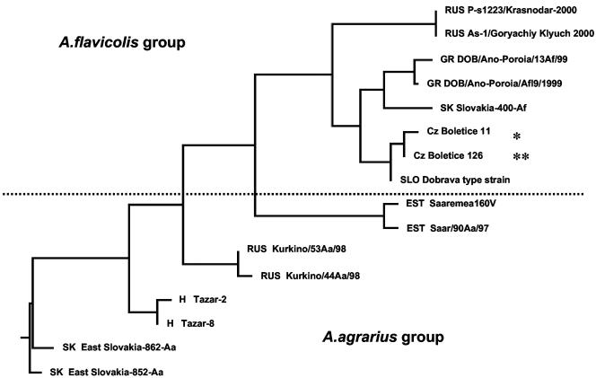
(A) Phylogram rooted to DOBV strain East Slovakia-856-Aa. Sequences were aligned in reference to nucleotide positions 720 to 1041 of the DOBV-Af type strain (GenBank accession number L41916). Branch distances correspond to sequence divergence. The GenBank accession numbers of the sequences used for the phylogram are (respectively, from top to bottom) AJ578082, AJ578081, AF442623, AF442622, DVI410619, DVI410615, and AJ269554, L41916, AJ009773, AJ009775, AJ131673, AJ131672, AF085336, AF085337, AJ269550, and AJ269549). *, sequence obtained in this study from M. musculus; **, sequence obtained in this study from A. sylvaticus. Country abbreviations are according to the international traffic code and precede the strain designations. The percent identities of the sequences in the DOBV-Af group to the original DOBV type strain are as follows: Cz Boletice 126, 99.1%; Cz Boletice 11, 98.8%; SK Slovakia-400-Af, 94.7%; GR DOBV/Ano-Poroia/AfI9/1999, 95.7%; GR DOBV/Ano-Poroia/13Af/1999, 96%; RUS As-1/Goryachiy Klyuch-2000, 90.4%; RUS P-s1223/Krasnodar-2000, 90.4%.
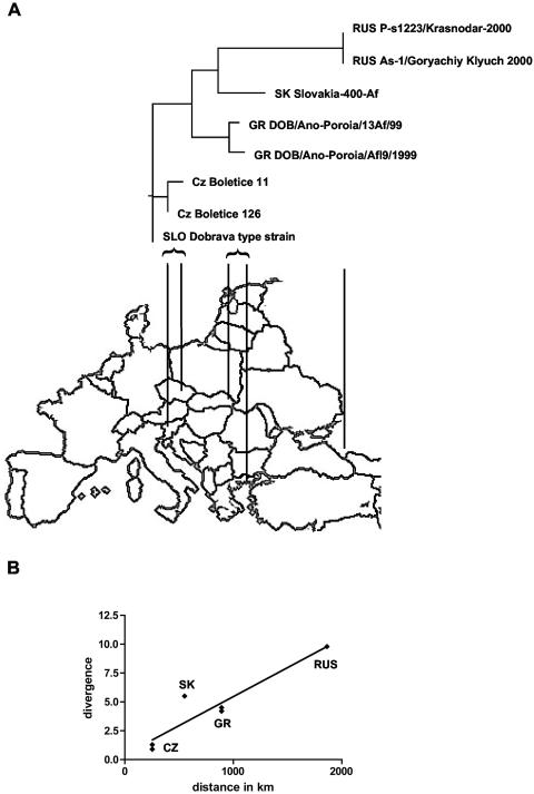
To plot divergence against geographical distance, divergence values were extracted from the alignment obtained with the ClustalW program, and the coordinates of the isolation sites were determined (http://wayhoo.com). Polar coordinate distances between the various isolation sites and the isolation site in Slovenia were calculated (http://jan.ucc.nau.edu/∼cvm/latlongdist.html).
Nucleotide sequence accession numbers.
The sequences obtained from A. sylvaticus and M. musculus have been submitted to GenBank and given the accession numbers AJ578081 and AJ578082, respectively.
RESULTS AND DISCUSSION
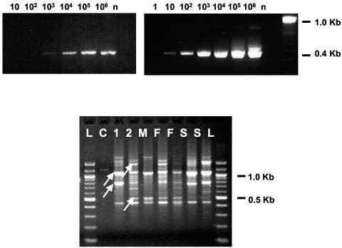
The DOBV TaqMan RT-PCR assay was tested with a synthetic RNA standard transcribed from a DOBV S segment ligated into pCRII (Invitrogen, Breda, The Netherlands). This assay had a sensitivity of detection of 10 molecules (standard range, 107 to 10 molecules; correlation coefficient, 0.99; efficiency, 0.84). We also established a second TaqMan RT-PCR to specifically detect the extreme 5′ end of the DOBV RNA transcript with a downstream primer and probe that bind to the trace plasmid sequence originating from between the T7 promoter and the 5′ end of the cloned DOBV S fragment (standard range, 105 to 102; correlation coefficient, 0.96; efficiency, 0.94). It had a sensitivity of detection of 100 molecules (Fig. 1, to panels). We tested the nested PCR with the synthetic RNA standard and found that we could increase the sensitivity of detection from 103 to 10 molecules by using the touchdown protocol instead of the protocol originally used for this PCR (Fig. 1, top panels). The TaqMan RT-PCR detected DOBV RNA in 7 of 474 rodent tissue samples. The nested PCR yielded a 389-bp band for 2 of 474 rodent tissue samples. These two samples also tested positive by the TaqMan RT-PCR (Table 1). The DOBV TaqMan assay additionally detected DOBV-Af in one A. flavicollis-positive sample and in four C. glareolus-positive samples. The DOBV TaqMan assay is specific for DOBV-Af and does not cross amplify DOBV-Aa, PUUV, or TULV sequences (data not shown). Several measures were adopted to avoid false-positive PCR signals. We did not culture DOBV-Af in a biosafety level 3 laboratory at the time of tissue preparation. To exclude false-positive results due to our synthetic DOBV-Af RNA standard, we tested all positive samples by the SRDOB TaqMan RT-PCR. All DOBV-positive samples tested negative with primer SRDOB (data not shown). We included one negative control for each TaqMan RT-PCR and three negative controls for each step of the nested PCR. The amplicons from the two nested PCRs were ligated into pCRII by TA cloning (Invitrogen). We sequenced the recombinant plasmids of two independent clones each and aligned the consensus sequence of the sequences obtained to other hantavirus sequences in GenBank.
FIG. 1.
(Top panels) Comparison of the sensitivity of the nested PCR by use of the published protocol (left) and a touchdown protocol (right). The values above each lane indicate the number of transcribed RNA molecules tested. n, negative control. (Bottom panel) RAPD analysis of mouse tissues. RAPD analysis was done with primers OPA01 and OPA08 (Operon Technologies). The results obtained with primer OPA01 are shown. The patterns in lane 1 and lanes S are identical, as are the patterns in lane 2 and lanes F. The arrows indicate prominent bands specifically identifying the species with the RAPD pattern. Lanes: L, 100-bp DNA ladder; C, negative control; S, positive control for A. sylvaticus; F, positive control for A. flavicollis; M, positive control for A. microps; 1, sample 126 from which the DOBV sequence was amplified; 2, sample 133, a DOBV-negative sample from A. flavicollis.
TABLE 1.
Results of hantavirus detection by nested PCR and TaqMan RT-PCR
| Sample no. | Rodent species | Rodent organ | DOBV-Af-specific TaqMan RT-PCR resulta | Nested PCR result |
|---|---|---|---|---|
| 126R | A. sylvaticus | Kidney | CP 15.85 | Positive |
| 144P | A. flavicollis | Spleen | CP 36.89 | |
| 11P | M. musculus | Spleen | CP 25.98 | Positive |
| 94R | C. glareolus | Kidney | CP 37.55 | |
| 157 P | C. glareolus | Spleen | CP 37.96 | |
| 55L | C. glareolus | Lung | CP 36.83 | |
| 190L | C. glareolus | Lung | CP 31.44 |
CP, crossing point.
Both sequences turned out to be closely related to DOBV-Af. One sequence originated from the kidney of a captured A. sylvaticus mouse and the other was amplified from the spleen of a captured M. musculus mouse. To confirm the identity of A. sylvaticus, whose phenotype can easily be mistaken for A. flavicollis in some parts of central Europe, we used a RAPD system to identify the samples from which the DOBV-Af sequence had been amplified (4). The RAPD analysis confirmed the identity of the mouse species implicated (Fig. 1, bottom panel), making this the first confirmed report of a DOBV-Af sequence identified in a captured A. sylvaticus mouse and a captured M. musculus rat. Excluding the primer binding sites, we aligned 319 bp of the sequences obtained to all published DOBV sequences that cover that region of the S fragment and to the respective sequence of HTNV type strain 76/118. In a weighted phylogram rooted to the DOBV sequence most closely related to the HTNV type strain, the new sequences group with the original DOBV-Af isolate and other DOBV-Af sequences (Fig. 2). The DOBV-Aa sequences group outside of the DOBV-Af clade. We also found in the GenBank data bank two entries (GenBank accession numbers AF442622 and AF442623) for sequences from A. sylvaticus isolates that as yet have not been published in a paper. These sequences and the one that we derived from A. sylvaticus group together in the DOBV-Af clade. The sequence that we have derived from M. musculus also grouped into this clade. In the DOBV-Af clade there seems to be an obvious correlation between the geographic distance from Slovenia and the percent identities of the sequences to the DOBV-Af type strain. These range from 99.1% for the closest sequence from the Czech Republic to 90.4% for the farthest sequence from Russia (Fig. 3A). In fact, the geographical distances of the sites of isolation of the DOBV-Af strains to the site of isolation of the DOBV-Af type strain plotted versus sequence divergence results in a correlation (r2) of 0.91 (P = 0.0008). Without the new sequences r2 value is reduced to 0.85 (P = 0.026) (Fig. 3B). A similar correlation has been observed and substantially analyzed for the tick-borne encephalitis virus cline in the Northern Hemisphere (27).
FIG. 3.
(A) Subtree of the DOBV-Af group rooted to the DOBV-Af type strain aligned to a partial map of central Europe; (B) correlation of the geographic distance of the sites of isolation of the divergent DOBV-Af from strains the site of isolation of the Slovenian DOBV-Af type strain to the divergence of DOBV-Af isolates from the DOBV-Af type strain.
In a study of the 1989 hemorrhagic fever with renal syndrome outbreak in Yugoslavia, 544 trapped rodents captured at various sites were studied (5). The investigators found hantavirus antigen-positive lung tissues (by immunofluorescence assay) and hantavirus antibody-positive sera (by immunofluorescence assay and enzyme-linked immunosorbent assay) in six rodent species (A. flavicollis, 46.5% [88 positive animals of 189 animals captured]; A. agrarius, 15.6% [10 of 64]; A. sylvaticus, 19.1% [28 of 146]; C. glareolus, 26.5% [36 of 63]; M. musculus, 48.3% [14 of 29]; and Rattus norvegicus, 66.6% [14 of 21]). In recent work (9), the lung tissues of A. flavicollis, A. sylvaticus, and C. glareolus rodents in Croatia have also been found to be hantavirus antigen positive. In Belgium a survey to study hantaviruses in wild, trapped rodents found C. glareolus, A. sylvaticus, and Mus arvalis rodents that were seropositive for PUUV by immunoglobulin G enzyme-linked immunosorbent assay and focus reduction neutralization test (8). Genetic information for TULV, which is carried by M. arvalis in central Europe, was amplified from the lung tissue of a Pitymys subterraneus rodent in Serbia (24).
Here we present the first genetic evidence of DOBV-Af in A. sylvaticus and M. musculus. This seems to confirm the findings from the earlier study by Gligic et al. (5), who indicated that these species were hantavirus positive by serological assays. In the Belgian study, A. sylvaticus was found to be PUUV seropositive. The investigators concluded that they had documented a spillover of PUUV from C. glareolus to A. sylvaticus, which share the same biotope. We seem to have observed a similar event for DOBV-Af spillover from A. flavicollis to A. sylvaticus, M. musculus, and C. glareolus, although spillover to C. glareolus has not yet been confirmed by sequencing data. The relatedness of the new DOBV-Af sequences to the DODV-Af type strain sequence argues for this possibility, especially since they also fit well into the observed geographic distribution of the DOBV-Af sequences. In fact, they are not deviant isolated lineages of their own but are most likely spillover strains from the local DOBV-Af stains usually present in A. flavicollis. Due to antigen cross-reactivity, which gives false-positive results, investigators may have been careful to interpret the antigen-positive results for rodent tissues as evidence for hantavirus infection in the past. However, in the light of the studies cited above and the data presented here, the view on the ecology of hantavirus transmission in rodent populations in central Europe may have to change. Spillover events seem to be far more common than expected. To study these spillover events in more detail, it will be necessary to examine the dynamics of the major host rodent populations. High population densities of host rodents, which fluctuate in 3- to 4-year cycles, have been directly linked to high hantavirus infection rates in the rodent population (13). Spillover from the rodent host populations to other rodents of the same biotopes should most likely occur at these time points. Future trapping programs should take these cycles into account.
Acknowledgments
This project was supported by grants InSanI 0598-V4301 and InSanI 030-V4304 of the Bundesministerium für Verteidigung, Bonn, Germany, and grant 0021620828 MSMT of the Ministry of Education, Youth and Sport of the Czech Republic.
REFERENCES
- 1.Antoniadis, A., A. Stylianakis, A. Papa, S. Alexiondaniel, A. N. Lampropoulos, C. J. Peters, and C. F. Spiropoulou. 1996. Direct genetic detection of Dobrava virus in Greek and Albanian patients with hemorrhagic fever with renal syndrome. J. Infect. Dis. 174:407-410. [DOI] [PubMed] [Google Scholar]
- 2.Avsic-Zupanc, T., K. Nemirov, M. Petrovec, T. Trilar, M. Poljak, A. Vaheri, and A. Plyusnin. 2000. Genetic analysis of wild-type Dobrava hantavirus in Slovenia: co-existence of two distinct genetic lineages within the same natural focus. J. Gen. Virol. 81(Pt 7):1747-1755. [DOI] [PubMed] [Google Scholar]
- 3.Avsic-Zupanc, T., S. Y. Xiao, R. Stojanovic, A. Gligic, G. van der Groen, and J. W. LeDuc. 1992. Characterization of Dobrava virus: a hantavirus from Slovenia, Yugoslavia. J. Med. Virol. 38:132-137. [DOI] [PubMed] [Google Scholar]
- 4.Bellinvia, E., P. Munclinger, and J. Flegr. 1999. Application of the RAPD technique for a study of the phylogenetic relationships among eight species of the genus Apodemus. Folia Zool. 48:241-248. [Google Scholar]
- 5.Gligic, A., R. Stojanovic, M. Obradovic, D. Hlaca, N. Dimkovic, G. Diglisic, V. Lukac, Z. Ler, R. Bogdanovic, B. Antonijevic, et al. 1992. Hemorrhagic fever with renal syndrome in Yugoslavia: epidemiologic and epizootiologic features of a nationwide outbreak in 1989. Eur. J. Epidemiol 8:816-825. [DOI] [PubMed] [Google Scholar]
- 6.Hughes, A. L., and R. Friedman. 2000. Evolutionary diversification of protein-coding genes of hantaviruses. Mol. Biol. Evol. 17:1558-1568. [DOI] [PubMed] [Google Scholar]
- 7.Klempa, B., H. A. Schmidt, R. Ulrich, S. Kaluz, M. Labuda, H. Meisel, B. Hjelle, and D. H. Kruger. 2003. Genetic interaction between distinct Dobrava hantavirus subtypes in Apodemus agrarius and A. flavicollis in nature. J. Virol. 77:804-809. [DOI] [PMC free article] [PubMed] [Google Scholar]
- 8.Klingstrom, J., P. Heyman, S. Escutenaire, K. B. Sjolander, F. De Jaegere, H. Henttonen, and A. Lundkvist. 2002. Rodent host specificity of European hantaviruses: evidence of Puumala virus interspecific spillover. J. Med. Virol. 68:581-588. [DOI] [PubMed] [Google Scholar]
- 9.Ledina, D., N. Bradaric, B. Borcic, B. Turkovic, I. Ivic, J. Bakic, M. Erceg, and N. Tvrtkovic. 2002. Dinara—new natural focus of hemorrhagic fever with renal syndrome in Croatia. Croat. Med. J. 43:576-580. [PubMed] [Google Scholar]
- 10.Lundkvist, A., M. Hukic, J. Horling, M. Gilljam, S. Nichol, and B. Niklasson. 1997. Puumala and Dobrava viruses cause hemorrhagic fever with renal syndrome in Bosnia-Herzegovina: evidence of highly cross-neutralizing antibody responses in early patient sera. J. Med. Virol. 53:51-59. [PubMed] [Google Scholar]
- 11.Lundkvist, A., V. Vasilenko, I. Golovljova, A. Plyusnin, and A. Vaheri. 1998. Human Dobrava hantavirus infections in Estonia. Lancet 352:369. [DOI] [PubMed] [Google Scholar]
- 12.Nemirov, K., H. Henttonen, A. Vaheri, and A. Plyusnin. 2002. Phylogenetic evidence for host switching in the evolution of hantaviruses carried by Apodemus mice. Virus Res. 90:207-215. [DOI] [PubMed] [Google Scholar]
- 13.Olsson, G. E., N. White, C. Ahlm, F. Elgh, A. C. Verlemyr, P. Juto, and R. T. Palo. 2002. Demographic factors associated with hantavirus infection in bank voles (Clethrionomys glareolus). Emerg. Infect. Dis. 8:924-929. [DOI] [PMC free article] [PubMed] [Google Scholar]
- 14.Papa, A., K. Nemirov, H. Henttonen, J. Niemimaa, A. Antoniadis, A. Vaheri, A. Plyusnin, and O. Vapalahti. 2001. Isolation of Dobrava virus from Apodemus flavicollis in Greece. J. Clin. Microbiol. 39:2291-2293. [DOI] [PMC free article] [PubMed] [Google Scholar]
- 15.Pejcoch, M., M. Heroldova, J. Zejda, F. Treml, and B. Kriz. 2003. Detection of hantavirus antigen in rodents in the Czech Republic. Epidemiol. Mikrobiol. Imunol. 52:18-24. [PubMed] [Google Scholar]
- 16.Plyusnin, A. 2002. Genetics of hantaviruses: implications to taxonomy. Arch. Virol. 147:665-682. [DOI] [PubMed] [Google Scholar]
- 17.Plyusnin, A., K. Nemirov, N. Apekina, A. Plyusnina, A. Lundkvist, and A. Vaheri. 1999. Dobrava hantavirus in Russia. Lancet 353:207. [DOI] [PubMed] [Google Scholar]
- 18.Plyusnin, A., A. Vaheri, and A. Lundkvist. 2003. Genetic interaction between Dobrava and Saaremaa hantaviruses: now or millions of years ago? J. Virol. 77:7156-7157. [DOI] [PMC free article] [PubMed] [Google Scholar]
- 19.Sachadyn, P., G. Sobiewska, and J. Kur. 1998. Thermal profile with alternately raised and lowered annealing temperature improves the PCR amplification using highly degenerate primers. Acta Biochim. Pol. 45:691-694. [PubMed] [Google Scholar]
- 20.Scharninghausen, J. J., H. Meyer, M. Pfeffer, D. S. Davis, and R. L. Honeycutt. 1999. Genetic evidence of Dobrava virus in Apodemus agrarius in Hungary. Emerg. Infect. Dis. 5:468-470. [DOI] [PMC free article] [PubMed] [Google Scholar]
- 21.Schmaljohn, C., and B. Hjelle. 1997. Hantaviruses: a global disease problem. Emerg. Infect. Dis. 3:95-104. [DOI] [PMC free article] [PubMed] [Google Scholar]
- 22.Sibold, C., H. Meisel, A. Lundkvist, A. Schulz, F. Cifire, R. Ulrich, O. Kozuch, M. Labuda, and D. H. Kruger. 1999. Short report: simultaneous occurrence of Dobrava, Puumala, and Tula hantaviruses in Slovakia. Am. J. Trop. Med. Hyg. 61:409-411. [DOI] [PubMed] [Google Scholar]
- 23.Sibold, C., R. Ulrich, M. Labuda, A. Lundkvist, H. Martens, M. Schutt, P. Gerke, K. Leitmeyer, H. Meisel, and D. H. Kruger. 2001. Dobrava hantavirus causes hemorrhagic fever with renal syndrome in central Europe and is carried by two different Apodemus mice species. J. Med. Virol. 63:158-167. [PubMed] [Google Scholar]
- 24.Song, J. W., A. Gligic, and R. Yanagihara. 2002. Identification of Tula hantavirus in Pitymys subterraneus captured in the Cacak region of Serbia-Yugoslavia. Int. J. Infect. Dis. 6:31-36. [DOI] [PubMed] [Google Scholar]
- 25.Vackova, M., P. Douda, J. Beran, P. Gal, and V. Radovnicky. 2002. Serologic detection of hantavirus antibodies. Epidemiol. Mikrobiol. Imunol. 51:74-77. [PubMed] [Google Scholar]
- 26.Weidmann, M., V. Rudaz, M. R. Nunes, P. F. Vasconcelos, and F. T. Hufert. 2003. Rapid detection of human pathogenic orthobunyaviruses. J. Clin. Microbiol. 41:3299-3305. [DOI] [PMC free article] [PubMed] [Google Scholar]
- 27.Zanotto, P. M., G. F. Gao, T. Gritsun, M. S. Marin, W. R. Jiang, K. Venugopal, H. W. Reid, and E. A. Gould. 1995. An arbovirus cline across the northern hemisphere. Virology 210:152-159. [DOI] [PubMed] [Google Scholar]


