

Abstract
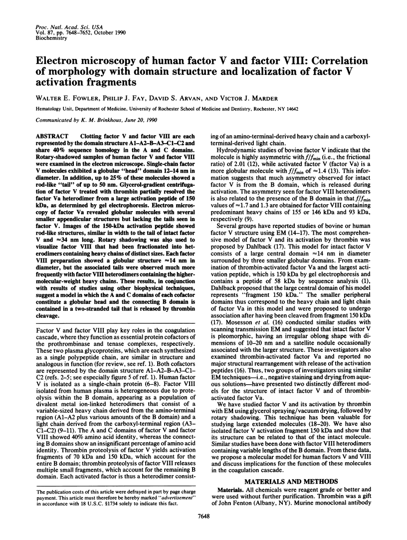
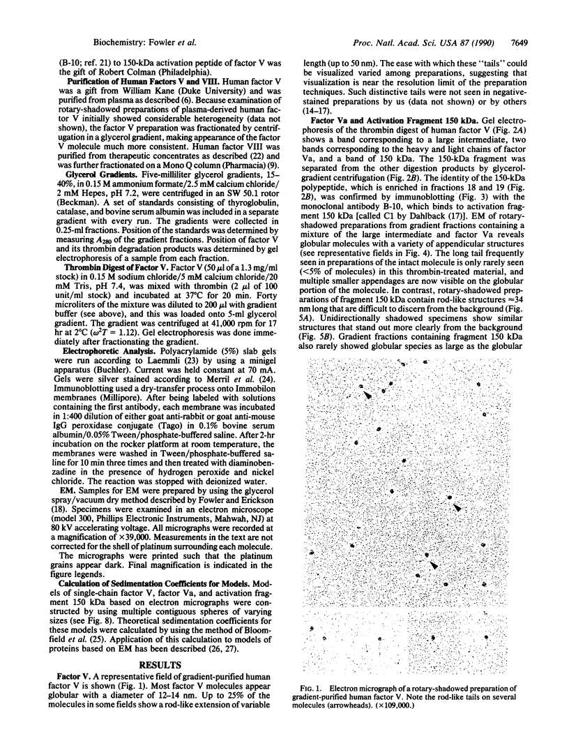
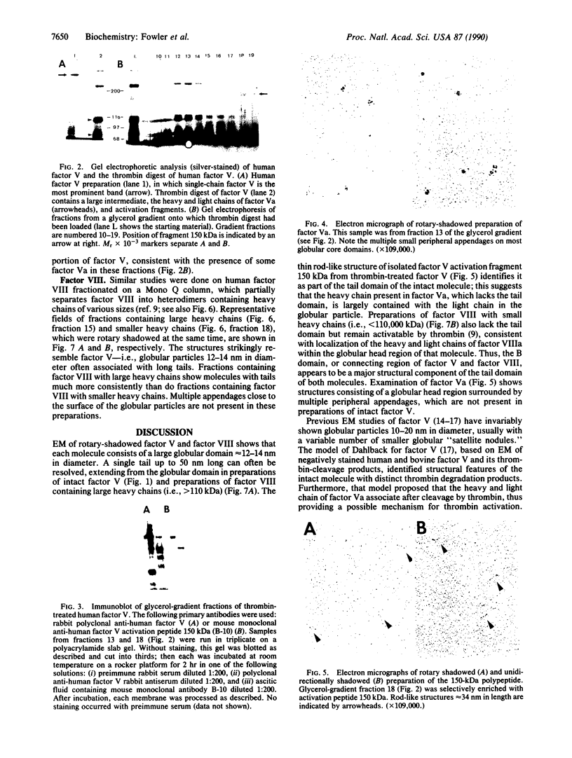
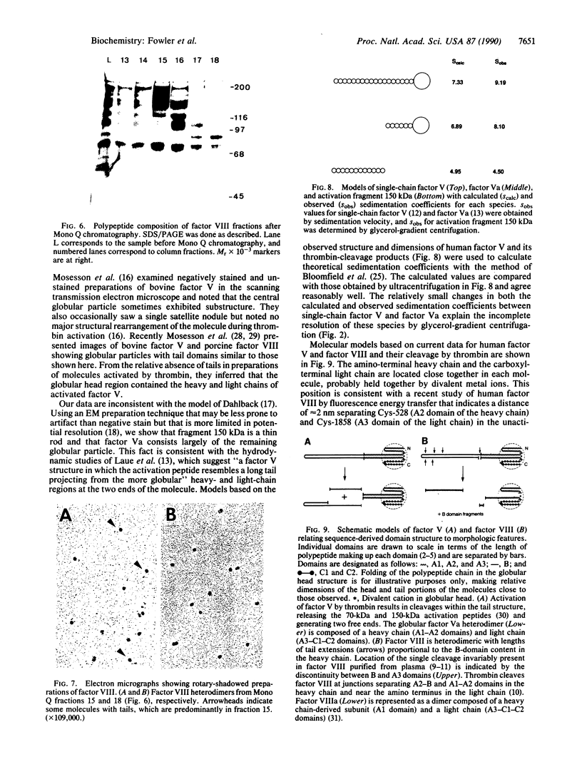
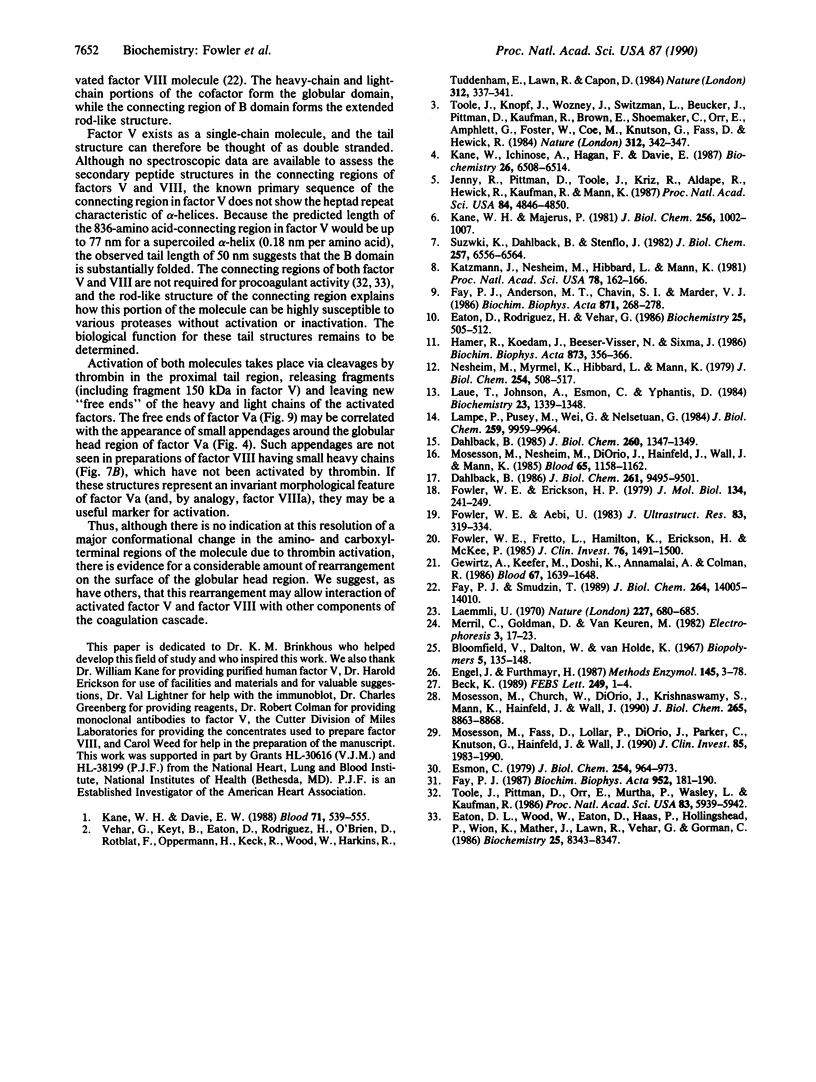
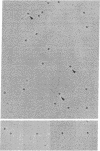
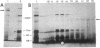
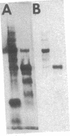
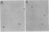
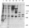
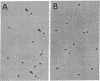
Clotting factor V and factor VIII are each represented by the domain structure A1-A2-B-A3-C1-C2 and share 40% sequence homology in the A and C domains. Rotary-shadowed samples of human factor V and factor VIII were examined in the electron microscope. Single-chain factor V molecules exhibited a globular "head" domain 12-14 nm in diameter. In addition, up to 25% of these molecules showed a rod-like "tail" of up to 50 nm. Glycerol-gradient centrifugation of factor V treated with thrombin partially resolved the factor Va heterodimer from a larger activation peptide of 150 kDa, as determined by gel electrophoresis. Electron microscopy of factor Va revealed globular molecules with several smaller appendicular structures but lacking the tails seen in factor V. Images of the 150-kDa activation peptide showed rod-like structures, similar in width to the tail of intact factor V and approximately 34 nm long. Rotary shadowing was also used to visualize factor VIII that had been fractionated into heterodimers containing heavy chains of distinct sizes. Each factor VIII preparation showed a globular structure approximately 14 nm in diameter, but the associated tails were observed much more frequently with factor VIII heterodimers containing the higher-molecular-weight heavy chains. These results, in conjunction with results of studies using other biophysical techniques, suggest a model in which the A and C domains of each cofactor constitute a globular head and the connecting B domain is contained in a two-stranded tail that is released by thrombin cleavage.
Full text
PDF
Images in this article
Selected References
These references are in PubMed. This may not be the complete list of references from this article.
- Beck K. Structural model of vinculin: correlation of amino acid sequence with electron-microscopical shape. FEBS Lett. 1989 May 22;249(1):1–4. doi: 10.1016/0014-5793(89)80002-x. [DOI] [PubMed] [Google Scholar]
- Bloomfield V., Dalton W. O., Van Holde K. E. Frictional coefficients of multisubunit structures. I. Theory. Biopolymers. 1967 Feb;5(2):135–148. doi: 10.1002/bip.1967.360050202. [DOI] [PubMed] [Google Scholar]
- Dahlbäck B. Bovine coagulation factor V visualized with electron microscopy. Ultrastructure of the isolated activated forms and of the activation fragments. J Biol Chem. 1986 Jul 15;261(20):9495–9501. [PubMed] [Google Scholar]
- Dahlbäck B. Ultrastructure of human coagulation factor V. J Biol Chem. 1985 Feb 10;260(3):1347–1349. [PubMed] [Google Scholar]
- Eaton D. L., Wood W. I., Eaton D., Hass P. E., Hollingshead P., Wion K., Mather J., Lawn R. M., Vehar G. A., Gorman C. Construction and characterization of an active factor VIII variant lacking the central one-third of the molecule. Biochemistry. 1986 Dec 30;25(26):8343–8347. doi: 10.1021/bi00374a001. [DOI] [PubMed] [Google Scholar]
- Eaton D., Rodriguez H., Vehar G. A. Proteolytic processing of human factor VIII. Correlation of specific cleavages by thrombin, factor Xa, and activated protein C with activation and inactivation of factor VIII coagulant activity. Biochemistry. 1986 Jan 28;25(2):505–512. doi: 10.1021/bi00350a035. [DOI] [PubMed] [Google Scholar]
- Engel J., Furthmayr H. Electron microscopy and other physical methods for the characterization of extracellular matrix components: laminin, fibronectin, collagen IV, collagen VI, and proteoglycans. Methods Enzymol. 1987;145:3–78. doi: 10.1016/0076-6879(87)45003-9. [DOI] [PubMed] [Google Scholar]
- Esmon C. T. The subunit structure of thrombin-activated factor V. Isolation of activated factor V, separation of subunits, and reconstitution of biological activity. J Biol Chem. 1979 Feb 10;254(3):964–973. [PubMed] [Google Scholar]
- Fay P. J., Anderson M. T., Chavin S. I., Marder V. J. The size of human factor VIII heterodimers and the effects produced by thrombin. Biochim Biophys Acta. 1986 Jun 23;871(3):268–278. doi: 10.1016/0167-4838(86)90208-6. [DOI] [PubMed] [Google Scholar]
- Fay P. J., Smudzin T. M. Intersubunit fluorescence energy transfer in human factor VIII. J Biol Chem. 1989 Aug 25;264(24):14005–14010. [PubMed] [Google Scholar]
- Fay P. J. Subunit structure of thrombin-activated human factor VIIIa. Biochim Biophys Acta. 1988 Jan 29;952(2):181–190. doi: 10.1016/0167-4838(88)90114-8. [DOI] [PubMed] [Google Scholar]
- Fowler W. E., Aebi U. Preparation of single molecules and supramolecular complexes for high-resolution metal shadowing. J Ultrastruct Res. 1983 Jun;83(3):319–334. doi: 10.1016/s0022-5320(83)90139-9. [DOI] [PubMed] [Google Scholar]
- Fowler W. E., Erickson H. P. Trinodular structure of fibrinogen. Confirmation by both shadowing and negative stain electron microscopy. J Mol Biol. 1979 Oct 25;134(2):241–249. doi: 10.1016/0022-2836(79)90034-2. [DOI] [PubMed] [Google Scholar]
- Fowler W. E., Fretto L. J., Hamilton K. K., Erickson H. P., McKee P. A. Substructure of human von Willebrand factor. J Clin Invest. 1985 Oct;76(4):1491–1500. doi: 10.1172/JCI112129. [DOI] [PMC free article] [PubMed] [Google Scholar]
- Gewirtz A. M., Keefer M., Doshi K., Annamalai A. E., Chiu H. C., Colman R. W. Biology of human megakaryocyte factor V. Blood. 1986 Jun;67(6):1639–1648. [PubMed] [Google Scholar]
- Hamer R. J., Koedam J. A., Beeser-Visser N. H., Sixma J. J. Human factor VIII: purification from commercial factor VIII concentrate, characterization, identification and radiolabeling. Biochim Biophys Acta. 1986 Oct 17;873(3):356–366. doi: 10.1016/0167-4838(86)90084-1. [DOI] [PubMed] [Google Scholar]
- Jenny R. J., Pittman D. D., Toole J. J., Kriz R. W., Aldape R. A., Hewick R. M., Kaufman R. J., Mann K. G. Complete cDNA and derived amino acid sequence of human factor V. Proc Natl Acad Sci U S A. 1987 Jul;84(14):4846–4850. doi: 10.1073/pnas.84.14.4846. [DOI] [PMC free article] [PubMed] [Google Scholar]
- Kane W. H., Davie E. W. Blood coagulation factors V and VIII: structural and functional similarities and their relationship to hemorrhagic and thrombotic disorders. Blood. 1988 Mar;71(3):539–555. [PubMed] [Google Scholar]
- Kane W. H., Ichinose A., Hagen F. S., Davie E. W. Cloning of cDNAs coding for the heavy chain region and connecting region of human factor V, a blood coagulation factor with four types of internal repeats. Biochemistry. 1987 Oct 6;26(20):6508–6514. doi: 10.1021/bi00394a033. [DOI] [PubMed] [Google Scholar]
- Kane W. H., Majerus P. W. Purification and characterization of human coagulation factor V. J Biol Chem. 1981 Jan 25;256(2):1002–1007. [PubMed] [Google Scholar]
- Katzmann J. A., Nesheim M. E., Hibbard L. S., Mann K. G. Isolation of functional human coagulation factor V by using a hybridoma antibody. Proc Natl Acad Sci U S A. 1981 Jan;78(1):162–166. doi: 10.1073/pnas.78.1.162. [DOI] [PMC free article] [PubMed] [Google Scholar]
- Laemmli U. K. Cleavage of structural proteins during the assembly of the head of bacteriophage T4. Nature. 1970 Aug 15;227(5259):680–685. doi: 10.1038/227680a0. [DOI] [PubMed] [Google Scholar]
- Lampe P. D., Pusey M. L., Wei G. J., Nelsestuen G. L. Electron microscopy and hydrodynamic properties of blood clotting factor V and activation fragments of factor V with phospholipid vesicles. J Biol Chem. 1984 Aug 10;259(15):9959–9964. [PubMed] [Google Scholar]
- Laue T. M., Johnson A. E., Esmon C. T., Yphantis D. A. Structure of bovine blood coagulation factor Va. Determination of the subunit associations, molecular weights, and asymmetries by analytical ultracentrifugation. Biochemistry. 1984 Mar 27;23(7):1339–1348. doi: 10.1021/bi00302a001. [DOI] [PubMed] [Google Scholar]
- Mosesson M. W., Church W. R., DiOrio J. P., Krishnaswamy S., Mann K. G., Hainfeld J. F., Wall J. S. Structural model of factors V and Va based on scanning transmission electron microscope images and mass analysis. J Biol Chem. 1990 May 25;265(15):8863–8868. [PubMed] [Google Scholar]
- Mosesson M. W., Fass D. N., Lollar P., DiOrio J. P., Parker C. G., Knutson G. J., Hainfeld J. F., Wall J. S. Structural model of porcine factor VIII and factor VIIIa molecules based on scanning transmission electron microscope (STEM) images and STEM mass analysis. J Clin Invest. 1990 Jun;85(6):1983–1990. doi: 10.1172/JCI114662. [DOI] [PMC free article] [PubMed] [Google Scholar]
- Mosesson M. W., Nesheim M. E., DiOrio J., Hainfeld J. F., Wall J. S., Mann K. G. Studies on the structure of bovine factor V by scanning transmission electron microscopy. Blood. 1985 May;65(5):1158–1162. [PubMed] [Google Scholar]
- Nesheim M. E., Myrmel K. H., Hibbard L., Mann K. G. Isolation and characterization of single chain bovine factor V. J Biol Chem. 1979 Jan 25;254(2):508–517. [PubMed] [Google Scholar]
- Suzuki K., Dahlbäck B., Stenflo J. Thrombin-catalyzed activation of human coagulation factor V. J Biol Chem. 1982 Jun 10;257(11):6556–6564. [PubMed] [Google Scholar]
- Toole J. J., Knopf J. L., Wozney J. M., Sultzman L. A., Buecker J. L., Pittman D. D., Kaufman R. J., Brown E., Shoemaker C., Orr E. C. Molecular cloning of a cDNA encoding human antihaemophilic factor. Nature. 1984 Nov 22;312(5992):342–347. doi: 10.1038/312342a0. [DOI] [PubMed] [Google Scholar]
- Toole J. J., Pittman D. D., Orr E. C., Murtha P., Wasley L. C., Kaufman R. J. A large region (approximately equal to 95 kDa) of human factor VIII is dispensable for in vitro procoagulant activity. Proc Natl Acad Sci U S A. 1986 Aug;83(16):5939–5942. doi: 10.1073/pnas.83.16.5939. [DOI] [PMC free article] [PubMed] [Google Scholar]