Abstract
Differentiated embryonic chondrocyte expressed gene 1 (DEC1) and differentiated embryonic chondrocyte expressed gene 2 (DEC2) belong to the Hairy/Enhancer of Split subfamily of basic helix-loop-helix factors. Previous studies have demonstrated that DEC proteins are involved in the regulation of circadian rhythms, response to hypoxia, and tumorigenesis. However, the roles of DEC1 and DEC2 in apoptosis of esophageal carcinoma remain unclear. In the present study, alterations in expression of apoptosis-related markers in human esophageal squamous cell carcinoma TE-11 cells treated with cisplatin were examined by western blot, while overall cell viability and apoptosis were analyzed by MTS assay and hematoxylin and eosin staining, respectively. Following cisplatin treatment, expression of DEC2 was downregulated, whereas expression of DEC1 was upregulated. DEC2 overexpression during cisplatin treatment markedly inhibited expression of the pro-apoptotic factor Bim and slightly increased the anti-apoptotic factor Bcl-xL. However, overexpression of DEC1 during cisplatin treatment failed to affect expression of these markers. Additionally, overexpression of DEC2 improved cell viability and decreased cell apoptosis induced by cisplatin. These results suggested that DEC2 exhibits anti-apoptotic effects in TE-11 esophageal squamous cell carcinoma cells. Inhibiting DEC2 may therefore have therapeutic potential for the treatment of esophageal cancer, in combination with cisplatin.
Keywords: differentiated embryonic chondrocyte expressed gene 1, differentiated embryonic chondrocyte expressed gene 2, apoptosis, esophageal squamous cell carcinoma, TE-11 cells
Introduction
Global cancer statistics suggest that ~455,800 new cases of esophageal cancer (EC) were diagnosed in 2012, and ~400,200 people succumbed to this disease worldwide (1). EC is ~3–4 times more common in males than females. EC is more malignant than other gastrointestinal cancers and is characterized by a variable geographic distribution of incidence and histological subtypes, including esophageal squamous cell carcinoma (ESCC) and esophageal adenocarcinoma (EAC) (1). ESCC is the most common subtype and accounts for over 90% of all EC cases in Asian countries, including China and Japan (2).
Cisplatin, a widely used platinum-containing chemotherapeutic drug, has been employed in the treatment of various types of cancer, and its primary function is induction of cell death by apoptosis (3). It is hypothesized that three different stages are involved in the apoptosis process: The initiation phase, the effector phase and the execution phase (4). The Bcl-2 protein family is a key regulator of the execution phase. There are >30 members of the Bcl-2 family of proteins reported in the literature, that are divided into the Bcl-2-like proteins, the Bcl2-associated X (Bax)-like proteins, and the Bcl-2 homology domain (BH3)-only proteins, according to their structure and function (5). When exposed to cytotoxic stimuli, BH3-only proteins initiate apoptosis by binding and inhibiting the anti-apoptotic Bcl-2-like proteins. This leads to the pro-apoptotic Bax-like proteins forming oligomers that permeabilize the mitochondrial outer membrane, resulting in the release of apoptogenic factors and activating the effector caspases to cause apoptosis (6). Bcl-2-interacting mediator of cell death (Bim), one of the BH3-only protein members, has been confirmed to have at least 6 isoforms (7). Among the various isoforms, BimEL (extra long form), BimL (long form) and BimS (short form) are the most well-characterized (5,8). Although all three isoforms promote apoptosis, they differ from each other in cytotoxicity and tissue expression distribution; BimS is the most cytotoxic but not universally expressed, while BimEL and BimL are expressed in various tissues and cell types (9).
Basic helix-loop-helix transcription factors differentiated embryonic chondrocyte expressed gene 1 [DEC1; officially known as basic helix-loop-helix family member e40 (BHLHE40)] and differentiated embryonic chondrocyte expressed gene 2 [DEC2; officially known as basic helix-loop-helix family member e41 (BHLHE41)] are regulators of circadian rhythms, response to hypoxia, mediators of epithelial-mesenchymal transition, and apoptosis (10–13). The function of DEC1 in apoptosis is controversial, however previous studies have revealed that DEC2 functions as an anti-apoptotic factor in several cancer cells, such as breast cancer MCF-7 cells and oral cancer HSC-3 cells (13–15). In the present study, the role of DEC1 and DEC2 in cisplatin-induced apoptosis was assessed in human esophageal TE-11 cancer cells.
Materials and methods
Cell culture and treatment
The human ESCC cell line TE-11 was purchased from the RIKEN BioResource Center (Tsukuba, Japan) through the National Bio-Resource Project of the Ministry of Education, Culture, Sports, Science and Technology, Japan. The cells were cultured in RPMI Medium 1640 (Gibco; Thermo Fisher Scientific, Inc., Waltham, MA, USA) supplemented with 10% fetal bovine serum (Gibco; Thermo Fisher Scientific, Inc.) at 37°C in a humidified atmosphere of 95% air and 5% CO2. Where noted, the cells were incubated with cisplatin (Sigma-Aldrich; Merck Millipore, Darmstadt, Germany) at various concentrations for 24 h.
DEC1 and DEC2 overexpression
The expression plasmids for human DEC1 and DEC2 were donated by Dr Katusmi Fujimoto (Hiroshima University, Japan) (11). TE-11 cells were seeded at 5×10⁴ cells per 35 mm well. DEC1 or DEC2 plasmid was transfected into the cells using Lipofectamine® LTX (Invitrogen; Thermo Fisher Scientific, Inc.) according to the manufacturer's instructions. Following transfection, the cells were incubated for 24 h and then treated with cisplatin at 20 or 40 µM for an additional 24 h. The cells were then used in the western blot analysis or the MTS assay.
Antibodies
Primary antibodies to DEC1 (cat. no. NB100-1800; Novus Biologicals, Ltd. Littleton, CO, USA; 1:5,000 dilution), DEC2 (H-72; cat. no. sc-32853; Santa Cruz Biotechnology, Inc., Dallas, TX, USA; 1:10,000 dilution), Bax (N-20; cat. no. sc-493; Santa Cruz Biotechnology, Inc.) and β-actin (cat. no. A5060; Sigma-Aldrich; Merck Millipore; 1:10,000 dilution) were used. Primary antibodies to poly ADP-ribose polymerase (PARP; cat. no. 9542; 1:5,000 dilution), cleaved caspase-3 (Asp175; cat. no. 9961; 1:1,000 dilution), cleaved caspase-8 (Asp391; cat. no. 9496; 1:1,5000 dilution), Bim (cat. no. 2819; 1:2,000 dilution), Bcl-2 (50E3; cat. no. 2870; 1:10,000 dilution) and Bcl-2-like 1 protein extra large (Bcl-xL; 54H6; cat. no. 2764; 1:10,000 dilution) were purchased from Cell Signaling Technology, Inc. (Danvers, MA, USA). Anti-rabbit IgG (cat. no. 17502) and anti-mouse IgG (cat. no. 17601) secondary antibodies were purchased from Immuno-Biological Laboratories Ltd. (Fujioka, Japan). Can Get Signal Immunoreaction Enhancer Solution (Toyobo Co., Ltd. Osaka, Japan) or Immunoshot Immunoreaction Enhancer Solution (Cosmobio Co., Ltd., Tokyo, Japan) were used to dilute the primary antibodies.
Western blot analysis
M-PER lysis buffer (Thermo Fisher Scientific, Inc.) was added to the cells cultured in a 6-well plate, and cells were incubated for 5 min at room temperature, with gentle agitation. The lysate was collected and transferred to a microcentrifuge tube and centrifuged at 12,000 × g for 10 min at 4°C. The supernatant was collected and transferred to a new tube for analysis. Protein concentration was determined using the bicinchoninic acid (BCA) assay. The purified protein (10 µg per lane) were subjected to 10% SDS-PAGE, and the proteins were transferred onto polyvinylidene difluoride membranes (Immobilon P; Merck Millipore), which were then probed with primary antibodies at 4°C overnight. The membranes were subsequently washed with TBS containing Tween 20, and were incubated with secondary antibodies for 1 h at room temperature with agitation. Proteins of interest were visualized with enhanced chemiluminescence (ECL) reagents using the ECL, ECL-Prime, or ECL-Select Western Blotting Detection system (Amersham; GE Healthcare Life Sciences, Chalfont, UK). Densitometry was performed using ImageJ version 1.48 (National Institutes of Health, Bethesda, MD, USA). Each experiment was repeated 3 times.
Cell viability assay
TE-11 cells were seeded at a density of 2.5×10³ into 96-well plates. The cells were transfected with an empty plasmid (pcDNA) or the expression plasmids for DEC1 or DEC2 (DEC1 pcDNA or DEC2 pcDNA, respectively). Following 18 h of transfection, the cells were cultured with or without 40 µM cisplatin for another 24 h. Cell viability was assessed with the MTS assay, as previously described (16).
Hematoxylin and eosin (H&E) staining
Apoptosis was evaluated by H&E staining. Briefly, TE-11 cells at 70% confluency were transfected with DEC2 plasmid DNA for 18 h, followed by treatment with 40 µM of cisplatin for 24 h. The cells were then fixed with 4% paraformaldehyde (Wako Pure Chemical Industries, Ltd., Osaka, Japan) in phosphate-buffered saline (PBS) for 15 min, permeabilized with 0.25% Triton X-100 (Sigma-Aldrich; Merck Millipore) in PBS for 20 min and finally stained by H&E.
Statistical analysis
Each experiment was repeated a minimum of three times. GraphPad Prism software version 7.02 (GraphPad Software, Inc., La Jolla, CA, USA) was used to perform one-way or two-way analyses of variance, followed by Dunnett's or Šidák's tests. Data are presented as the mean ± standard deviation. P<0.05 was considered to indicate a statistically significant difference.
Results
Effects of cisplatin on the expression of DEC1 and DEC2 in TE-11 cells
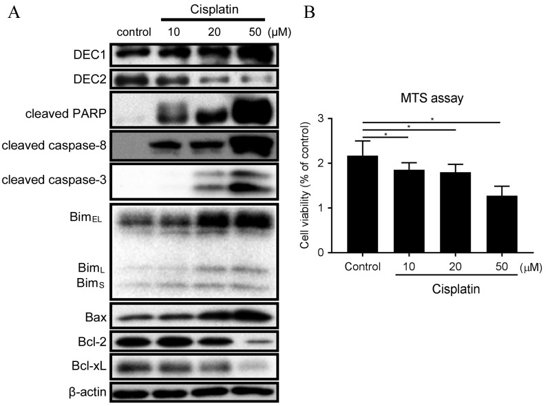
Cisplatin treatment resulted in different outcomes on the endogenous expression of DEC1 and DEC2 (Fig. 1A). Expression of DEC2 was decreased with 20 and 50 µM cisplatin, whereas expression of DEC1 was increased in the same conditions (Fig. 1A). Treatment with 10 µM cisplatin induced expression of cleaved PARP, cleaved caspase-8, BimEL, BimL and BimS (Fig. 1A). Treatment with 20 and 50 µM cisplatin further increased the amounts of cleaved PARP, cleaved caspase-8, cleaved caspase-3, Bim and Bax, whereas it decreased the expression of Bcl-2 and Bcl-xL (Fig. 1A). In addition, the ratio of Bax/Bcl-2 protein expression was strongly increased with 50 µM cisplatin (Fig. 1A). Treatment of TE-11 cells with 10, 20 and 50 µM cisplatin was demonstrated to significantly reduce cell viability (Fig. 1B).
Figure 1.
Cisplatin treatment affects expression of DEC1, DEC2 and apoptotic markers in TE-11 cells. (A) TE-11 cells were treated with 10, 20 and 50 µM cisplatin for 24 h. Cell lysates were subjected to western blot analysis for protein expression of DEC1, DEC2, cleaved PARP, cleaved caspase-8, cleaved caspase-3, Bim isoforms, Bax, Bcl-2, Bcl-xL and β-actin. (B) TE-11 cells were treated with 10, 20 and 50 µM cisplatin for 24 h, and then cell viability was measured using the MTS-assay. Data are presented as the mean ± standard deviation, from three independent experiments (*P<0.05). DEC, differentiated embryonic chondrocyte expressed gene; PARP, poly ADP-ribose polymerase; Bim, Bcl-2-interacting mediator of cell death; EL, extra long; L, long; S, short; Bax, Bcl-2 associated protein X apoptosis regulator; Bcl-2, B-cell lymphoma 2 apoptosis regulator; BcL-xL, Bcl-2-like 1 protein extra large.
DEC2 inhibits Bim expression and apoptosis induced by cisplatin in TE-11 cells
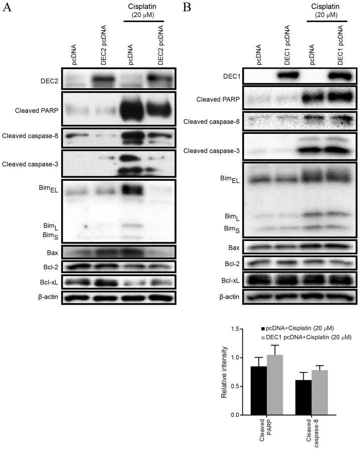
The effect of DEC1 or DEC2 overexpression on apoptosis was examined by transient transfection of expression plasmids into TE-11 cells. DEC2 overexpression in the presence of 20 µM cisplatin visibly decreased the amounts of cleaved PARP, cleaved caspase-3, cleaved caspase-8 and Bim (EL, L, and S) in TE-11 cells, compared with cells transfected with empty vector control (Fig. 2A). Conversely, DEC2 overexpression slightly increased the expression of Bcl-xL compared with control, but had little effect on the expression of Bcl-2 (Fig. 2A). Cisplatin augmented DEC1 expression in a dose dependent manner (Fig. 1A); however, DEC1 overexpression had no significant effect on cleaved PARP (1.24-fold those of pcDNA-trasnsfected group) or cleaved caspase-8 (1.28-fold those of pcDNA-transfected group) protein expression levels under cisplatin treatment in TE-11 cells (Fig. 2B).
Figure 2.
DEC2 inhibits expression of apoptotic markers following cisplatin treatment. TE-11 cells were transfected for 24 h with pcDNA, (A) DEC2 pcDNA or (B) DEC1 pcDNA. Transfected cells were incubated with 20 µM cisplatin for an additional 24 h, and then lysed. Cell lysates were subjected to western blot analysis for protein expression of DEC1, DEC2, cleaved PARP, cleaved caspase-8, cleaved caspase-3, Bim isoforms, Bax, Bcl-2, Bcl-xL and β-actin. Data are presented as the mean ± standard deviation, from three independent experiments. DEC, differentiated embryonic chondrocyte expressed gene; pcDNA, empty vector control; DEC2 pcDNA, DEC2 overexpression vector; DEC1 pcDNA, DEC1 overexpression vector; PARP, poly ADP-ribose polymerase; Bim, Bcl-2-interacting mediator of cell death; EL, extra long; L, long; S, short; Bax, Bcl-2 associated protein X apoptosis regulator; Bcl-2, B-cell lymphoma 2 apoptosis regulator; BcL-xL, Bcl-2-like 1 protein extra large.
DEC2 overexpression inhibits cisplatin-induced cell death in TE-11 cells
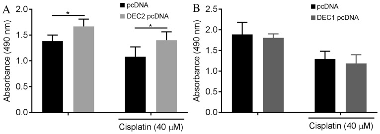
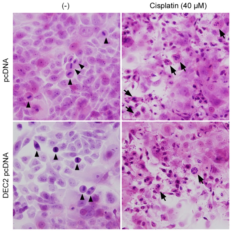
Overexpression of DEC1 or DEC2 and cisplatin treatment were combined in TE-11 cells to investigate whether DEC proteins affect cell viability (Fig. 3). DEC2 overexpression significantly increased the number of live cells in the absence or presence of cisplatin compared with pcDNA control (Fig. 3A), whereas DEC1 overexpression had little effect (Fig. 3B). Finally, the apoptotic changes in TE-11 cells were examined by H&E staining (Fig. 4). Cell mitoses were observed in the pcDNA- or DEC2 pcDNA-transfected cells without cisplatin treatment (Fig. 4, arrowheads), however, cisplatin significantly increased the number of apoptotic TE-11 cells (Fig. 4, arrows) and decreased the number of cell mitoses.
Figure 3.
DEC2 overexpression inhibits cell death induced by cisplatin. TE-11 cells were transfected for 24 h with pcDNA, (A) DEC2 pcDNA or (B) DEC1 pcDNA. Transfected cells were treated with 40 µM cisplatin for an additional 24 h, and then cell viability was measured using the MTS-assay. Data are presented as the mean ± standard deviation, from three independent experiments (*P<0.05). DEC, differentiated embryonic chondrocyte expressed gene; pcDNA, empty vector control; DEC2 pcDNA, DEC2 overexpression vector; DEC1 pcDNA, DEC1 overexpression vector.
Figure 4.
DEC2 overexpression inhibits apoptosis. Apoptosis was visualized by hematoxylin and eosin staining in TE-11 cells transfected with either pcDNA or DEC2 pcDNA, following treatment with either DMSO or 40 µM cisplatin for 24 h. Arrow heads indicate mitotic cells and arrows indicate apoptotic cells. DEC, differentiated embryonic chondrocyte expressed gene; pcDNA, empty vector control; DEC2 pcDNA, DEC2 overexpression vector.
Discussion
In the present study, the roles of DEC1 and DEC2 in the process of cisplatin-induced apoptosis of human esophageal carcinoma cells were analyzed. Cisplatin treatment increased expression of DEC1, but decreased expression of DEC2 in TE-11 cells. DEC2 overexpression in the presence of cisplatin markedly inhibited multiple apoptotic markers, including all three splice variants of Bim, BimEL, BimL and BimS, as well as cleaved caspase-3, cleaved caspase-8, and cleaved PARP. DEC2 had been demonstrated to inhibit apoptosis in the breast adenocarcinoma cell line MCF-7 (13) and the squamous cell carcinoma cell line HSC-3 (14). However, in another squamous cell carcinoma cell line, CA9-22, which demonstrates high endogenous expression of epidermal growth factor receptor (EGFR), DEC2 failed to affect cisplatin-induced apoptosis (14). In order to address this discrepancy, EGFR expression was analyzed in TE-11 cells. A431 cells, which are known to express high levels of EGFR, were used as a positive control. TE-11 cells exhibited significantly lower amount of EGFR expression than A431 cells (data not shown), which might explain why DEC2 functions as an inhibitor of apoptosis in TE-11 cells, but not in the previously reported CA9-22 cells.
DEC protein expression is modulated in normal and cancerous cells by several types of stimulation, such as cytokines (15), anticancer reagents (13), and hypoxia (12). DEC2 has also been identified as one of the members of the circadian genes, which exhibit rhythmic expression during the day, not only in normal cells but also in cancer cells (11,17). As a result, scientists and clinicians are beginning to take chronochemotherapy into consideration, which is a time-dependent manner of treatment administration to cancer patients. Cisplatin is one of the most commonly used chemotherapeutic drugs for the treatment of a variety of tumors. However, side effects including tumor resistance and nephrotoxicity greatly limit its use. It has been reported that cisplatin transporter molecules exhibit varying activity levels over a 24-h period, with greater expression in the evening compared with the morning (18). Future studies are required into the association between DEC2 and these transporter molecules, and the functions and underlying mechanisms of DEC2 in regulating apoptosis, to evaluate its full potential as a target in esophageal carcinoma chemotherapy.
Acknowledgements
This work was supported by JSPS KAKENHI, Grants-in-Aid for Young Scientists from the Ministry of Education, Culture, Sports, Science, and Technology of Japan (grant no. 26870028) and a Grant for Hirosaki University Institutional Research (grant no. 5310139303).
References
- 1.Torre LA, Bray F, Siegel RL, Ferlay J, Lortet-Tieulent J, Jemal A. Global cancer statistics, 2012. CA Cancer J Clin. 2015;65:87–108. doi: 10.3322/caac.21262. [DOI] [PubMed] [Google Scholar]
- 2.Lin Y, Totsuka Y, He Y, Kikuchi S, Qiao Y, Ueda J, Wei W, Inoue M, Tanaka H. Epidemiology of esophageal cancer in Japan and China. J Epidemiol. 2013;23:233–242. doi: 10.2188/jea.JE20120162. [DOI] [PMC free article] [PubMed] [Google Scholar]
- 3.Dasari S, Tchounwou PB. Cisplatin in cancer therapy: Molecular mechanisms of action. Eur J Pharmacol. 2014;740:364–378. doi: 10.1016/j.ejphar.2014.07.025. [DOI] [PMC free article] [PubMed] [Google Scholar]
- 4.Eastman A. The Mechanism of Action of Cisplatin: From Adducts to Apoptosis. Cisplatin Verlag Helvetica Chimica Acta. 2006:111–134. [Google Scholar]
- 5.Cory S, Adams JM. The Bcl2 family: Regulators of the cellular life-or-death switch. Nat Rev Cancer. 2002;2:647–656. doi: 10.1038/nrc883. [DOI] [PubMed] [Google Scholar]
- 6.Czabotar PE, Lessene G, Strasser A, Adams JM. Control of apoptosis by the BCL-2 protein family: Implications for physiology and therapy. Nat Rev Mol Cell Biol. 2014;15:49–63. doi: 10.1038/nrm3722. [DOI] [PubMed] [Google Scholar]
- 7.Marani M, Tenev T, Hancock D, Downward J, Lemoine NR. Identification of novel isoforms of the bh3 domain protein bim which directly activate bax to trigger apoptosis. Mol Cell Biol. 2002;22:3577–3589. doi: 10.1128/MCB.22.11.3577-3589.2002. [DOI] [PMC free article] [PubMed] [Google Scholar]
- 8.O'Connor L, Strasser A, O'Reilly LA, Hausmann G, Adams JM, Cory S, Huang DC. Bim: A novel member of the Bcl-2 family that promotes apoptosis. EMBO J. 1998;17:384–395. doi: 10.1093/emboj/17.2.384. [DOI] [PMC free article] [PubMed] [Google Scholar]
- 9.O'Reilly LA, Cullen L, Visvader J, Lindeman GJ, Print C, Bath ML, Huang DC, Strasser A. The Proapoptotic BH3-only protein bim is expressed in hematopoietic, epithelial, neuronal, and germ cells. Am J Pathol. 2000;157:449–461. doi: 10.1016/S0002-9440(10)64557-9. [DOI] [PMC free article] [PubMed] [Google Scholar]
- 10.Wu Y, Sato F, Yamada T, Bhawal UK, Kawamoto T, Fujimoto K, Noshiro M, Seino H, Morohashi S, Hakamada K, et al. The BHLH transcription factor DEC1 plays an important role in the epithelial-mesenchymal transition of pancreatic cancer. Int J Oncol. 2012;41:1337–1346. doi: 10.3892/ijo.2012.1559. [DOI] [PubMed] [Google Scholar]
- 11.Honma S, Kawamoto T, Takagi Y, Fujimoto K, Sato F, Noshiro M, Kato Y, Honma K. Dec1 and Dec2 are regulators of the mammalian molecular clock. Nature. 2002;419:841–844. doi: 10.1038/nature01123. [DOI] [PubMed] [Google Scholar]
- 12.Miyazaki K, Kawamoto T, Tanimoto K, Nishiyama M, Honda H, Kato Y. Identification of functional hypoxia response elements in the promoter region of the DEC1 and DEC2 genes. J Biol Chem. 2002;277:47014–47021. doi: 10.1074/jbc.M204938200. [DOI] [PubMed] [Google Scholar]
- 13.Wu Y, Sato F, Bhawal UK, Kawamoto T, Fujimoto K, Noshiro M, Morohashi S, Kato Y, Kijima H. Basic helix-loop-helix transcription factors DEC1 and DEC2 regulate the paclitaxel-induced apoptotic pathway of MCF-7 human breast cancer cells. Int J Mol Med. 2011;27:491–495. doi: 10.3892/ijmm.2011.617. [DOI] [PubMed] [Google Scholar]
- 14.Wu Y, Sato F, Bhawal UK, Kawamoto T, Fujimoto K, Noshiro M, Seino H, Morohashi S, Kato Y, Kijima H. BHLH transcription factor DEC2 regulates pro-apoptotic factor Bim in human oral cancer HSC-3 cells. Biomed Res. 2011;33:75–82. doi: 10.2220/biomedres.33.75. [DOI] [PubMed] [Google Scholar]
- 15.Liu Y, Sato F, Kawamoto T, Fujimoto K, Morohashi S, Akasaka H, Kondo J, Wu Y, Noshiro M, Kato Y, Kijima H. Anti-apoptotic effect of the basic helix-loop-helix (bHLH) transcription factor DEC2 in human breast cancer cells. Genes Cells. 2010;15:315–325. doi: 10.1111/j.1365-2443.2010.01381.x. [DOI] [PubMed] [Google Scholar]
- 16.Wu Y, Sato H, Suzuki T, Yoshizawa T, Morohashi S, Seino H, Kawamoto T, Fujimoto K, Kato Y, Kijima H. Involvement of c-Myc in the proliferation of MCF-7 human breast cancer cells induced by bHLH transcription factor DEC2. Int J Mol Med. 2015;35:815–820. doi: 10.3892/ijmm.2014.2042. [DOI] [PubMed] [Google Scholar]
- 17.Sato F, Bhawal UK, Kawamoto T, Fujimoto K, Imaizumi T, Imanaka T, Kondo J, Koyanagi S, Noshiro M, Yoshida H, et al. Basic-helix-loop-helix (bHLH) transcription factor DEC2 negatively regulates vascular endothelial growth factor expression. Genes Cells. 2008;13:131–144. doi: 10.1111/j.1365-2443.2007.01153.x. [DOI] [PubMed] [Google Scholar]
- 18.Sorensen DN, Dakup PP, Gaddameedhi S. Chronopharmacology of cisplatin: Role of the circadian rhythm in modulating the cisplatin transporters levels. The FASEB Journal. 2016;30:S935. (1 Suppl) [Google Scholar]


