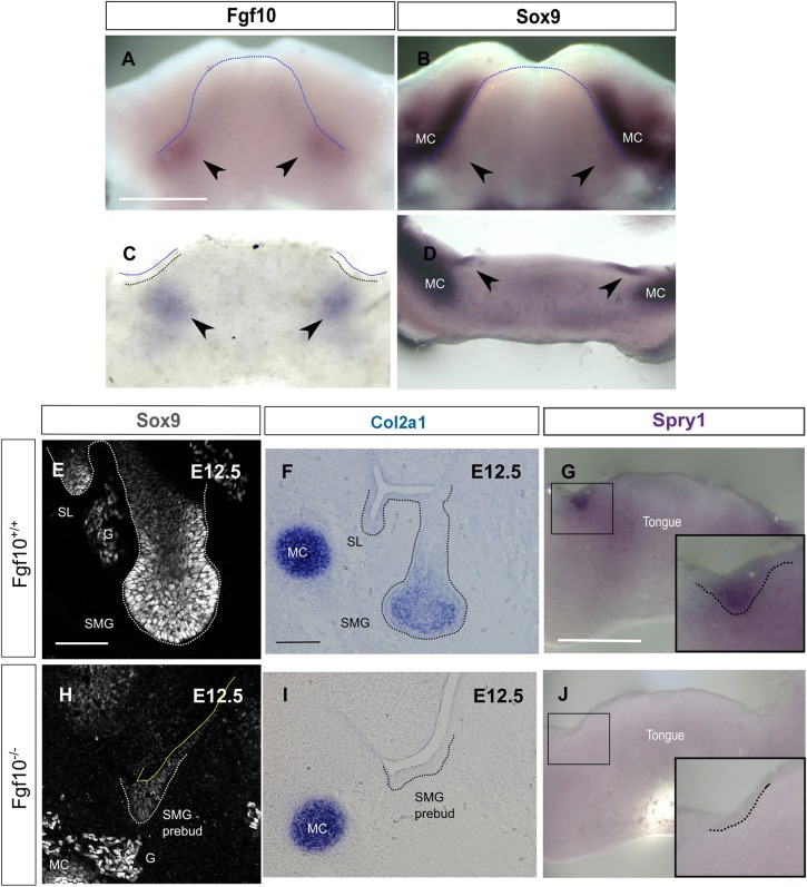
Fig. 8.
Fgf10 maintains Sox9 expression during the initial stages of salivary gland development. (A-D) In situ hybridisation for Fgf10 (A,C) and Sox9 (B,D) on E11.0 mandibles (A,B) and frontal mandibular slices (C,D). Arrowheads indicate the site of expression in submandibular glands. (E,H) Immunofluorescence for Sox9 in Fgf10+/+ and Fgf10−/− submandibular glands at E12.5. (F-J) In situ hybridisation for Col2a1 (F,I) and Spry1 (G,J) in Fgf10+/+ (F,G) and Fgf10−/− (I,J) submandibular glands. Dotted lines delineate the tongue (A,B), the placode of the salivary glands (C,G,J) or the salivary gland epithelium (E,F,H,I). Boxes (G,J) indicate the placode of the developing submandibular glands, as magnified in insets. G, ganglion; MC, Meckel's cartilage; SL, sublingual gland; SMG, submandibular gland. Scale bars: 500 μm (A-D,G,J); 50 μm (E,H); 100 μm (F,I).

