

Abstract
An 86-year-old man presented with severe pain in the upper abdomen along with fever. On physical examination, we found an arterial blood pressure of 84/43 mm Hg, a heart rate of 80 bpm and a temperature of 38.3°C. The abdomen was painful and peristalsis was absent. Empiric antibiotic therapy for sepsis was started with amoxicillin/clavulanate and gentamicin. CT scan of the abdomen revealed an emphysematous cholecystitis. Percutaneous ultrasound-guided cholecystostomy was applied. Bile cultures revealed Clostridium perfringens. Emphysematous cholecystitis is a life-threatening form of acute cholecystitis that occurs as a consequence of ischaemic injury to the gallbladder, followed by translocation of gas-forming bacteria (ie, C. perfringens, Escherichia coli, Klebsiella and Streptococci). The mortality associated with emphysematous cholecystitis is higher than in non-emphysematous cholecystitis (15% vs 4%). Therefore, early diagnosis with radiological imaging is of vital importance.
Background
We present a patient with an emphysematous cholecystitis caused by Clostridium perfringens. Emphysematous cholecystitis is a rare and life-threatening condition. In contrast to the findings in physical examination and laboratory testing, radiological examination shows characteristic findings.
We decided to write this case report because it represents a rare medical problem in which early diagnosis is of vital importance. We believe that this case report might be helpful in the diagnosis and treatment of this condition in future cases.
Case presentation
An 86-year-old man was admitted to the department of internal medicine, with severe pain in the upper abdomen, radiating to the right shoulder. The pain had been present for 3 days and had increased in the last hours. The patient had a fever and reported of nausea and vomiting. His medical history consisted mainly of chronic heart failure based on ischaemic cardiomyopathy.
On physical examination, the patient had an arterial blood pressure of 84/43 mm Hg and a heart rate of 80 bpm (while using a β-blocker). His temperature was 38.3°C. The abdomen was painful (predominantly in the right upper quadrant) and peristalsis was absent.
Investigations
Laboratory testing revealed leukocytosis (20.500/µL (normal range: 3.500–11.000/µL)), an elevation in serum kreatinin (389 µmol/L (normal range: 60–115 µmol/L)) and C reactive protein (237 mg/L (normal range:<10 mg/L)). Arterial blood gas analysis showed metabolic acidosis with respiratory compensation with normal lactate levels. Further laboratory testing showed no abnormalities. The CT scan of the abdomen revealed an emphysematous cholecystitis with intramural emphysema of the gallbladder and gas in the intrahepatic bile ducts (figure 1).
Figure 1.

CT image of emphysematous cholecystitis. There is intramural emphysema involving the gallbladder (arrow left), as well as gas in the intrahepatic bile ducts (arrow right). There were no gallstones.
Treatment
According to our local protocol, empiric antibiotic therapy for sepsis was started with amoxicillin/clavulanate and gentamicin.
The patient’s clinical condition deteriorated and he became haemodynamically unstable. Cholecystectomy was not performed because of his poor medical condition. Source control with percutaneous ultrasound-guided cholecystostomy was applied and approximately 100 mL of black fluid was drained from the gallbladder.
Outcome and follow-up
Despite our treatment, the patient's condition deteriorated. He remained haemodynamically unstable and died the next day. Bile cultures were positive for C. perfringens, sensitive to amoxicillin/clavulanate (figure 2).
Figure 2.
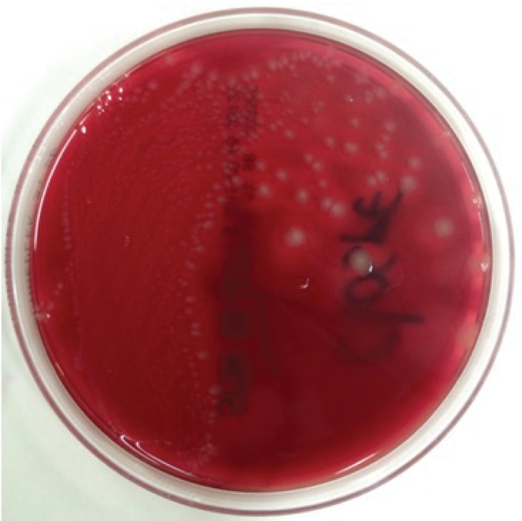
A photograph of the bile culture shows moderate growth of bacteria (anaerobic agar plate). MALDI/TOF analysis identified the bacteria as Clostridium perfringens.
Discussion
Emphysematous cholecystitis is a rare diagnosis and a life-threatening form of acute cholecystitis. Its pathophysiology differs from cholecystitis caused by abnormal biliary-enteric communication (ie, gallstones).1 2 Emphysematous cholecystitis occurs as a consequence of ischaemic injury to the gallbladder.2 3 The main predisposing factors are atherosclerosis, systemic hypoperfusion, arterial embolism and vasculitis. Emphysematous cholecystitis is also associated with diabetes mellitus.3 Ischaemic necrosis is followed by translocation of gas-forming bacteria and subsequent accumulation of gas in the wall and the lumen of the gallbladder.2
Emphysematous cholecystitis, unlike non-emphysematous cholecystitis, is more commonly observed in elderly men (male–female ratio 4:1). Many of the affected individuals have diabetes mellitus.1 2 Commonly reported causative organisms include C. perfringens, Escherichia coli, Klebsiella and Streptococci.2
The infection can be complicated by gangrene, perforation and sepsis. The mortality associated with emphysematous cholecystitis is higher than in non-emphysematous cholecystitis (15% vs 4%).2 3 Therefore, early diagnosis with radiological imaging, such as CT, is of vital importance—given the non-specific findings in physical examination and laboratory testing—and the characteristic findings in radiological examination.4 It is important to consider that the ultrasound report may note “presence of overlying bowel gas, which makes the visualisation of the gallbladder difficult”, while in fact this might reflect the presence of air in the gallbladder wall.5
Treatment of choice, besides antibiotic therapy aimed at the causative organisms, is cholecystectomy. Early surgical intervention might prevent the development of gangrene and perforation, which in turn will reduce mortality.1 Cholecystectomy is traditionally performed by laparotomy. Laparoscopic intervention is less invasive, but high conversion rates are reported in emergency settings.2 In elderly patients in poor condition, in whom cholecystectomy is not possible, cholecystostomy can alternatively be used as a temporary means of source control.2 6
Learning points.
Emphysematous cholecystitis is a rare diagnosis and a life-threatening form of acute cholecystitis. Mainly due to septic shock, gangrene and perforation, mortality rates are higher than that of non-emphysematous cholecystitis.
Emphysematous cholecystitis occurs as a consequence of ischaemic injury to the gallbladder, leading to translocation of gas-forming bacteria.
Early diagnosis is of vital importance. CT scan of the abdomen shows characteristic findings.
Abdominal ultrasound may be inconclusive, because the presence of gas in the gallbladder wall might be mistaken for overlying bowel gas.
Footnotes
Competing interests: None declared.
Patient consent: Obtained.
Provenance and peer review: Not commissioned; externally peer reviewed.
References
- 1.Rosenberg AA, Cherry-Bukowiec JR, Li SH et al. Emphysematous cholecystitis. Surg Infect (Larchmt) 2013;14:483–5. 10.1089/sur.2012.157 [DOI] [PubMed] [Google Scholar]
- 2.Bouras G, Lunca S, Vix M et al. A case of emphysematous cholecystitis managed by laparoscopic surgery. JSLS 2005;9:478–80. [PMC free article] [PubMed] [Google Scholar]
- 3.Carrascosa MF, Salcines-Caviedes JR. Clinical images: emphysematous cholecystitis. CMAJ 2012;10:184. [DOI] [PMC free article] [PubMed] [Google Scholar]
- 4.Shrestha Y, Trottier S. Images in clinical medicine. Emphysematous cholecystitis. N Engl J Med 2007;357:1238 10.1056/NEJMicm063675 [DOI] [PubMed] [Google Scholar]
- 5.Zakko SF, Afdhal NH, Chopra S et al. Acute cholecystitis: pathogenesis, clinical features and diagnosis. UpToDate Chopra S (Ed). http://www.uptodate.com/contents/acute-cholecystitis-pathogenesis-clinical-features-and-diagnosis?source=search_result&search=acute+cholecystitis&selectedTitle=1~67#H249918905. Accessed December 2015. [Google Scholar]
- 6.Katagiri H, Yoshinaga Y, Kanda Y et al. Emphysematous cholecystitis successfully treated by laparoscopic surgery. J Surg Case Rep 2014;2014:pii: rju027 10.1093/jscr/rju027 [DOI] [PMC free article] [PubMed] [Google Scholar]