Summary
Background
Inertial measurement unit (IMU) sensor‐based techniques are becoming more popular in horses as a tool for objective locomotor assessment.
Objectives
To describe, evaluate and validate a method of stride detection and quantification at walk and trot using distal limb mounted IMU sensors.
Study design
Prospective validation study comparing IMU sensors and motion capture with force plate data.
Methods
A total of seven Warmblood horses equipped with metacarpal/metatarsal IMU sensors and reflective markers for motion capture were hand walked and trotted over a force plate. Using four custom built algorithms hoof‐on/hoof‐off timing over the force plate were calculated for each trial from the IMU data. Accuracy of the computed parameters was calculated as the mean difference in milliseconds between the IMU or motion capture generated data and the data from the force plate, precision as the s.d. of these differences and percentage of error with accuracy of the calculated parameter as a percentage of the force plate stance duration.
Results
Accuracy, precision and percentage of error of the best performing IMU algorithm for stance duration at walk were 28.5, 31.6 ms and 3.7% for the forelimbs and −5.5, 20.1 ms and −0.8% for the hindlimbs, respectively. At trot the best performing algorithm achieved accuracy, precision and percentage of error of −27.6/8.8 ms/−8.4% for the forelimbs and 6.3/33.5 ms/9.1% for the hindlimbs.
Main limitations
The described algorithms have not been assessed on different surfaces.
Conclusions
Inertial measurement unit technology can be used to determine temporal kinematic stride variables at walk and trot justifying its use in gait and performance analysis. However, precision of the method may not be sufficient to detect all possible lameness‐related changes. These data seem promising enough to warrant further research to evaluate whether this approach will be useful for appraising the majority of clinically relevant gait changes encountered in practice.
Keywords: horse, inertial measurement unit, gait analysis, stride events, kinematics
Introduction
Many gait events can be effectively evaluated through subjective visual lameness appraisal by an experienced clinician 1. However, although it has been shown that experienced clinicians are more repeatable and have a higher interobserver agreement in detection of gait asymmetries when compared with inexperienced observers 2, even the most experienced clinicians are reliant on the limitations of the human visual perception of motion 3. Inertial measurement unit (IMU) sensor‐based technology for objective gait analysis in horses has been under constant development. These systems often rely on sensors mounted in the midline of the horse (e.g. pelvis and head) that make use of a signal decomposition routine 4 to describe and record motion in the vertical plane. This method provides reliable and objective quantification of head and pelvis movement asymmetry which is related to changes in weightbearing and propulsion due to lameness. Quantification of limb kinematics or spatial and temporal stride variables might be a useful addition to the current technology, providing extra information that might be related to specific gait changes due to lameness. To achieve this, characterisation of the gait and motion patterns of each individual limb, which are known to influence trunk and head motion symmetry 5, 6, is necessary.
The accurate and precise detection of the stride, i.e. the accurate detection of hoof‐on/off events, is a crucial prerequisite for the proper determination of temporal and spatial stride characteristics and the currently used IMU‐based systems can only achieve this partially. Since these sensors are commonly mounted on the upper body of the horse they can only obtain limited information regarding limb gait events like hoof‐on/off at trot 7 or at walk 8, thereby severely limiting their application for detection of limb kinematics.
The purpose of this study was to describe, evaluate and validate a new method of stride detection and characterisation using limb mounted IMU sensors at walk and trot. It was hypothesised that the use of IMU sensors with a higher G range than usual and using multi‐level algorithms would enable accurate and precise detection of hoof‐on and hoof‐off gait events that would be comparable to the performance of motion capture as the other alternative to the use of a force plate, the use of which principally is limited to laboratory situations.
Materials and methods
Horses
A total of seven Warmblood horses (six mares and one gelding) with a body mass range of 506–608 kg (mean 564.4 kg), height at the withers range 1.58–1.75 m (mean 1.65 m) and age range of 5–21 years (mean 7.5 years) were used for this study. All subjects had no history of lameness and no lameness was observed during visual examination at walk and trot on a straight line prior to data collection.
Data collection
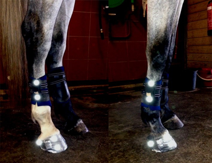
All subjects were instrumented with one IMU sensor (Promove‐mini)a on each limb. Each sensor (mass 20 g) was firmly attached to the lateral aspect of each metacarpal/metatarsal bone using a custom made holster (Fig 1). Additionally, IMUs were attached to the right front and hind hooves for data collection for another, unrelated study. The sensors were set to a sampling rate of 200 Hz, with the low g accelerometer set at ±16 g and the high g accelerometer set at ±200 g. Data was stored using the internal memory of each sensor (2 Gb microSD). Synchronisation between all sensors was guaranteed with an error of less than 100 ns. Reflective markers (12.5 mm Ø, spherical passive marker)b were glued proximally and distally to each of the limb mounted IMUs and three additional markers attached to each right hoof (lateral heel, lateral toe area and lateral coronet). Motion capture data was recorded at 200 Hz using 6 infrared cameras (ProReflex 240)b, positioned along the y axis of a force plate (Z4852C)c. The force plate was covered with a 5 mm rubber mat. The analogue force plate signal was fed in to an A/D converterb, processed at 1 kHz with 12 bit resolution and sampled by the motion capture software (QTM)b at 200 Hz ensuring that the sampling moments between measurements of the two systems have a fixed and known timing relationship. Speed over the force plate was measured using two pairs of photoelectric sensors spaced 2 m apart, centred over the force plate. All trials were videorecorded using standard equipment for retrospective analysis of the collected data. Prior to data collection, all instruments were calibrated according to manufacturers’ instructions.
Figure 1.

Reflective motion capture markers and inertial measurement unit (IMU) sensors attached to a horse in standardised locations. The IMU sensors were attached to the right lateral metatarsal and metacarpal bones, one reflective marker was placed above and beneath each sensor. Also, three markers were attached to the right fore‐ and right hind hooves (heel, lateral toe and lateral coronet), but only the lateral heel and lateral toe markers were used for the motion capture detection algorithm in the current study.
All subjects were fitted with the instruments and were led over the force plate by an experienced handler in walk and trot for 5 min as a warm‐up exercise before the data collection. A minimum of five valid force plate impacts of the right front hoof and right hind hoof were collected at walk and trot from all subjects. In order to collect five valid measurements for each limb, an average of 25 trials were needed for trot and 19 for walk. A trial was considered to be valid if the impact of the hoof was located within the force plate measuring limits; only one limb was in contact with the plate at a time, if the horse was trotting straight at a constant pace and within a preset speed range of 0.8–1.4 m/s at walk and 1.7–2.7 m/s at trot.
Kinematic analysis
All collected data was processed and analysed in custom written scripts using Matlab (r2015a)d. The IMU collected data was frame synchronised with the motion capture system by evaluating the cross correlation function between the metacarpal/metatarsal angular velocity measured by the IMU and the motion capture system. A threshold of 30 N was used for the force plate calculations. The hoof‐on event was determined as the moment when the load along the vertical axis (z) passed 30 N and hoof‐off as the first moment after hoof‐on, when the force along the z axis dropped below 30 N. Detailed information on the synchronisation method used is provided as Supplementary Item 1.
Inertial measurement unit event detection algorithm
A total of four algorithms were used to calculate the hoof‐on/off events. Algorithm 1 is a novel algorithm based on direct inertial measures (acceleration and rate of turn), requiring very low computing power and suitable for running in a real‐time configuration. Algorithm 2 is an implementation of a previously described algorithm 8. A post‐processing routine based on the vector magnitude of the acceleration signal (acceleration magnitude peak) calculated by the IMU accelerometer was developed as an attempt to increase the accuracy and precision of algorithms 1 and 2. This routine was applied to algorithms 1 and 2 resulting in two new algorithms, 3 and 4, respectively. A detailed description of the IMU algorithms is provided as Supplementary Item 2.
Motion capture event detection algorithm
Using the data from heel and toe reflective markers, an algorithm was designed to detect the toe/heel‐on/off events. The algorithm is based on direct kinematic data collected by the motion capture cameras 9. A detailed description of the motion capture algorithms is provided as Supplementary Item 3.
Data analysis
Using the force plate as the gold standard reference for stride characterisation timing, accuracy of the computed parameters (i.e. hoof‐on/off, toe‐on/off, heel‐on/off) was calculated as the difference in milliseconds between the IMU or motion capture generated data and the data from the force plate and precision as the s.d. of the accuracy. A positive accuracy indicates an overestimation (i.e. IMU or motion capture detection of event later than force plate) of the parameter calculated by the IMU or motion capture system and a negative accuracy indicates an underestimation of that parameter (i.e. IMU or motion capture detection of event before force plate). Performance of the algorithms was judged based on primarily demonstrating the best precision (closest to zero) combined with the best accuracy (closest to zero).
For further comparison of the algorithms’ performance, the accuracy of IMU stance duration was determined as described above for hoof‐on/off, with the force plate calculated stance duration as a reference and with stance duration defined as the time between hoof‐on and the subsequent hoof‐off. Combination of algorithms for stance duration calculation were also evaluated and tested to identify the combination that obtained the best overall accuracy and precision. For the motion capture data, stance duration was calculated for both limbs and gaits, based on four possible combinations (i.e. toe‐on toe‐off; toe‐on heel‐off; heel‐on heel‐off and heel‐on toe‐off). Accuracy and precision of motion capture stance duration were calculated as described above for the IMU. The percentage of error in stance duration was calculated with the accuracy as a percentage of the force plate measured stance duration. If any of the IMU or motion capture algorithms failed to perform detection of an event (e.g. hoof‐on/off), the calculation of that specific event for that trial was not included in the final calculations. This was verified in a number of trials in our experiment for algorithms 1, 3 and algorithm combination (n = 2; n = 3, respectively) and in eight events for the motion capture detection. However, the remaining calculated parameters not related to that specific event for that trial were kept.
Open software (R version 3.2.3)e was used for statistical analysis using the package ‘nlme’ (version 3.1–121) for linear mixed effects model and ‘BlandAltmanLeh’ (version 0.1.0) for calculation of IMU and motion capture stance duration limits of agreement. For statistical comparison of the 4 different IMU algorithms and the algorithm combination, the calculated square root transformed absolute accuracy for the stance duration was used as the outcome variable. Horse Id was used as random effect to account for the correlated observations within horse; explanatory variables are algorithm, gait, limb and the interaction between limb and algorithm. A constant variance function (varIdent) for algorithms was added to the model to take the different variances between algorithms into account. Model adequacy (normality and constancy of variance) was confirmed using visualisation of the scatter plot residuals vs. fitted values and explanatory variables respectively and QQ‐plots. The Akaike's information criterion was used to select the best model.
Results
A total of 150 stance phases were collected; 77 for the front limb (37 for walk; 40 at trot) and 73 for the hindlimb (36 at walk and 37 at trot). Visual assessment of the force plate ground reaction forces data showed the typical patterns for each specific gait and three measurements were excluded from the final analysis due to abnormal ground reaction force curves.
Inertial measurement unit‐based determination of hoof‐on/hoof‐off events compared with force plate data (descriptive statistics)
The four different tested algorithms performed differently for both limbs and gaits as demonstrated in Table 1. Algorithm 3 had the best performance for hoof‐on, at walk in the front limb (accuracy: 0.3 ms; precision: 11.5 ms). For the hoof‐on detection algorithm 2 had an overall negative accuracy that after the acceleration magnitude peak post‐processing routine (algorithm 4) becomes closer to zero in the front limb and in the hindlimb becomes positive; however, precision hardly changes. The hoof‐off detection for most algorithms shows a negative accuracy. Algorithm 1, after the post‐processing acceleration magnitude peak routine (algorithm 3), for hoof‐off detection, shows an improvement in accuracy for both front and hindlimb at walk and trot. When comparing within the same algorithm, the hoof‐off and hoof‐on moments, the latter had better accuracy and precision than the former.
Table 1.
Tabular representation of the descriptive statistics for Inertial measurement unit (IMU) hoof‐on and hoof‐off detection vs. the ‘gold standard’ force plate (FP)
| Algorithm | Hoof‐on | Hoof‐off | ||||
|---|---|---|---|---|---|---|
| Accuracy (ms) | Precision (ms) | Accuracy (ms) | Precision (ms) | |||
| Walk | Front | 1 | 10.9 | 27.2 | 28.8 | 26.0 |
| 2 | −71.0 | 31.1 | −45.2 | 51.5 | ||
| 3 | 0.3 | 11.5 | 14.2 | 31.0 | ||
| 4 | −58.8 | 46.1 | −40.5 | 53.2 | ||
| Hind | 1 | 14.1 | 8.1 | −42.7 | 12.9 | |
| 2 | −18.3 | 13.5 | −15.1 | 21.2 | ||
| 3 | 2.0 | 11.5 | −5.4 | 14.3 | ||
| 4 | 0.1 | 14.8 | −9.8 | 26.6 | ||
| Trot | Front | 1 | −3.8 | 23.9 | 28.8 | 17.5 |
| 2 | −99.2 | 58.0 | −26.8 | 19.2 | ||
| 3 | 7.9 | 6.7 | −3.7 | 35.4 | ||
| 4 | −82.6 | 61.8 | −19.7 | 7.5 | ||
| Hind | 1 | 16.3 | 10.1 | 17.6 | 29.1 | |
| 2 | −10.8 | 12.5 | −17.9 | 46.7 | ||
| 3 | 11.3 | 9.1 | −2.3 | 46.9 | ||
| 4 | 11.3 | 9.1 | −19.9 | 32.7 | ||
n = 7 horses. Accuracy, mean difference in milliseconds between IMU and FP; precision, the s.d. of the accuracy between IMU and FP. Accuracy and precision are deemed better if closer to zero.
Motion capture‐based determination of hoof‐on/off events compared with force plate (descriptive statistics)
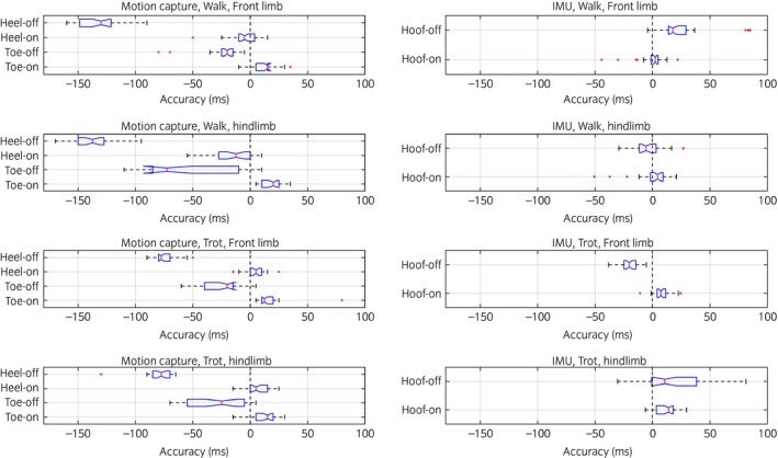
The performance of our motion capture‐based detection is summarised in Table 2 and a descriptive comparison with the IMU algorithms is illustrated in Figure 2. The hoof‐on event defined as heel‐on measured by the motion capture system showed the best overall performance, except at walk for the hindlimbs where the toe‐on moment appears to be the most accurate and precise method for this purpose. The hoof‐off moment defined as the toe‐off measured by the motion capture system shows the best overall accuracy and precision combination. Similar to the IMU‐based determination of hoof‐on/off events, the hoof‐on detection has a better overall performance when compared with hoof‐off detection. However, the IMU approach performed overall better for hoof‐off detection.
Table 2.
Descriptive statistics for motion capture determined hoof‐on and hoof‐off detection vs. force plate (FP) measured stance duration events
| Toe‐on | Heel‐on | Toe‐off | Heel‐off | ||||||
|---|---|---|---|---|---|---|---|---|---|
| Accuracy (ms) | Precision (ms) | Accuracy (ms) | Precision (ms) | Accuracy (ms) | Precision (ms) | Accuracy (ms) | Precision (ms) | ||
| Walk | Front | 4.0 | 51.9 | −5.1 | 13.0 | −27.1 | 20.7 | −132.1 | 18.3 |
| Hind | 18.4 | 8.1 | −15.8 | 17.4 | −57.7 | 39.1 | −136.4 | 16.9 | |
| Trot | Front | 15.5 | 12.4 | 3.9 | 8.3 | −23.2 | 17.0 | −75.8 | 10.0 |
| Hind | 14.5 | 7.4 | 5.6 | 10.0 | −27.0 | 25.8 | −78.0 | 7.8 | |
Accuracy, mean difference in milliseconds (ms) between the motion capture calculated and the FP measured stance duration; precision, s.d. of the mean difference between the motion capture calculated and FP measured stance duration (accuracy). Accuracy and precision are deemed better if closer to zero.
Figure 2.

Horizontal box plot of motion capture‐based detection accuracy (left) and inertial movement unit (algorithm combination as described in Table 3) based detection accuracy (right). A positive accuracy indicates an over estimation of the detected event [i.e. inertial measurement unit (IMU) or motion capture detection of event later than force plate] and a negative accuracy indicates an under estimation of the event (i.e. IMU or motion capture detection of event before force plate). Box represents the interquartile range, whiskers represent 75th percentile + 1.5 *interquartile range (IQR) and 25th percentile −1.5 *IQR, respectively. Notch represents the 95% confidence interval of the median.
Inertial measurement unit stance duration accuracy, precision, limits of agreement and model estimates
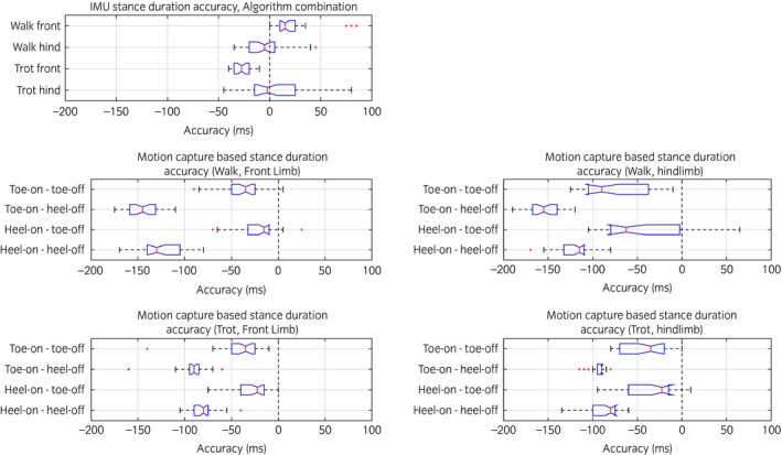
The stance duration calculation performance of the IMU algorithms and the algorithm combination is resumed in Table 3 and illustrated in Figure 3. Bland‐Altman limits of agreement can be found in Table 3, Figure 4 and model estimates are resumed in Supplementary Item 4.
Table 3.
Descriptive statistics of stance duration of the different Inertial measurement unit (IMU) algorithms vs. ‘gold standard’ force plate (FP)
| Algorithm | Accuracy (ms) | Precision (ms) | Lower limits of agreement | Upper limits of agreement | Error (%) | ||
|---|---|---|---|---|---|---|---|
| Walk | Front | 1 | 17.9 | 35.7 | −52.1 | 87.9 | 2.3 |
| 2 | 25.8 | 51.2 | −74.5 | 126.2 | 3.4 | ||
| 3 | 13.9 | 31.49 | −47.8 | 75.6 | 1.8 | ||
| 4 | 18.3 | 60.3 | −99.8 | 136.5 | 2.4 | ||
| Combination (3 + 1) | 28.5 | 31.6 | −33.5 | 90.4 | 3.7 | ||
| Hind | 1 | −56.8 | 12.5 | −81.1 | −32.3 | −7.4 | |
| 2 | 3.2 | 23.6 | −43.1 | 49.5 | 0.5 | ||
| 3 | −7.4 | 17.8 | −42.3 | 27.4 | −1 | ||
| 4 | −9.8 | 29.9 | −68.5 | 48.8 | −1.2 | ||
| Combination (4 + 3) | −5.5 | 20.1 | −44.8 | 33.8 | −0.8 | ||
| Trot | Front | 1 | 32.6 | 28.1 | −22.4 | 87.6 | 10.2 |
| 2 | 72.4 | 55.7 | −36.8 | 181.6 | 21.9 | ||
| 3 | −11.6 | 34.6 | −79.4 | 56.2 | −3.8 | ||
| 4 | 62.9 | 64.0 | −62.5 | 188.3 | 18.8 | ||
| Combination (3 + 4) | −27.6 | 8.8 | −44.8 | −10.4 | −8.4 | ||
| Hind | 1 | 1.3 | 34.4 | −66.1 | 68.8 | 1.3 | |
| 2 | −7.1 | 50.1 | −105.3 | 91.1 | −2.2 | ||
| 3 | −13.6 | 48.9 | −109.4 | 82.3 | −4.2 | ||
| 4 | −31.2 | 31.6 | −93.1 | 30.8 | −10.6 | ||
| Combination (3 + 1) | 6.3 | 33.5 | −59.4 | 72.0 | 3 |
n = 7 horses. Accuracy, mean difference in milliseconds (ms) between IMU and FP stance duration; precision, the s.d. of the accuracy; error, the relative mean difference between IMU and FP stance duration (accuracy) as a percentage of the FP stance duration. Accuracy, precision and error are deemed better if closer to zero.
Figure 3.

Horizontal box plot of the accuracy of stance duration. Top left, accuracy with inertial measurement unit (IMU) sensors with algorithm combination as described in Table 3. Remaining plots represent the stance duration calculated using the motion capture data and based on toe/heel‐on/off moments and possible combinations thereof. Positive accuracy indicates over estimation of the detected event (i.e. IMU or motion capture detection of the event later than force plate) and negative accuracy indicates under estimation of the event (i.e. IMU or motion capture detection of the event before force plate). Accuracy is higher closer to zero. The box represents the interquartile range, whiskers represent 75th percentile + 1.5 *interquartile range (IQR) and 25th percentile −1.5 *IQR, respectively. Notch represents the 95% confidence interval of the median.
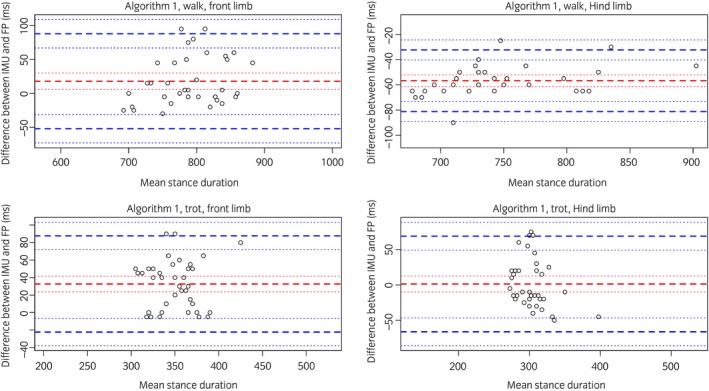
Figure 4.

Bland‐Altman plots of the stance duration calculated by the Z (algorithm 1) vs. force plate calculated stance duration. Top left, data calculated at walk for the front limb; top right, data calculated at walk for the hindlimb; bottom left, data calculated at trot for the front limb; bottom right, data calculated at trot for the hindlimb.
When taking the algorithm combination as a reference and with a 95% confidence interval (CI), our model shows that for the front limbs, the difference in predicted absolute accuracy of the algorithm combination (reference) is not significantly different from algorithm 1 (P = 0.7), but the differences between algorithm 2 (P<0.05), algorithm 3 (P = 0.05) and algorithm 4 (P<0.05) are significant. For the hindlimbs, the difference in predicted absolute accuracy of the algorithm combination (reference) with algorithms 1, 2 and 4 is significant (P<0.05), but this difference is not significant for algorithm 3 (P = 0.7). A significantly higher (further away from zero) estimated absolute stance duration accuracy (5.45, 95% CI: 4.67–6.23) was also observed for trot when compared with walk (P<0.001). Also, a significantly lower (closer to zero) estimated absolute accuracy (3.69, 95% CI: 2.59–4.80) was observed for the hindlimbs when compared with the front limbs (P = 0.02).
Motion capture stance duration accuracy, precision, limits of agreement and percentage of error (descriptive statistics)
The stance duration calculation performance (accuracy, precision, percentage of error and limits of agreement) of the algorithms and their combinations is resumed in Supplementary Item 5) and illustrated in Figure 3 for the algorithm combination. Using stance duration defined by heel‐on/toe‐off moments, we achieved the best overall performance for stance duration calculation. The best performance was achieved for the front limbs at walk (accuracy: −22.0 ms, precision: 21.6 ms, percentage of error: 3.1%) and at trot (accuracy: −27.2 ms, precision: 19.7 ms, percentage of error: 8.0%).
Discussion
Our study addressed four different challenges related to stride detection and calculation of stride parameters based on IMU sensors placed on the lower limbs. 1) Different functional and kinematic properties between front and back limb 10, 11, 2) different kinematic features between walk and trot 12, 3) detection of hoof events, based on sensors placed on an adjacent body segment, and 4) interindividual variations in limb kinematics 13.
We limited ourselves to collect data from the right limbs since all quadrupedal vertebrates exhibit bilateral movement symmetry between limb pairs (i.e. front pair and hind pair) 14, reducing the number of force plate trials demanded for each subject. We have assumed that the IMU represented the absolute 3D motion of the cannon bone, deliberately ignoring the artefact caused by skin displacement since previous research demonstrated that this effect is minimal in the equine lower limb 15.
Stance duration accuracy was used as the reference parameter for comparison of the four algorithms and the algorithm combinations since the parameter intrinsically depends on both the hoof‐on/off moments, can be easily and with great reliability calculated using a force plate and motion capture 9, 16 and is known to be significantly influenced by lameness 17.
A lower percentage of error in IMU stance duration calculation is observed for walk (3.7 and 1.9%) when compared with trot (8.4 and 9.1%). This can be attributed to the shorter stance duration at trot, since the overall precision of the algorithms is comparable for both gaits. The combination of different algorithms demonstrated to be a successful method to improve the overall performance of our IMU calculations, as has been demonstrated previously 9. Our implementation of the previously described algorithm (algorithm 2) showed comparable performances to the initially reported algorithm, proving a good reproducibility of this approach 8.
During hoof impact there is a pronounced acceleration peak that can be measured at the level of the hoof 18, 19 and also of the metacarpal bone 8, 20. Previous research demonstrated that an attenuation of up to 87% of the initial impact vibrations can be expected at the level of the third metacarpal bone 21, with most of the attenuation happening at the interface between the hoof wall and distal phalanx. The signal will thus be measured at the level of the cannon bone after a time lapse, when compared with the first registration of the impact peak by the force plate 20. This attenuation and retardation effect can help explain the positive IMU accuracy observed in some instances for the detection of the hoof‐on moment in the algorithms dependent on the acceleration magnitude peak feature (i.e. algorithms 3 and 4). The algorithms dependent on this acceleration feature were able to effectuate significant improvement. However, this presumes that the primary algorithms were capable of performing successful initial processing. Failure of the primary algorithm may result in inappropriate AMP detection, as a local acceleration peak not related to hoof‐on/off may be selected resulting in poor event detection.
When evaluating the accuracy of the hoof events calculated by the motion capture system, it becomes clear that the heel‐on moment is the closest event to force plate hoof‐on and toe‐off is the closest event to force plate hoof‐off. Also, our IMU approach demonstrated a better overall accuracy in detecting the hoof‐off moment. We suggest that this can be explained by the fact that the motion capture detection algorithm is more affected by hoof conformation and variation in foot placement 22, since the algorithm depends on how the toe/heel are elevated from the ground and this can be affected by hoof conformation, whereas the IMU measures the actual limb movement resulting from the hoof‐off event. Recent research reported that there was a high degree of variation in foot placement in horses 22, 23 and this is thus more likely to affect our described motion capture algorithm than the IMU method.
Our motion capture algorithm also shows a promising accuracy and precision for determining the hoof events (i.e. heel/toe‐on/off), although further development of this technique is needed in order to achieve further improvements. Motion capture technology has been previously used, based on different algorithmic approaches 9, 16 for the determination of stance duration but to the authors’ knowledge, not for the differentiation of the toe/heel events and this might provide important insights when evaluating balance, rolling and conformation of the hoof. These observations stress the value of using both techniques in conjunction (IMU and motion capture) for biomechanical research and clinical applications in horses. However, the advantages of the IMU system (most importantly its low cost and versatility), make the IMU the best single alternative for daily use in practice.
Previous research demonstrated that in lame horses the stance duration, as a proportion of stride duration, increases significantly (3% in the forelimbs and 2% in the hindlimbs) in the lame limb 24, 25, as does absolute stance duration 26. These reported changes in stance duration are small and might in certain cases fall below the limits of detection of the techniques here proposed, as reflected in the wide limits of agreement observed (Fig 4). However, further research into the actual effects of lameness on the calculated parameters is needed to assess this, since most research so far was either performed on a treadmill forcing the horse to a constant speed, or by using stationary force plates permitting evaluation of single individual strides only and thus susceptible to effects of intertrial variation.
Conclusions
This study shows that IMU sensors placed on the lower limbs of horses can be used for the accurate and fairly precise stride detection and characterisation in horses at walk and trot. The quantitative establishment of temporal and spatial gait variables, such as stride duration and stance time, based on IMU technology that offers the great advantages of easy applicability in the field and relatively low costs, then becomes possible. Although stance duration calculation using our proposed IMU‐based approach is quite accurate, it is less precise, as reflected in the wide limits of agreement observed. Further research is needed in order to investigate to what extent the proposed algorithmic approaches are capable of detecting kinematic changes due to lameness and other abnormal gaits (e.g. ataxia). Nevertheless, the presented approach can serve as an easy method of determining the necessary gait events (i.e. hoof‐on/off), justifying further development of this modern technology.
Authors’ declaration of interests
None.
Ethical animal research
The study was approved by the local ethics committee in compliance with the Dutch Act on Animal Experimentation.
Sources of funding
This study was funded by STW Valorisation Grant 13448.
Authorship
F. Braganca contributed to data processing, statistics and the writing of the manuscript. S. Bosch, M. Marin‐Parianu and B.J. Van der Zwaag contributed to algorithm and scripts development and data processing. J. Voskamp contributed to data collection, algorithm and scripts development and data processing. R. van Weeren and W. Back contributed to writing the manuscript. J. Vernooij contributed to statistical analysis.
Supporting information
Supplementary Item 1: Inertial measurement unit and motion capture synchronisation.
Supplementary Item 2: Inertial measurement unit algorithms.
Supplementary Item 3: Camera event detection.
Supplementary Item 4: Linear mixed model estimates and confidence intervals (square root transformed).
Supplementary Item 5: Stance duration descriptive statistics of motion capture stance duration calculation.
Acknowledgements
Special thanks to the technical assistance of the students and animal care takers of the department.
Manufacturers' addresses
Inertia Technology B.V, AG Enschede, The Netherlands.
Qualisys AB, Motion Capture Systems, Göteborg, Sweden.
Kistler, Winterthur, Switzerland.
MathWorks, Natick, Massachusetts, USA.
R‐Studio, Boston, Massachusetts, USA.
References
- 1. Dyson, S. (2014) Recognition of lameness: man vs. machine. Vet. J. 201, 245–248. [DOI] [PubMed] [Google Scholar]
- 2. Hammarberg, M. , Egenvall, A. , Pfau, T. and Rhodin, M. (2015) Rater agreement of visual lameness assessment in horses during lungeing. Equine Vet. J. 48, 78–82. [DOI] [PMC free article] [PubMed] [Google Scholar]
- 3. Parkes, R.S.V. , Weller, R. , Groth, A.M. , May, S. and Pfau, T. (2009) Evidence of the development of “domain‐restricted” expertise in the recognition of asymmetric motion characteristics of hindlimb lameness in the horse. Equine Vet. J. 41, 112–117. [DOI] [PubMed] [Google Scholar]
- 4. Pfau, T. (2005) A method for deriving displacement data during cyclical movement using an inertial sensor. J. Exp. Biol. 208, 2503–2514. [DOI] [PubMed] [Google Scholar]
- 5. Buchner, H.H. , Savelberg, H.H. , Schamhardt, H.C. and Barneveld, A. (1996) Head and trunk movement adaptations in horses with experimentally induced fore‐ or hindlimb lameness. Equine Vet. J. 28, 71–76. [DOI] [PubMed] [Google Scholar]
- 6. Uhlir, C. , Licka, T. , Kübber, P. , Peham, C. , Scheidl, M. and Girtler, D. (1997) Compensatory movements of horses with a stance phase lameness. Equine Vet. J. 29, Suppl 23, 102–105. [DOI] [PubMed] [Google Scholar]
- 7. Starke, S.D. , Witte, T.H. , May, S. and Pfau, T. (2012) Accuracy and precision of hind limb foot contact timings of horses determined using a pelvis‐mounted inertial measurement unit. J. Biomech. 45, 1522–1528. [DOI] [PubMed] [Google Scholar]
- 8. Olsen, E. , Andersen, P.H. and Pfau, T. (2012) Accuracy and precision of equine gait event detection during walking with limb and trunk mounted inertial sensors. Sensors 12, 8145–8156. [DOI] [PMC free article] [PubMed] [Google Scholar]
- 9. Boye, J.K. , Thomsen, M.H. , Pfau, T. and Olsen, E. (2014) Accuracy and precision of gait events derived from motion capture in horses during walk and trot. J. Biomech. 47, 1220–1224. [DOI] [PubMed] [Google Scholar]
- 10. Back, W. , Schamhardt, H.C. , Savelberg, H.H. , van den Bogert, A.J. , Bruin, G. , Hartman, W. and Barneveld, A. (1995) How the horse moves: 1. Significance of graphical representations of equine forelimb kinematics. Equine Vet. J. 27, 31–38. [DOI] [PubMed] [Google Scholar]
- 11. Back, W. , Schamhardt, H.C. , Savelberg, H.H. , van den Bogert, A.J. , Bruin, G. , Hartman, W. and Barneveld, A. (1995) How the horse moves: 2. Significance of graphical representations of equine hind limb kinematics. Equine Vet. J. 27, 39–45. [DOI] [PubMed] [Google Scholar]
- 12. Roach, J.M. , Pfau, T. , Bryars, J. , Unt, V. , Channon, S.B. and Weller, R. (2014) Sagittal distal limb kinematics inside the hoof capsule captured using high‐speed fluoroscopy in walking and trotting horses. Vet. J. 202, 94–98. [DOI] [PubMed] [Google Scholar]
- 13. Degueurce, C. , Pourcelot, P. , Audigié, F. , Denoix, J.M. and Geiger, D. (1997) Variability of the limb joint patterns of sound horses at trot. Equine Vet. J. 29, Suppl 23, 89–92. [DOI] [PubMed] [Google Scholar]
- 14. Abourachid, A. (2003) A new way of analysing symmetrical and asymmetrical gaits in quadrupeds. C. R. Biol. 326, 625–630. [DOI] [PubMed] [Google Scholar]
- 15. van Weeren, P.R. , van den Bogert, A.J. and Barneveld, A. (1990) A quantitative analysis of skin displacement in the trotting horse. Equine Vet. J. 22, Suppl 9, 101–109. [DOI] [PubMed] [Google Scholar]
- 16. Starke, S.D. and Clayton, H.M. (2015) A universal approach to determine footfall timings from kinematics of a single foot marker in hoofed animals. PeerJ 3, e783. [DOI] [PMC free article] [PubMed] [Google Scholar]
- 17. Weishaupt, M.A. (2008) Adaptation strategies of horses with lameness. Vet. Clin. North Am. Equine Pract. 24, 79–100. [DOI] [PubMed] [Google Scholar]
- 18. Witte, T.H. (2004) Determination of peak vertical ground reaction force from duty factor in the horse (Equus caballus). J. Exp. Biol. 207, 3639–3648. [DOI] [PubMed] [Google Scholar]
- 19. Witte, T.H. , Hirst, C.V. and Wilson, A.M. (2006) Effect of speed on stride parameters in racehorses at gallop in field conditions. J. Exp. Biol. 209, 4389–4397. [DOI] [PubMed] [Google Scholar]
- 20. Gustås, P. , Johnston, C. , Roepstorff, L. and Drevemo, S. (2001) In vivo transmission of impact shock waves in the distal forelimb of the horse. Equine Vet. J. 33, Suppl 33, 11–15. [DOI] [PubMed] [Google Scholar]
- 21. Willemen, M.A. , Jacobs, M.W. and Schamhardt, H.C. (1999) In vitro transmission and attenuation of impact vibrations in the distal forelimb. Equine Vet. J. 31, Suppl 30, 245–248. [DOI] [PubMed] [Google Scholar]
- 22. Wilson, A. , Agass, R. , Vaux, S. , Sherlock, E. , Day, P. , Pfau, T. and Weller, R. (2016) Foot placement of the equine forelimb: relationship between foot conformation, foot placement and movement asymmetry. Equine Vet. J. 48, 90–96. [DOI] [PubMed] [Google Scholar]
- 23. van Heel, M.C.V. , Kroekenstoel, A.M. , van Dierendonck, M.C. , van Weeren, P.R. and Back, W. (2006) Uneven feet in a foal may develop as a consequence of lateral grazing behaviour induced by conformational traits. Equine Vet. J. 38, 646–651. [DOI] [PubMed] [Google Scholar]
- 24. Weishaupt, M.A. , Wiestner, T. , Hogg, H.P. , Jordan, P. and Auer, J.A. (2006) Compensatory load redistribution of horses with induced weight‐bearing forelimb lameness trotting on a treadmill. Vet. J. 171, 135–146. [DOI] [PubMed] [Google Scholar]
- 25. Weishaupt, M.A. , Wiestner, T. , Hogg, H.P. , Jordan, P. and Auer, J.A. (2010) Compensatory load redistribution of horses with induced weightbearing hindlimb lameness trotting on a treadmill. Equine Vet. J. 36, 727–733. [DOI] [PubMed] [Google Scholar]
- 26. Ishihara, A. , Bertone, A.L. and Rajala‐Schultz, P.J. (2005) Association between subjective lameness grade and kinetic gait parameters in horses with experimentally induced forelimb lameness. Am. J. Vet. Res. 66, 1805–1815. [DOI] [PubMed] [Google Scholar]
Associated Data
This section collects any data citations, data availability statements, or supplementary materials included in this article.
Supplementary Materials
Supplementary Item 1: Inertial measurement unit and motion capture synchronisation.
Supplementary Item 2: Inertial measurement unit algorithms.
Supplementary Item 3: Camera event detection.
Supplementary Item 4: Linear mixed model estimates and confidence intervals (square root transformed).
Supplementary Item 5: Stance duration descriptive statistics of motion capture stance duration calculation.


