Abstract
Theiler's murine encephalomyelitis virus (TMEV) infection of the brain induces a virus-specific CD8+ T-cell response in genetically resistant mice. The peak of the immune response to the virus occurs 7 days after infection, with an immunodominant CD8+ T-cell response against a VP2-derived capsid peptide in the context of the Db molecule. The process of activation of antigen-specific T cells that migrate to the brain in the TMEV model has not been defined. The site of antigenic challenge in the TMEV model is directly into the brain parenchyma, a site that is considered immune privileged. We investigated the hypothesis that antiviral CD8+ T-cell responses are initiated in situ upon intracranial inoculation with TMEV. To determine whether a brain parenchymal antigen-presenting cell is responsible for the activation of virus-specific CD8+ T cells, we evaluated the CD8+ T-cell response to the VP2 peptide in bone marrow chimeras and mutant mice lacking peripheral lymphoid organs. The generation of the anti-TMEV CD8+ T-cell response in the brain requires priming by a bone marrow-derived antigen-presenting cell and the presence of peripheral lymphoid organs. Although our results show that activation of TMEV-specific CD8+ T cells occurs in the peripheral lymphoid compartment, they do not exclude the possibility that the immune response to TMEV is initiated by a brain-resident, bone marrow-derived, antigen-presenting cell.
The brain is considered an immune-privileged site, protected by physical barriers isolating the organ from circulating immune cells and soluble factors. Most cells in the central nervous system (CNS) normally do not express the major histocompatibility-encoded antigen-presenting molecules that mediate communication between cells in the body and T cells of the immune system. Little is known about how an immune response is mobilized in the brain, particularly because a well-defined lymphatic system is not present in the CNS. To delineate how T-cell-mediated immune responses develop in the brain, we have investigated how an effective antiviral T-cell response to Theiler's murine encephalitis virus (TMEV) becomes established, defining the nature of the antigen-presenting cells (APCs) initiating the response and the anatomical requirements for the T-cell response to develop.
TMEV is a mouse picornavirus that causes a biphasic disease in the central nervous system of susceptible mice (39, 54). During the acute phase, the virus selectively infects the gray matter of the brain and spinal cord. By day 3, infection is associated with a massive influx of inflammatory cells into the brain, including CD4+ and CD8+ T cells. In susceptible mice, the ensuing immune response is ineffective and the virus infection persists in glial cells and macrophages for the life of the animal, resulting in extensive demyelination in the brain and spinal cord. In mice expressing the H-2Db class I antigen-presenting molecule, a dominant population of CD8+ T cells specific for the viral peptide VP2121-130 appears in the brain by day 5 after infection and vastly expands by day 7 (33). The virus infection is subsequently cleared. Specific depletion of the virus-specific CD8+ T cells prevents virus clearance, demonstrating that the CD8+ T-cell response is responsible for clearing the virus from the brain (45). Curiously, we find that activated CD8+ T cells bearing specificity for the immune-dominant peptide antigen are not evident in the peripheral secondary lymphoid compartments prior to their appearance in the brain. This raises important questions about the process leading to the mobilization of an immune response against antigens in the central nervous system.
A number of reports suggest that the brain might play an unusual role in the development of an antigen-specific immune response. In particular, microglia with macrophage-like properties may be important antigen-presenting cells in this process. We found, however, that the brain is not the primary site of T-cell activation in TMEV infection; rather, the development of a robust antiviral immune response in the brain is dependent on the participation of bone marrow-derived antigen-presenting cells and peripheral secondary lymphoid organs.
MATERIALS AND METHODS
Mice.
FVB/Cr (H-2q) mice were obtained from Jackson Laboratories (Bar Harbor, Maine) and bred at the Mayo Clinic barrier facility. FVB/Db transgenic mice were generated at the Mayo Transgenic Core Facility by using an 8-kb HindIII genomic DNA fragment containing the complete endogenous promoter and coding sequences. C57BL/6 and B6 129S-Ltatm1Dch (lymphotoxin alpha knockout, or Ltα−/−) mice were obtained from Jackson Laboratories. Six- to 8-week-old mice were used in the experiments. All animal protocols were performed according to the National Institutes of Health guidelines and with the approval of the Mayo Institutional Animal Care and Use Committee (IACUC).
Virus.
Mice were infected intracranially with 2 × 10⁶ PFU of the DA strain of Theiler's virus in a volume of 10 μl. In the case of mice that received adoptive transfer of splenocytes, virus was injected 1 day after the adoptive transfer.
Tetramers and antibodies.
Db/VP2 (FHAGSLLVFM) (10, 20) and Db/E7 (RAHYNIVTF) (44) fluorescent tetramers were prepared as described elsewhere (23). Anti-CD44-APC, anti-CD44-fluorescein isothiocyanate (FITC), anti-CD45-APC, and anti-CD8α-FITC were obtained from BD/Pharmingen (San Diego, Calif.).
Isolation of tissue-infiltrating lymphocytes.
Brains, spleens, and cervical lymph nodes from virus-infected mice were homogenized in 1 ml of RPMI 1640. The brain homogenates were centrifuged in a 30% Percoll mix (20 ml of brain homogenate, 9 ml of Percoll, 1 ml of 10× phosphate-buffered saline [PBS]) at 10,000 rpm for 30 min at 4°C in a Beckman Coulter Allegra 6R centrifuge. Brain-infiltrating lymphocytes (BILs) recovered from the bottom of the gradient were washed in 50 ml of RPMI by centrifugation at 1,500 rpm for 5 min at 4°C in a Beckman Coulter Allegra 6R centrifuge. BILs and tissue-infiltrating lymphocytes were resuspended in RPMI and depleted of red blood cells by using a hypotonic solution and were washed again for use in flow cytometry analysis.
Antibody staining and flow cytometry.
Tissue-infiltrating lymphocytes were resuspended in 50 μl of fluorescence-activated cell sorter (FACS) medium (1% bovine serum albumin [BSA] and 0.02% sodium azide in Hank's balanced salts solution [HBSS]) containing a 1:50 dilution of VP2-phycoerythrin (PE) or E7-PE tetramer and were incubated on ice for 40 min. These cells were then stained for surface molecules for an additional 20 min. Cells were washed three times with FACS medium and were fixed in 2% paraformaldehyde. Flow cytometry was performed using a FACScalibur cytometer (Becton Dickinson, San Diego, Calif.). Flow cytometry data was analyzed using Cell Quest (Becton Dickinson). Dead cells were excluded from the analysis based on the scatter profile. Data is shown in a 104 log scale.
Splenectomy.
Mice were anesthetized by intraperitoneal injection of a 1:15 dilution of a 100-mg Ketaset-100-mg Rompun mix in PBS. Wild-type B6 mice received 15 μl of the Ketaset-Rompun mix/g, and Ltα−/− mice received 12 μl/g. A dorsoventral incision was made on the skin as well as on the peritoneal membrane on the costal border of the thorax, on the left side of the mouse. The spleen was removed by cutting around the splenic vessels proximal to the spleen. The peritoneal cavity and skin were stitched with a silk suture and needle. Splenectomized mice were monitored daily for 4 days and were allowed to recover for 2 weeks prior to infection with Theiler's virus. Only male mice of both B6 and Ltα−/− strains were used in the splenectomy experiments.
Bone marrow transplants.
Bone marrow was obtained from the tibia and femur of donor mice. The marrow was cleared of red blood cells using a hypotonic solution consisting of 0.8% NH4Cl and 0.1 and 0.003% EDTA. Mature T cells were depleted by positive selection with CD4 and CD8 paramagnetic bead antibodies (MACS magnetic isolation system; Miltenyi Biotec, Auburn, Calif.). Bone marrow recipient mice were treated with tetracycline for 1 week before and 1 week after the bone marrow transplant was performed. Recipient mice received a lethal dose of irradiation of 900 rads, followed by intravenous injection of 5 × 10⁶ bone marrow cells. Mice were allowed to recover for 8 weeks before other procedures were done.
Adoptive transfer of splenocytes.
Spleens from FVB and FVB/Db mice were removed and homogenized in RPMI. The splenocytes were devoid of red blood cells, washed, and resuspended in PBS. Recipient mice were irradiated at 400 rads a day prior to intravenous injection of 108 splenocytes from donor mice.
Statistics.
Data were analyzed using t tests and analysis of variance.
RESULTS
Activation of virus-specific CD8+ T cells upon Theiler's virus infection requires antigen presentation by a bone marrow-derived APC.
FVB/Cr (H-2q) mice develop an ineffective inflammatory response following intracranial challenge with TMEV. The virus infection persists over the lifetime of the animal and is accompanied by a chronic demyelinating disease (5, 53). FVB/Db transgenic mice, on the other hand, develop an effective inflammatory defense mediated by an immunodominant CD8+ T-cell response specific for the TMEV peptide VP121-130 presented in the context of the Db class I antigen-presenting molecule (33). The virus is cleared from the CNS by the T cells, and no demyelinating disease ensues (5, 7, 45). Genetic studies indicate that the ability to clear TMEV infection from the CNS maps to the D locus of the major histocompatibility complex (14, 48, 52, 53). Alleles of the homologous K locus have little effect on disease outcome (6, 7, 45). The locus specificity of the disease phenotype suggests that the K and D class I genes may be differentially expressed within tissues that are determining the effectiveness of the antiviral immune response (4). Therefore, we sought to determine whether the D locus was functioning in the CNS at the level of induction of the immune response and/or by mediating the targeting of immune effector functions.
To identify the origin of the antigen-presenting cell responsible for the activation of antigen-specific T cells during TMEV infection in the brain, we investigated whether a cell of brain origin (including microglial cells) or bone marrow origin might be necessary for the activation of virus-specific CD8+ T cells. Accordingly, we performed reciprocal bone marrow transplants between lethally irradiated mice that are genetically identical except for the expression of the Db class I gene. These chimeric animals were repopulated with donor bone marrow-derived, antigen-presenting cells, but they retained radiation-resistant host parenchyma within the brain.
To determine whether T-cell-activating antigen-presenting cells responsible for an effective immune response against TMEV within the CNS were radiation sensitive and derived from the bone marrow, FVB/Db hosts received bone marrow from FVB mice after lethal irradiation. In this model, class I Db expression is restricted to tissue-resident APCs, including the brain. If radiation-resistant cells within the brain were able to sustain activation of virus-specific T cells, CD8+ T cells staining positive for fluorescently tagged VP2/Db tetramers should be detected by flow cytometry. As a positive control, FVB/Db mice that had received syngeneic bone marrow were also tested.
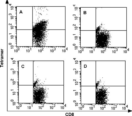
Eight weeks after bone marrow transplantation, the recipient mice and appropriate controls were infected intracranially with Theiler's virus. Seven days after infection, brain-infiltrating lymphocytes were isolated from the FVB/Db hosts and analyzed for VP2121-130-specific T cells by tetramer staining. The presence of VP2-specific CD8+ T cells in the brain was used as evidence of antigen-specific activation of T cells. While nonirradiated FVB/Db mice normally mount an immunodominant VP2121-130-specific T-cell response in the brain by day 7 postinfection, irradiated FVB/Db mice are nonresponsive (Table 1 and data not shown). Positive control mice (irradiated FVB/Db hosts reconstituted with syngeneic bone marrow) expressing H-2Db in both peripheral and brain cells developed the characteristic immunodominant T-cell response specific for the VP2121-130 antigen (47.2% ± 2.6% [n = 2] of the CD8+ T-cell response) (Fig. 1A and B). However, FVB/Db mice that received bone marrow from FVB mice and consequently expressing the Db molecule only in cells not derived from the bone marrow grafts were not able to generate the response (no staining above background) (Fig. 1C and D). Microglial cells, marked by intermediate staining levels when probed with CD45-specific antibody, expressed the H-2Db antigen-presenting molecule in these animals. These results indicate that a bone marrow-derived APC expressing the relevant antigen-presenting molecule is required for activating VP2-specific CD8+ T cells and that radiation-resistant and brain parenchymal cells expressing the Db molecule cannot substitute for this function.
TABLE 1.
Summary of experiments performed to determine the origin of APC that primes the VP2-specific CD8+ T-cell response to TMEV
| BIL examined | Class I expression (H-2Db) in:
|
H-2Bb-restricted T cells | VP2 response in the brain | |
|---|---|---|---|---|
| Brain | BM-derived APCs | |||
| Experimental group | ||||
| FVB(BM) → FVB/Db | + | − | Yes | No |
| FVB/Db(BM) → FVB/Db(spleen) → FVBa | − | + | Yes | Yes |
| Controla | ||||
| FVB/Db(BM) → FVB/Db(spleen) → FVB/Db | + | + | Yes | Yes |
| FVB/Db(BM) → FVB/Db | + | + | Yes | Yes |
| FVB(BM) → FVB/Db(spleen) → FVB/Db | + | ?b | Yes | No |
| FVB/Db(BM) → FVB | − | + | No | No |
| Irradiated FVB/Db/no adoptive transfer | + | − | No | No |
Spleen cells were adoptively transferred 8 weeks after bone marrow grafts.
Status of APCs not known.
FIG. 1.
Activation of virus-specific CD8+ T cells upon Theiler's virus infection does not occur through antigen presentation by a brain parenchymal cell. BILs from FVB/Db mice that received bone marrow from FVB or FVB/Db (control) donors were analyzed for the presence of antiviral CD8+ T cells at day 7 after TMEV infection. Only live CD8+ cells are shown. (A) VP2121-130 tetramer staining of FVB/Db→FVB/Db BILs. The average percentage of CD8+ T cells positive for VP2 tetramer staining after subtraction of E7 background staining is 47.2% ± 2.6% (n = 2). (B) Negative control E7 tetramer staining of FVB/Db→FVB/Db BILs. (C) VP2121-130 tetramer staining of FVB→FVB/Db BILs. No staining above E7 background staining was detected (n = 4). (D) Negative control staining of FVB→FVB/Db BILs with E7 tetramer. (Panel A compared to panel C: 47.2% ± 2.6% versus 0%; P < 0.001).
Activation of virus-specific CD8+ T cells upon Theiler's virus infection does not require antigen presentation by a brain parenchymal cell.
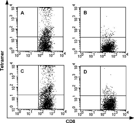
To determine whether activation of antiviral CD8+ T cells in the TMEV model requires antigen presentation by brain cells, animals expressing H-2Db on bone marrow-derived cells of the immune system but not in brain parenchymal cells were generated. FVB host mice received FVB/Db-derived bone marrow after lethal irradiation. Eight weeks after the transplant, the FVB hosts received an adoptive transfer of FVB/Db splenocytes in order to provide T cells able to recognize antigen in the context of the Db molecule. A day after the adoptive transfer of spleen cells, the FVB hosts were infected intracranially with TMEV. FVB/Db control mice received bone marrow and splenocytes from FVB/Db donors. Brain-infiltrating lymphocytes from FVB hosts and FVB/Db control animals were isolated at day 7 postinfection and were analyzed by flow cytometry using fluorescent tetramer probes. FVB/Db hosts that received the FVB/Db bone marrow generated a VP2-specific CD8+ T-cell response (26.2% ± 2.5% [n = 4] of the CD8+ T cells) (Fig. 2A and B). FVB animals reconstituted with FVB/Db bone marrow and spleen cells responded similarly (25.3% ± 7.8% [n = 5]; P = 0.924) (Fig. 2C and D). However, when FVB mice were reconstituted with FVB/Db bone marrow in the absence of adoptively transferred FVB/Db spleen cells, the chimeric FVB hosts were not able to generate the VP2 response, a finding consistent with the hypothesis that Db-restricted T cells (positively selected only in Db-expressing thymus [26]) in the periphery are required for the development of the anti-VP2121-130-specific antiviral response (Table 1 and data not shown). This result demonstrates that the antigen-presenting cell responsible for the activation of virus-specific CD8+ T cells when viral antigen is injected directly into the brain is of bone marrow origin and does not require expression of class I antigen-presenting molecules by brain parenchymal cells. However, this result does not exclude a role for a bone marrow-derived, brain-resident, antigen-presenting cell, such as a perivascular macrophage or dendritic cell, in initiating the immune response to intracranially delivered Theiler's virus.
FIG. 2.
Activation of virus-specific CD8+ T cells upon Theiler's virus infection requires antigen presentation by a bone marrow-derived, antigen-presenting cell. BILs from FVB or FVB/Db (control) mice that received bone marrow and splenocytes from FVB/Db donors were analyzed for the presence of antiviral CD8+ T cells at day 7 postinfection with TMEV. Only live CD8+ cells are shown. (A) VP2121-130 tetramer staining of FVB/Db→FVB/Db BILs. The average percentage of CD8+ T cells positive for VP2 tetramer staining after subtraction of E7 background staining is 26.2% ± 2.5% (n = 4). (B) Negative control E7 tetramer staining of FVB/Db→FVB/Db BILs. (C) VP2121-130 tetramer staining of FVB/Db→FVB BILs. The average percentage of CD8+ T cells positive for VP2 tetramer staining after subtraction of E7 background staining is 25.3% ± 7.8% (n = 5). (D) Staining of FVB/Db→FVB BILs with negative control E7 tetramer. (Panel A compared to panel C: 26.2% ± 2.5% versus 25.3% ± 7.8%; P = 0.924).
Virus-specific CD8+ T cells are recruited to the brain in mice lacking peripheral lymph nodes.
Altogether, our data demonstrate that the antigen-presenting cell responsible for activation of antiviral CD8+ T cells in the TMEV model is a cell of hematopoietic origin. Two nonexclusive possibilities exist regarding how and where an effective T-cell response develops. One is that the immune response develops in peripheral lymph nodes in the traditional manner; the other is that exceptional events are involved in the generation of immunity directly in the brain. Foreign antigen drains to regional lymph nodes, where naïve T cells can be activated given the appropriate stimulatory signals. Although the brain does not have a defined lymphatic system or a putative regional lymph node, cervical lymph nodes have been implicated previously as a site for developing T-cell responses for antigens derived from the brain (16, 19, 27, 41, 49, 55). In normal mice we find that at days 3 and 5 after intracranial infection with Theiler's virus, 0.58% ± 0.04% (n = 5) and 3.5% ± 0.6% (n = 4) of CD8+ T cells in the cervical lymph nodes are specific for virus by tetramer staining, respectively. While the response to the virus is not detected in the brain at day 3, the frequency of VP2-specific CD8+ T cells is 24.0% ± 5.5% (n = 6) 5 days after infection. Similarly, absolute numbers of virus-specific CD8+ T cells obtained from the cervical lymph nodes and brain following infection at day 3 and day 5 do not reflect major expansion events occurring in the lymph nodes (data not shown).
To identify the site of activation of virus-specific CD8+ T cells when a pathogenic antigen is directly introduced into the central nervous system, we studied the response to Theiler's virus in mice deficient in secondary lymphoid organs. To determine whether a peripheral lymph node is the site of activation of TMEV-specific CD8+ T cells, lymphotoxin alpha knockout mice (LTα−/−) were intracranially infected with Theiler's virus. Lymphotoxin alpha knockout mice have been shown to lack peripheral lymph nodes, but T-cell responses in the spleen are normal (18). Dissection of the mice after viral infection confirmed that these mice lack cervical, axillary, mesenteric, and popliteal lymph nodes.
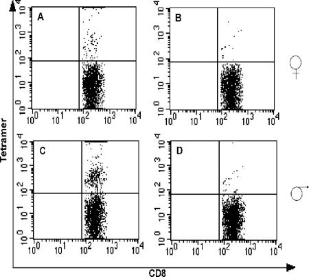
Lymphotoxin alpha-deficient mice that lack peripheral lymph nodes generated VP2-specific CD8+ T cells in the brain at day 7 after infection (Fig. 3A and C), although they appeared in smaller numbers than in normal B6 or 129 mice (16.4% ± 1.4% [n = 4] versus 61.5% ± 3.7% [n = 2] of CD8+ T cells; P = <0.001) (33). Also, virus-specific cell levels were lower in the female mice (9.4% ± 0.2% versus 16.4% ± 1.4%; P = 0.029) (Fig. 3A). This result indicates that peripheral lymph nodes play an important function in the development of the CD8+ antiviral response that unfolds in the brain, but that in the absence of lymph nodes some TMEV-specific CD8+ T cells can still be activated and found in the brain.
FIG. 3.
Virus-specific CD8+ T cells are recruited to the brain in mice lacking peripheral lymph nodes. Ltα−/− mice were infected with Theiler's virus, and BILs were analyzed for the presence of VP2-specific CD8+ T cells by flow cytometry at day 7 postinfection. Only live CD8+ cells are shown. Percentages reflect numbers after subtraction of E7 tetramer control background staining. (A) VP2 tetramer staining of BILs from female Ltα−/− mice. The percentage of CD8+ T cells positive for tetramer is 9.4% ± 0.2% (n = 2). (B) Negative control E7 tetramer staining of BILs from female mice. (C) VP2 tetramer staining of BILs from male Ltα−/− mice. The percentage of CD8+ T cells positive for tetramer is 16.4% ± 1.4% (n = 4). (D) Staining of BILs from male mice with E7 negative control tetramer.
The spleen is not required for activation and recruitment of virus-specific CD8+ T cells to the brain of mice infected with Theiler's virus.
The spleen is a large secondary lymphoid organ and a major source of B and T lymphocytes. Anti-TMEV CD8+ T cells were detected in the spleen as early as 3 days after infection in the brain (8.3 × 10⁴ tetramer-positive cells; n = 3). The number of virus-specific CD8+ T cells in the spleen peaked at day 5 postinfection (7.6 × 10⁵ tetramer-positive cells; n = 3). These numbers were at least 5 and 50 times higher at day 3 and day 5, respectively, in the spleen than in the cervical lymph nodes and the brain (data not shown), indicating that the spleen may be the source of anti-TMEV CD8+ T cells.
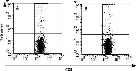
To investigate the role of the spleen as a site of activation of TMEV-specific CD8+ T lymphocytes, C57BL/6 mice were splenectomized prior to intracranial infection with the virus. At different times after inoculation with TMEV, the brain, superficial cervical lymph nodes, blood, and bone marrow were analyzed for the presence of VP2-specific CD8+ T cells using the fluorescent Db:VP2121-130 tetramer. No difference in the frequency of VP2-specific CD8+ T cells was found in the brain of splenectomized and nonsplenectomized mice at day 3, 5, or 7 postinfection (P = 0.861; n = 5 animals/group) (Fig. 4B and C and data not shown). Similarly, the response to TMEV in the superficial cervical lymph nodes in splenectomized animals at time points that precede the peak of the response in the brain (days 3 and 5) was similar to that of nonsplenectomized mice (data not shown). Because the virus-specific CD8+ T-cell response remained relatively unchanged in the brain and cervical lymph nodes of splenectomized animals, it indicated the possibility that activation events may occur in the brain.
FIG. 4.
VP2-specific CD8+ T cells are recruited to the brain of splenectomized B6 mice in numbers comparable to those for nonsplenectomized mice. B6 mice were splenectomized and allowed to recover for 2 weeks prior to infection with Theiler's virus. Seven days postinfection, lymphocytes from the brain were isolated and analyzed by flow cytometry for the presence of VP2 tetramer-specific CD8+ T cells. Only CD8+ live cells are shown. (A) BILs from a sham-infected mouse. (B) Brain-infiltrating CD8+ T cells from a TMEV-infected, nonsplenectomized mouse. (C) CD8+ T cells isolated from the brain of a splenectomized mouse infected with TMEV.
Theiler's virus-specific CD8+ T cells are activated in a peripheral lymphoid organ.
To determine if the spleen compensates for missing lymph nodes in this model, male LTα knockout mice were splenectomized 2 weeks before intracranial infection with Theiler's virus. Seven days postinfection the bone marrow, blood, and brain were analyzed for the presence of VP2-specific CD8+ T cells by flow cytometry. Dissection of the mice confirmed that peripheral lymph nodes were absent. Flow cytometry analysis for the CD8 T-cell marker showed that the splenectomized mice have a considerable pool of circulating lymphocytes in the blood and bone marrow, despite the absence of the spleen (data not shown). Splenectomized LTα−/− mice had a substantial number of CD8+ lymphocytes at day 7 after infection (14.6% ± 1.0% and 10.5% ± 0.5% of live cells analyzed from blood and bone marrow, respectively; n = 3) but had a very low frequency of VP2-specific T cells (Fig. 5A). These responses were reduced significantly, from 16.4% ± 1.4% (n = 4) in nonsplenectomized male mice to 2.5% ± 0.4% (n = 3) in splenectomized LTα−/− males (P = <0.001). Tetramer-positive cells were also absent in the blood and bone marrow (data not shown).
FIG. 5.
VP2-specific CD8+ T-cell numbers are reduced to background levels in splenectomized Ltα−/− mice. Male Ltα−/− mice were splenectomized 2 weeks prior to infection with Theiler's virus. Brain-infiltrating lymphocytes were analyzed for the presence of VP2-specific CD8+ T cells by flow cytometry at day 7 postinfection. Only gated CD8+ T cells are shown. Percentages reflect numbers after subtraction of E7 tetramer control background staining. (A) VP2 tetramer staining of BILs from splenectomized Ltα−/− mice. CD8+ T cells specific for Db/VP2 are reduced to near background levels in these mice (2.5% ± 0.4%; n = 3). (B) Staining of BILs from splenectomized Ltα−/− mice with negative control E7 tetramer.
The activation phenotype of CD8+ T cells in the brain of splenectomized LTα−/− mice was evaluated by staining for the CD44 activation marker and compared to that of nonsplenectomized LTα−/− mice and wild-type B6 mice at day 7 after infection. Analysis of the gated CD8+ T-cell populations showed that more than 90% of the CD8+ T cells in the brain of infected B6 wild-type mice have an activated phenotype (CD44hi) (Fig. 6A). However, the number of CD44hi CD8+ T cells is decreased in nonsplenectomized LTα−/− mice to 71.5% ± 0.6% (n = 2; P = 0.017) and is dramatically decreased in splenectomized LTα−/− mice to 57.3% ± 5.1% (n = 3; P = 0.001) (Fig. 6B and C). At face value, we observed an increase in the number of CD44lo CD8+ T cells in the splenectomized LTα−/− mice compared to levels for B6 wild-type mice. These results indicate that in the absence of peripheral secondary lymphoid organs, there is migration of CD8+ T cells to the brain upon infection, but these cells are not virus specific as determined by tetramer staining and have not undergone appropriate activation. Therefore, the secondary lymphoid organs are critical for mounting an antigen-specific antiviral response in the brain, and the brain by itself cannot sustain T-cell activation upon in situ challenge.
FIG. 6.
Brain-infiltrating CD8+ T cells from splenectomized Ltα−/− mice are not properly activated in response to Theiler's virus infection. Analysis of BILs from B6 wild-type, Ltα−/−, and splenectomized Ltα−/− TMEV-infected mice stained for CD8+ and the activation marker CD44. Only gated CD8+ T cells are shown. (A) CD44 profile of CD8+ T cells from wild-type B6 mice. The percentage of cells in M2 (CD44hi) was 93.9% ± 3.6% (n = 3). (B) CD44 profile of CD8+ T cells from nonsplenectomized Ltα−/− mice. The percentage of cells in M2 (CD44hi) was 71.5% ± 0.6% (n = 2). (C) CD44 profile of CD8+ T cells from splenectomized Ltα−/− mice. The percentage of cells in M2 (CD44hi) was 57.3% ± 5.1% (n = 3; P = 0.003 by one-way analysis of variance test).
DISCUSSION
Following intracranial inoculation with Theiler's virus, small numbers of CD8+ T cells begin appearing in the brain by day 3, bearing an activated phenotype as defined by CD44 expression. The number of activated CD8+ T cells increases with time, and antigen-specific CD8+ T cells can be detected in the brain for the first time at day 5 postinfection, peaking at day 7. In contrast to other virus models, we have not observed substantial antiviral responses in peripheral lymphoid organs, such as the spleen and cervical lymph nodes, prior to the appearance of the immunodominant CD8+ T-cell population in the brain. This observation led to the hypothesis that the brain itself may support activation and proliferation of TMEV-specific T cells upon intracranial antigenic challenge.
Others have argued that CNS-resident glial cells can present antigen to T cells. Microglia and other types of brain-resident macrophages express class II and class I molecules, as well as costimulatory molecules, both in vitro and in vivo upon activation (1, 2, 12, 17, 24, 29, 31, 47, 50, 59). Moreover, in vitro studies suggest that the brain may be capable of activating T cells. These studies show that activated microglia can present both virus and myelin antigen and can activate clonal T-cell populations to proliferate (3, 13, 21, 22, 43, 60). On the other hand, astrocytes and microglia have been shown to secrete or express immunomodulatory molecules that can inhibit T-helper-cell proliferation, such as nitric oxide (NO), CTLA-4, and granulocyte-macrophage colony-stimulating factor (2, 25, 28, 34, 42). However, the effects of these regulatory mechanisms in CD8+ T-cell responses have not been studied.
The hypothesis that the brain may be a site of activation and expansion of antiviral T cells in the Theiler's virus model is supported by findings from Kang et al. (35) and Musette et al. (46), which show by T-cell receptor spectratyping that T cells expand in the CNS in Theiler's virus-infected mice. Observations from our laboratories also suggest that T-cell proliferation in response to Theiler's virus infection may occur in the brain (51). To establish whether the activation of antiviral CD8+ T cells in the Theiler's virus model occurs in situ upon intracranial infection, we sought to identify the nature and origin of the antigen-presenting cell that primes the CD8+ T-cell response to TMEV. Using a bone marrow transplant strategy, we demonstrated that the antigen-presenting cells that mediate activation of CD8+ T cells in the Theiler's virus model are derived from the bone marrow. Our results exclude the possibility that brain parenchymal cells, namely microglia, are responsible for activating TMEV-specific CD8+ T cells. However, it remains possible that brain-resident, bone marrow-derived macrophages, such as perivascular macrophages or pericytes (30, 58), participate in the activation of anti-TMEV T cells.
As demonstrated here, the spleen is not required for the activation and migration of VP2, virus-specific CD8+ T cells to the brain of mice infected with TMEV. Previously it was reported that VP2-specific responses are functional and present in significant numbers in mice lacking the CD62L molecule, a homing molecule required for circulation of naïve T cells through the lymph nodes (61). In agreement with this finding, we have shown that mice lacking lymph nodes due to a deficiency in lymphotoxin alpha expression generate VP2-specific responses in the brain as well, though in smaller numbers than those found in wild-type mice. Decreased CD8+ T-cell numbers and/or function have also been observed in lymphotoxin alpha-deficient mice during infection with lymphocytic choriomeningitis virus (LCMV) and during herpes simplex virus (HSV) and influenza virus infection of the brain (37, 40, 57). Our results indicate that the lymph nodes are not required for the generation of anti-TMEV responses but that their presence has some influence. The finding that the VP2-specific response is eliminated in mice lacking both the spleen and lymph nodes indicates that the spleen compensates for the absence of lymph nodes in the lymphotoxin alpha-deficient mice.
The T-cell response to influenza virus in mice deficient in lymphotoxin alpha expression is delayed by 2 to 3 days (40). Thus, it is possible that the peak of the anti-TMEV response occurs after day 7 in LTα−/− mice and that the absence or decrease in the magnitude of the antiviral response at day 7 may reflect a delay in the activation of the CD8+ T cells. However, LTα−/− mice are susceptible to TMEV-induced demyelination (38), indicating that these mice have an impaired anti-TMEV CD8+ T-cell response.
Although the CNS is not associated with a conventional lymphatic pathway, antigen present in the brain parenchyma is able to migrate from the CNS into the cervical lymph nodes through the lymph (11, 15). Thus, upon intracranial infection with Theiler's virus, T cells may be specifically activated in the cervical lymph nodes. Activation may occur through priming by a brain-resident, bone marrow-derived APC, such as a perivascular macrophage or pericyte, that is able to migrate from the brain to the cervical lymph nodes.
Because intracranial infection breaks the blood-brain barrier, two alternative routes of antigen delivery to the lymph nodes should be considered. Monocytoid dendritic cells are recruited to the brain as early as 3 days after infection with Theiler's virus (data not shown). Dendritic cells in perivascular spaces or dendritic cell precursors that are recruited from the bloodstream into the site of infection may phagocytose viral antigen, upregulate costimulatory molecules, and migrate to the lymph nodes, where they are able to present viral peptides to naïve T cells. It has been recently demonstrated that when dendritic cells are introduced in the brain together with ovalbumin (OVA) protein, these cells are able to process and present ovalbumin peptides (36). Moreover, this study showed that the generation of a peripheral T-cell response to ovalbumin and recruitment of OVA-specific T cells to the brain requires the migration of dendritic cells carrying ovalbumin antigen from the brain into the cervical lymph nodes.
Another alternative mechanism for delivery of viral antigen to the lymph nodes for antigen presentation by a bone marrow-derived APC is through viremia. As a consequence of breaking the blood-brain barrier, infectious virus may drain to the cervical lymph nodes, where antigen would be presented to naïve T cells by bone marrow-derived professional APCs residing in the lymph nodes. These two mechanisms for antigen delivery from the brain may occur either concomitantly or in succession, as has been demonstrated for peripheral tissues. Jenkins and colleagues have demonstrated that antigen is delivered to the lymph nodes in two waves (32). The first wave involves drainage of antigen through lymphatics and presentation by lymph node APCs, while the second event involves migration of tissue-associated dendritic cells.
Antiviral T-cell responses in the brain have been studied during infection with the neurotropic strain of mouse hepatitis virus (MHV). Similar to our model, a prominent cytotoxic response to the nucleocapsid protein of MHV is observed in the brain during acute encephalitis, but these responses are undetectable in peripheral lymphoid organs (8, 9, 56). A recent study looked at the kinetics of virus-specific CD8+ T-cell expansion and trafficking to the brain during infection with MHV. Virus-specific CD8+ T cells were detected in the cervical lymph nodes and spleen prior to their appearance in the brain, as determined by tetramer staining and gamma-interferon secretion (41). This study concluded that the cervical lymph nodes are the primary site of virus-specific T-cell proliferation. Similar to the MHV model, we conclude that the antiviral response to TMEV in the brain occurs in the lymph nodes. However, under certain circumstances, we have observed that the increase in the number of virus-specific cells in the brain cannot be accounted for by the number of virus-specific cells in the periphery, suggesting that an amplification step may take place in the brain. In situ proliferation has been observed in the CNS of TMEV-infected mice (35, 46, 51). Furthermore, we have found that brain-infiltrating CD8+ T cells isolated from TMEV-infected mice at days 3 and 5 postinfection incorporate BrdU in vitro in the absence of further antigenic stimulation (data not shown).
Altogether, these results form the basis of our view that activation of antigen-specific T cells by intracranial TMEV infection occurs in the peripheral lymphoid compartment, is dependent on an APC recruited from the bone marrow, and is followed by migration and possibly further expansion of virus-specific CD8+ T cells in the brain.
Acknowledgments
We thank the Mayo Flow Cytometry and Transgenic Core Facilities and Allen Bieber for assistance with splenectomies.
This work was supported by NIH grants PO1 NS38468 and NSR01-34189.
REFERENCES
- 1.Aloisi, F. 1999. Antigen presentation in the central nervous system, p. 89-100. In L. A. G. Martino (ed.), From basic immunology to immune-mediated demyelination. Springer, New York, N.Y.
- 2.Aloisi, F., F. Ria, and L. Adorini. 2000. Regulation of T-cell responses by CNS antigen-presenting cells: different roles for microglia and astrocytes. Immunol. Today 21:141-147. [DOI] [PubMed] [Google Scholar]
- 3.Aloisi, F., F. Ria, G. Penna, and L. Adorini. 1998. Microglia are more efficient than astrocytes in antigen processing and in Th1 but not Th2 cell activation. J. Immunol. 160:4671-4680. [PubMed] [Google Scholar]
- 4.Altintas, A., Z. Cai, L. R. Pease, and M. Rodriguez. 1993. Differential expression of H-2K and H-2D in the central nervous system of mice infected with Theiler's virus. J. Immunol. 151:2803-2812. [PubMed] [Google Scholar]
- 5.Azoulay, A., M. Brahic, and J. F. Bureau. 1994. FVB mice transgenic for the H-2Db gene become resistant to persistent infection by Theiler's virus. J. Virol. 68:4049-4052. [DOI] [PMC free article] [PubMed] [Google Scholar]
- 6.Azoulay-Cayla, A., S. Dethlefs, B. Perarnau, E. L. Larsson-Sciard, F. A. Lemonnier, M. Brahic, and J. F. Bureau. 2000. H-2Db−/− mice are susceptible to persistent infection by Theiler's virus. J. Virol. 74:5470-5476. [DOI] [PMC free article] [PubMed] [Google Scholar]
- 7.Azoulay-Cayla, A., S. Syan, M. Brahic, and J. F. Bureau. 2001. Roles of the H-2D(b) and H-K(b) genes in resistance to persistent Theiler's murine encephalomyelitis virus infection of the central nervous system. J. Gen. Virol. 82:1043-1047. [DOI] [PubMed] [Google Scholar]
- 8.Bergmann, C. C., J. D. Altman, D. Hinton, and S. A. Stohlman. 1999. Inverted immunodominance and impaired cytolytic function of CD8+ T cells during viral persistence in the central nervous system. J. Immunol. 163:3379-3387. [PubMed] [Google Scholar]
- 9.Bergmann, C. C., C. Ramakrishna, M. Kornacki, and S. A. Stohlman. 2001. Impaired T cell immunity in B cell-deficient mice following viral central nervous system infection. J. Immunol. 167:1575-1583. [DOI] [PubMed] [Google Scholar]
- 10.Borson, N. D., C. Paul, X. Lin, W. K. Nevala, M. A. Strausbauch, M. Rodriguez, and P. J. Wettstein. 1997. Brain-infiltrating cytolytic T lymphocytes specific for Theiler's virus recognize H2Db molecules complexed with a viral VP2 peptide lacking a consensus anchor residue. J. Virol. 71:5244-5250. [DOI] [PMC free article] [PubMed] [Google Scholar]
- 11.Bradbury, M. W., H. F. Cserr, and R. J. Westrop. 1981. Drainage of cerebral interstitial fluid into deep cervical lymph of the rabbit. Am. J. Physiol. 240:F329—F336. [DOI] [PubMed] [Google Scholar]
- 12.Cannella, B., and C. S. Raine. 1995. The adhesion molecule and cytokine profile of multiple sclerosis lesions. Ann. Neurol. 37:424-435. [DOI] [PubMed] [Google Scholar]
- 13.Cash, E., and O. Rott. 1994. Microglial cells qualify as the stimulators of unprimed CD4+ and CD8+ T lymphocytes in the central nervous system. Clin. Exp. Immunol. 98:313-318. [DOI] [PMC free article] [PubMed] [Google Scholar]
- 14.Clatch, R. J., R. W. Melvold, S. D. Miller, and H. L. Lipton. 1985. Theiler's murine encephalomyelitis virus (TMEV)-induced demyelinating disease in mice is influenced by the H-2D region: correlation with TEMV-specific delayed-type hypersensitivity. J. Immunol. 135:1408-1414. [PubMed] [Google Scholar]
- 15.Cserr, H. F., C. J. Harling-Berg, and P. M. Knopf. 1992. Drainage of brain extracellular fluid into blood and deep cervical lymph and its immunological significance. Brain Pathol. 2:269-276. [DOI] [PubMed] [Google Scholar]
- 16.Cserr, H. F., and P. M. Knopf. 1992. Cervical lymphatics, the blood-brain barrier and the immunoreactivity of the brain: a new view. Immunol. Today 13:507-512. [DOI] [PubMed] [Google Scholar]
- 17.De Simone, R., A. Giampaolo, B. Giometto, P. Gallo, G. Levi, C. Peschle, and F. Aloisi. 1995. The costimulatory molecule B7 is expressed on human microglia in culture and in multiple sclerosis acute lesions. J. Neuropathol. Exp. Neurol. 54:175-187. [DOI] [PubMed] [Google Scholar]
- 18.De Togni, P., J. Goellner, N. H. Ruddle, P. R. Streeter, A. Fick, S. Mariathasan, S. C. Smith, R. Carlson, L. P. Shornick, J. Strauss-Schoenberger, et al. 1994. Abnormal development of peripheral lymphoid organs in mice deficient in lymphotoxin. Science 264:703-707. [DOI] [PubMed] [Google Scholar]
- 19.De Vos, A. F., M. van Meurs, H. P. Brok, L. A. Boven, R. Q. Hintzen, P. van der Valk, R. Ravid, S. Rensing, L. Boon, B. A. t Hart, and J. D. Laman. 2002. Transfer of central nervous system autoantigens and presentation in secondary lymphoid organs. J. Immunol. 169:5415-5423. [DOI] [PubMed] [Google Scholar]
- 20.Dethlefs, S., N. Escriou, M. Brahic, S. van der Werf, and E. L. Larsson-Sciard. 1997. Theiler's virus and Mengo virus induce cross-reactive cytotoxic T lymphocytes restricted to the same immunodominant VP2 epitope in C57BL/6 mice. J. Virol. 71:5361-5365. [DOI] [PMC free article] [PubMed] [Google Scholar]
- 21.Dhib-Jalbut, S., N. Gogate, H. Jiang, H. Eisenberg, and G. Bergey. 1996. Human microglia activate lymphoproliferative responses to recall viral antigens. J. Neuroimmunol. 65:67-73. [DOI] [PubMed] [Google Scholar]
- 22.Frei, K., C. Siepl, P. Groscurth, S. Bodmer, C. Schwerdel, and A. Fontana. 1987. Antigen presentation and tumor cytotoxicity by interferon-gamma-treated microglial cells. Eur. J. Immunol. 17:1271-1278. [DOI] [PubMed] [Google Scholar]
- 23.Garboczi, D. N., D. T. Hung, and D. C. Wiley. 1992. HLA-A2-peptide complexes: refolding and crystallization of molecules expressed in Escherichia coli and complexed with single antigenic peptides. Proc. Natl. Acad. Sci. USA 89:3429-3433. [DOI] [PMC free article] [PubMed] [Google Scholar]
- 24.Gerritse, K., J. D. Laman, R. J. Noelle, A. Aruffo, J. A. Ledbetter, W. J. Boersma, and E. Claassen. 1996. CD40-CD40 ligand interactions in experimental allergic encephalomyelitis and multiple sclerosis. Proc. Natl. Acad. Sci. USA 93:2499-2504. [DOI] [PMC free article] [PubMed] [Google Scholar]
- 25.Gimsa, U., A. O. Ren, P. Pandiyan, D. Teichmann, I. Bechmann, R. Nitsch, and M. C. Brunner-Weinzierl. 2004. Astrocytes protect the CNS: antigen-specific T helper cell responses are inhibited by astrocyte-induced upregulation of CTLA-4 (CD152). J. Mol. Med. 82:364-372. [DOI] [PubMed] [Google Scholar]
- 26.Hare, K. J., E. J. Jenkinson, and G. Anderson. 2001. Specialisation of the thymic epithelial cells for positive selection of CD4+8+ thymocytes. Cell. Mol. Biol. 47:119-127. [PubMed] [Google Scholar]
- 27.Harling-Berg, C., P. M. Knopf, J. Merriam, and H. F. Cserr. 1989. Role of cervical lymph nodes in the systemic humoral immune response to human serum albumin microinfused into rat cerebrospinal fluid. J. Neuroimmunol. 25:185-193. [DOI] [PubMed] [Google Scholar]
- 28.Hayashi, M., M. E. Dorf, and S. Abromson-Leeman. 1993. Granulocyte-macrophage colony stimulating factor inhibits class II major histocompatibility complex expression and antigen presentation by microglia. J. Neuroimmunol. 48:23-32. [DOI] [PubMed] [Google Scholar]
- 29.Hayes, G. M., M. N. Woodroofe, and M. L. Cuzner. 1987. Microglia are the major cell type expressing MHC class II in human white matter. J. Neurol. Sci. 80:25-37. [DOI] [PubMed] [Google Scholar]
- 30.Hickey, W. F., K. Vass, and H. Lassmann. 1992. Bone marrow-derived elements in the central nervous system: an immunohistochemical and ultrastructural survey of rat chimeras. J. Neuropathol. Exp. Neurol. 51:246-256. [DOI] [PubMed] [Google Scholar]
- 31.Issazadeh, S., V. Navikas, M. Schaub, M. Sayegh, and S. Khoury. 1998. Kinetics of expression of costimulatory molecules and their ligands in murine relapsing experimental autoimmune encephalomyelitis in vivo. J. Immunol. 161:1104-1112. [PubMed] [Google Scholar]
- 32.Itano, A. A., S. J. McSorley, R. L. Reinhardt, B. D. Ehst, E. Ingulli, A. Y. Rudensky, and M. K. Jenkins. 2003. Distinct dendritic cell populations sequentially present antigen to CD4 T cells and stimulate different aspects of cell-mediated immunity. Immunity 19:47-57. [DOI] [PubMed] [Google Scholar]
- 33.Johnson, A. J., M. K. Njenga, M. J. Hansen, S. T. Kuhns, L. Chen, M. Rodriguez, and L. R. Pease. 1999. Prevalent class I-restricted T-cell response to the Theiler's virus epitope Db:VP2121-130 in the absence of endogenous CD4 help, tumor necrosis factor alpha, gamma interferon, perforin, or costimulation through CD28. J. Virol. 73:3702-3708. [DOI] [PMC free article] [PubMed] [Google Scholar]
- 34.Juedes, A. E., and N. H. Ruddle. 2001. Resident and infiltrating central nervous system APCs regulate the emergence and resolution of experimental autoimmune encephalomyelitis. J. Immunol. 166:5168-5175. [DOI] [PubMed] [Google Scholar]
- 35.Kang, J. A., M. Mohindru, B. S. Kang, S. H. Park, and B. S. Kim. 2000. Clonal expansion of infiltrating T cells in the spinal cords of SJL/J mice infected with Theiler's virus. J. Immunol. 165:583-590. [DOI] [PubMed] [Google Scholar]
- 36.Karman, J., C. Ling, M. Sandor, and Z. Fabry. 2004. Initiation of immune responses in brain is promoted by local dendritic cells. J. Immunol. 173:2353-2361. [DOI] [PubMed] [Google Scholar]
- 37.Kumaraguru, U., I. A. Davis, S. Deshpande, S. S. Tevethia, and B. T. Rouse. 2001. Lymphotoxin alpha−/− mice develop functionally impaired CD8+ T-cell responses and fail to contain virus infection of the central nervous system. J. Immunol. 166:1066-1074. [DOI] [PubMed] [Google Scholar]
- 38.Lin, X., X. Ma, M. Rodriguez, X. Feng, L. Zoecklein, Y. X. Fu, and R. P. Roos. 2003. Membrane lymphotoxin is required for resistance to Theiler's virus infection. Int. Immunol. 15:955-962. [DOI] [PubMed] [Google Scholar]
- 39.Lipton, H. L. 1975. Theiler's virus infection in mice: an unusual biphasic disease process leading to demyelination. Infect. Immun. 11:1147-1155. [DOI] [PMC free article] [PubMed] [Google Scholar]
- 40.Lund, F. E., S. Partida-Sanchez, B. O. Lee, K. L. Kusser, L. Hartson, R. J. Hogan, D. L. Woodland, and T. D. Randall. 2002. Lymphotoxin-alpha-deficient mice make delayed, but effective, T and B cell responses to influenza. J. Immunol. 169:5236-5243. [DOI] [PubMed] [Google Scholar]
- 41.Marten, N. W., S. A. Stohlman, J. Zhou, and C. C. Bergmann. 2003. Kinetics of virus-specific CD8+ T-cell expansion and trafficking following central nervous system infection. J. Virol. 77:2775-2778. [DOI] [PMC free article] [PubMed] [Google Scholar]
- 42.Matsumoto, Y., H. Hanawa, M. Tsuchida, and T. Abo. 1993. In situ inactivation of infiltrating T cells in the central nervous system with autoimmune encephalomyelitis. The role of astrocytes. Immunology 79:381-390. [PMC free article] [PubMed] [Google Scholar]
- 43.Matsumoto, Y., K. Ohmori, and M. Fujiwara. 1992. Immune regulation by brain cells in the central nervous system: microglia but not astrocytes present myelin basic protein to encephalitogenic T cells under in vivo-mimicking conditions. Immunology 76:209-216. [PMC free article] [PubMed] [Google Scholar]
- 44.Melero, I., M. C. Singhal, P. McGowan, H. S. Haugen, J. Blake, K. E. Hellstrom, G. Yang, C. H. Clegg, and L. Chen. 1997. Immunological ignorance of an E7-encoded cytolytic T-lymphocyte epitope in transgenic mice expressing the E7 and E6 oncogenes of human papillomavirus type 16. J. Virol. 71:3998-4004. [DOI] [PMC free article] [PubMed] [Google Scholar]
- 45.Mendez-Fernandez, Y. V., A. J. Johnson, M. Rodriguez, and L. R. Pease. 2003. Clearance of Theiler's virus infection depends on the ability to generate a CD8+ T-cell response against a single immunodominant viral peptide. Eur. J. Immunol. 33:2501-2510. [DOI] [PubMed] [Google Scholar]
- 46.Musette, P., J. F. Bureau, G. Gachelin, P. Kourilsky, and M. Brahic. 1995. T lymphocyte repertoire in Theiler's virus encephalomyelitis: the nonspecific infiltration of the central nervous system of infected SJL/J mice is associated with a selective local T cell expansion. Eur. J. Immunol. 25:1589-1593. [DOI] [PubMed] [Google Scholar]
- 47.Olson, J. K., A. M. Girvin, and S. D. Miller. 2001. Direct activation of innate and antigen-presenting functions of microglia following infection with Theiler's virus. J. Virol. 75:9780-9789. [DOI] [PMC free article] [PubMed] [Google Scholar]
- 48.Patick, A. K., L. R. Pease, C. S. David, and M. Rodriguez. 1990. Major histocompatibility complex-conferred resistance to Theiler's virus-induced demyelinating disease is inherited as a dominant trait in B10 congenic mice. J. Virol. 64:5570-5576. [DOI] [PMC free article] [PubMed] [Google Scholar]
- 49.Phillips, M. J., M. Needham, and R. O. Weller. 1997. Role of cervical lymph nodes in autoimmune encephalomyelitis in the Lewis rat. J. Pathol. 182:457-464. [DOI] [PubMed] [Google Scholar]
- 50.Pope, J. G., C. L. Vanderlugt, S. M. Rahbe, H. L. Lipton, and S. D. Miller. 1998. Characterization of and functional antigen presentation by central nervous system mononuclear cells from mice infected with Theiler's murine encephalomyelitis virus. J. Virol. 72:7762-7771. [DOI] [PMC free article] [PubMed] [Google Scholar]
- 51.Rodriguez, M. 1991. Immunoglobulins stimulate central nervous system remyelination: electron microscopic and morphometric analysis of proliferating cells. Lab. Investig. 64:358-370. [PubMed] [Google Scholar]
- 52.Rodriguez, M., and C. S. David. 1985. Demyelination induced by Theiler's virus: influence of the H-2 haplotype. J. Immunol. 135:2145-2148. [PubMed] [Google Scholar]
- 53.Rodriguez, M., J. Leibowitz, and C. S. David. 1986. Susceptibility to Theiler's virus-induced demyelination. Mapping of the gene within the H-2D region. J. Exp. Med. 163:620-631. [DOI] [PMC free article] [PubMed] [Google Scholar]
- 54.Rodriguez, M., E. Oleszak, and J. Leibowitz. 1987. Theiler's murine encephalomyelitis: a model of demyelination and persistence of virus. Crit. Rev. Immunol. 7:325-365. [PubMed] [Google Scholar]
- 55.Stevenson, P. G., C. R. Bangham, and S. Hawke. 1997. Recruitment, activation and proliferation of CD8+ memory T cells in an immunoprivileged site. Eur. J. Immunol. 27:3259-3268. [DOI] [PubMed] [Google Scholar]
- 56.Stohlman, S. A., S. Kyuwa, J. M. Polo, D. Brady, M. M. Lai, and C. C. Bergmann. 1993. Characterization of mouse hepatitis virus-specific cytotoxic T cells derived from the central nervous system of mice infected with the JHM strain. J. Virol. 67:7050-7059. [DOI] [PMC free article] [PubMed] [Google Scholar]
- 57.Suresh, M., G. Lanier, M. K. Large, J. K. Whitmire, J. D. Altman, N. H. Ruddle, and R. Ahmed. 2002. Role of lymphotoxin alpha in T-cell responses during an acute viral infection. J. Virol. 76:3943-3951. [DOI] [PMC free article] [PubMed] [Google Scholar]
- 58.Thomas, W. E. 1999. Brain macrophages: on the role of pericytes and perivascular cells. Brain Res. Brain Res. Rev. 31:42-57. [DOI] [PubMed] [Google Scholar]
- 59.Ulvestad, E., K. Williams, L. Bo, B. Trapp, J. Antel, and S. Mork. 1994. HLA class II molecules (HLA-DR, -DP, -DQ) on cells in the human CNS studied in situ and in vitro. Immunology 82:535-541. [PMC free article] [PubMed] [Google Scholar]
- 60.Williams, K., Jr., E. Ulvestad, L. Cragg, M. Blain, and J. P. Antel. 1993. Induction of primary T-cell responses by human glial cells. J. Neurosci. Res. 36:382-390. [DOI] [PubMed] [Google Scholar]
- 61.Zhang, X., L. Brewer, B. Walcheck, A. Johnson, L. R. Pease, and M. K. Njenga. 2001. Theiler's virus-infected L-selectin-deficient mice have decreased infiltration of CD8(+) T lymphocytes in central nervous system but clear the virus. J. Neuroimmunol. 116:178-187. [DOI] [PubMed] [Google Scholar]


