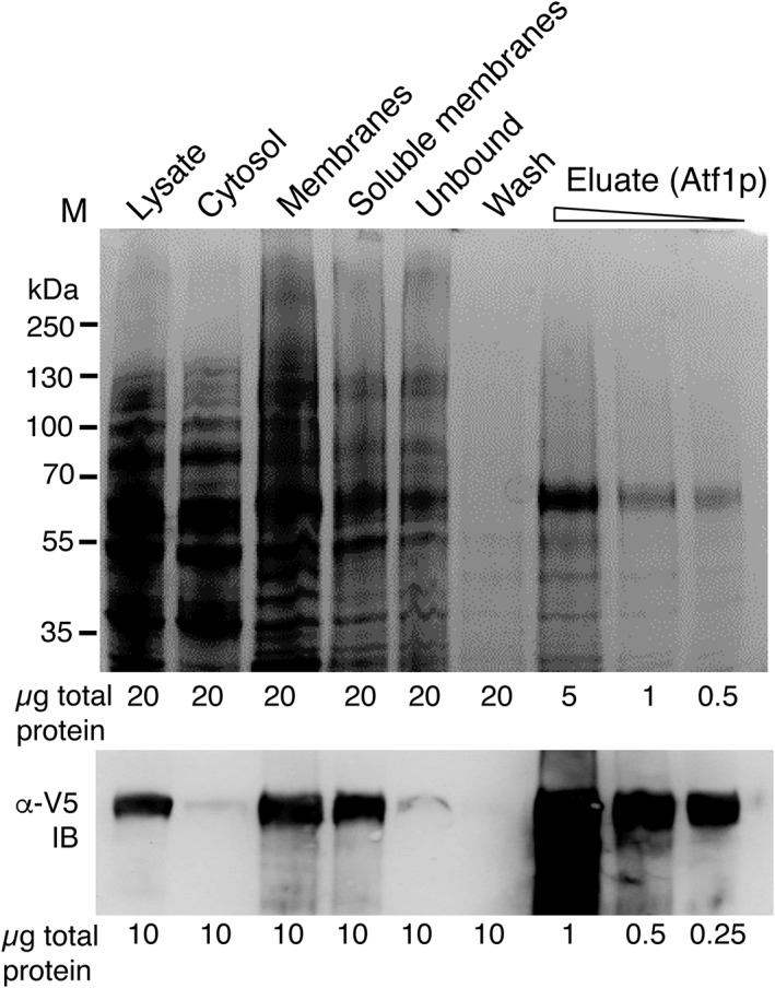
Figure 1.

Expression of Atf1p and purification after solubilization with the detergent Thesit. Top panel, SDS‐PAGE gel of cell fractions and column purification fractions as shown, stained with Coomassie Brilliant Blue. Bottom panel, western blot (IB) of the same samples with anti‐V5‐HRP (α‐V5) confirming both the membrane localization of recombinant Atf1p and the success of detergent solubilization and purification. The protein loadings used for Coomassie staining and western blotting are shown
