

Abstract
Background: The aim of this review was to further define the clinical condition triquetrohamate (TH) impaction syndrome (THIS), an entity underreported and missed often. Its presentation, physical findings, and treatment are presented. Methods: Between 2009 and 2014, 18 patients were diagnosed with THIS. The age, sex, hand involved, activity responsible for symptoms, and defining characteristics were recorded. The physical findings, along with ancillary studies, were reviewed. Delay in diagnosis and misdiagnoses were assessed. Treatment, either conservative or surgical, is presented. Follow-up outcomes are presented. Results: There were 15 male and 3 females, average age of 42 years. Two-handed sports such as golf and baseball accounted for more than 60% of the cases, and these cases were the only ones that involved the lead nondominant hand, pain predominantly at impact. Delay in diagnosis averaged greater than 7 months, with triangular fibrocartilage (TFCC) and extensor carpi ulnaris (ECU) accounting for more than 50% of misdiagnoses. Physical findings of note included pain over the TH joint, worse with passive dorsiflexion and ulnar deviation. Radiographic findings are described. Instillation of lidocaine with the wrist in radial deviation under fluoroscopic imaging with relief of pain helped to confirm the diagnosis. Conservative treatment was successful in 9 of 18 patients (50%), whereas in the remaining, surgical intervention allowed approximately 80% return to full activities without limitation. Conclusion: Triquetrohamate impaction syndrome remains an underreported and often unrecognized cause of ulnar-sided wrist pain. In this report, the largest series to date, its presentation, defining characteristics, and treatment options are further elucidated.
Keywords: triquetrohamate impaction, ulnar-sided wrist pain
Introduction
The successful treatment of ulnar-sided wrist pain requires a thorough knowledge of the many causes that contribute to this condition. The differential diagnosis can include injury to the triangular fibrocartilage (TFCC), the extensor carpi ulnaris (ECU), the lunotriquetral ligament (LT) joint, as well as ulnocarpal impaction, fractures, and arthritic conditions. Incorrect diagnosis can lead to delayed treatment and ultimate significant dysfunction. Shin et al,5 Sachar,4 and others have added much knowledge to this diagnostic dilemma. In 1996, Nakao et al,3 in a case report, described triquetrohamate (TH) impaction and introduced this diagnosis as a cause of ulnar-sided wrist pain. Scant attention to this diagnosis has persisted, mostly discussed in review articles only.1 Since then, the senior authors have treated a series of patients with TH impaction syndrome (THIS), and this review represents a retrospective analysis of their presentation and treatment. This study represents the largest series to date and confirms its validity as an important cause of ulnar-sided wrist pain.
Materials and Methods
Between 2009 and 2014, 18 patients were seen with ulnar-sided wrist pain with the diagnosis of TH impaction made. All patients were seen by 1 of 2 hand fellowship-trained orthopedic surgeons. The hand involved dominance, sport involved, mechanism of injury, and symptom presentation were documented. The physical findings with specific attention to differential diagnosis were recorded. Radiographic findings along with the use of ancillary studies are presented. Treatment including conservative and surgical intervention is outlined. Follow-up outcomes included chart review with compilation of pain relief (Visual Analog Scale [VAS]), range of motion, and return to preinjury activity. Discussion emphasizes acknowledgment of this entity, THIS as an important cause of ulnar-sided wrist pain previously underreported.
Results
There were 18 patients treated with a final diagnosis of THIS, with15 males and 3 females. The range in age was 18 to 64 years, the average age being 41.6. The average time to delay in diagnosis averaged 7.4 months (range, 1-14 months). The initial misdiagnosis included TFCC tear (n = 7), ECU tendinitis (n = 3), lunotriquetral pathology (n = 3), ganglion (n = 1), and nondescript ulnar-sided wrist pain (n = 4). Symptoms were present in the dominant hand in 11 patients, and the nondominant in 7. Dominant hand involvement was seen in all activities from racquet sports, such as tennis, squash, and racquetball, to club sports such as golf and baseball, as well as weight lifting (n = 11). However, the nondominant or lead hand was only involved in golf and batting sports (n = 7).
The findings on exam often overlapped with other diagnoses, that is, pain to palpation over the TFCC worse with ulnar deviation, pain to palpation over lunotriquetral joint worse with ballottement, and pain with radioulnar joint compression. However, with direct and detailed inspection, all patients demonstrated pain with palpation over the TH joint that was increased with forced passive dorsiflexion and ulnar deviation of the wrist, a novel exam technique. With detailed attention, the pain elicited to palpation was 1 to 2 cm distal to the TFCC and located at the distal aspect of the triquetrum.
All patients initially underwent standard radiographs including posteroanterior (PA), lateral, and obliques, with only 2 patients revealing definite pathology at the TH joint. One patient, a previous college football player and low handicap golfer, exhibited significant joint space narrowing at the TH joint. Another patient, a professional baseball player, demonstrated a loose body in the TH joint. All remaining patients did not show any pathology on standard radiographs; however, as this series progressed, it was determined that a PA in neutral, but also in radial and ulnar deviation, allowed better visualization of this articulation, and 7 remaining patients showed pathology, ranging from loose bodies, osteophytes, narrowing of the joint, and osteochondral defect from where the distal aspect of the triquetrum impinged into the ulnar aspect of the hamate. Furthermore, this PA view in radial deviation not only opened up this articulation for better visualization but also allowed for fluoroscopic imaging to inject 12, cc of lidocaine and assess pain relief and better confirmation of this joint as the source of symptoms. The last 7 patients treated in this series underwent this diagnostic maneuver with positive results.
The use of computed tomography (CT) and magnetic resonance imaging (MRI) helped to document TH pathology. In 11 of 14 patients who received MRIs, studies revealed a spectrum of findings including synovitis, a ganglion cyst arising from the pisotriquetral joint extending to the hook of the hamate, osseous edema, chondromalacia, and increased signal on T2 imaging suggestive of inflammation localized where the distal triquetrum impacted on the reciprocal hamate articular interface. Computed tomography was performed on 7 patients, and 5 scans documented joint space narrowing, sclerosis of subchondral bone, and loose bodies. In summary, all patients showed evidence of radiographic abnormality, either on plain films or advanced imaging (CT or MRI). The use of standard PA radiographs in both radial and ulnar deviation better allowed the clinician to confirm minimal findings.
Initial treatment was conservative, and if this failed, operative interventions were utilized; the different procedures are outlined in Table 1. Follow-up ranged from 13 to 101 months, with an average of 4.4 years. Final follow-up success was determined by history, exam, and radiographs in 13 of 18 patients and in the remaining 5 through telephone survey. Fifty percent (9 patients) responded to conservative treatment including rest, nonsteroidal anti-inflammatory drugs (NSAIDs), selective corticosteroid injections, splinting, and directed rehabilitation with emphasis on core strengthening and sport-specific optimization of technique. This refinement includes batting mechanics in baseball and forearm position in tennis. The remaining 9 of 18 patients required operative intervention. Of the operative candidates, 3 patients had arthroscopic procedures with all 3 undergoing synovectomy, 2 having removal of loose bodies, 1 undergoing microfracture, and 2 had removal of hypertrophic triquetrum and/or hamate. The radial midcarpal portal was used for visualization with the camera, and the ulnar midcarpal portal was accessed for instrumentation and debridement. Open procedures for the remaining 6 patients were utilized when soft tissue interposition was indicated, or the extent of partial excision of the carpal bone precluded arthroscopic removal. Different surgical procedures were utilized depending on surgeon preference and findings at surgery. The decision to proceed with partial excision of either the triquetrum or hamate was made intraoperatively, and was based on the observation of which carpal bone was responsible for the impingement. Specific procedures are found in Table 1. As described in the table, the particular treatment instituted correlated with MRI findings when the study documented synovitis; that is, usual treatment was synovectomy. In more advanced cases where radiographs showed significant hypertrophic changes, treatment usually correlated with these findings; that is, usual treatment was bony debridement. Posttreatment outcomes were as follows: in the nonoperative conservative group (9 patients), 5 of 9 (56%) had complete relief of symptoms and returned to all activities without pain. The remaining 4 patients (44%) experienced partial relief of symptoms and returned to previous activities but with intermittent discomfort. Time to return to activity in the conservative group was immediate return to at most 1 month after treatment, with average return to activity being 0.82 months. In the operative group (9 patients), 7 of 9 (78%) described complete relief of symptoms and returned to all preinjury activities, whereas the remaining 2 (22%) still experienced mild pain with preinjury activities but were satisfied with their result. The time to return to activity in the operative group ranged from 1 to 4 months, with an average return time of 2.67 months. As shown, the average return to activity was longer in the operative group when compared with the nonoperative/conservative group. Including all 18 patients, treatment for TH impaction yielded 12 of 18 (67%) success in returning all patients to pain-free status, whereas the remaining 6 of 18 (33%) experienced moderate improvement in pain with limited return to activity. Still, this group was satisfied with their results and would recommend this form of treatment for this condition (THIS).
Table 1.
THIS Patient Summary Table.
| Patient | Sex | Age | Side | Dominant hand | MOI | Time to diagnosis (mo) | First diagnosis | Surgery (Y/N) | Procedure |
|---|---|---|---|---|---|---|---|---|---|
| 1 | M | 49 | Left | Right | Golf | 6 | Nonspecific wrist pain | N | NA |
| 2 | M | 53 | Right | Right | Tennis | 3 | TFCC tear | N | NA |
| 3 | F | 19 | Left | Right | Softball | 9 | LT tear | Y | Synovectomy, soft tissue interposition |
| 4 | M | 23 | Left | Right | Golf | 6 | TFCC tear | N | NA |
| 5 | M | 41 | Left | Right | Baseball | 5 | Nonspecific wrist pain | Y | Excision of triquetral fragment, limited microfracture |
| 6 | M | 20 | Left | Right | Baseball | 9 | Nonspecific wrist pain | Y | Arthroscopic synovectomy, removal of triquetral osteophyte, soft tissue interposition |
| 7 | M | 27 | Left | Right | Golf | 3 | Nonspecific wrist pain | N | NA |
| 8 | M | 63 | Left | Right | Golf | 6 | ECU tendinitis | N | NA |
| 9 | M | 49 | Left | Right | Golf | 6 | Nonspecific wrist pain | N | NA |
| 10 | M | 31 | Left | Right | Golf | 8 | Ulnar-sided wrist pain | N | NA |
| 11 | M | 47 | Left | Right | Golf | 1 | Nonspecific wrist pain | Y | Distal hamate shelf resected which consisted of area of impingement |
| 12 | M | 51 | Right | Right | Labor | 12 | Ganglion cyst | Y | Excision of ganglion cyst and hamate bony spur |
| 13 | F | 20 | Right | Right | Weight training | 12 | Nonspecific wrist pain | Y | Excision of SL ganglion and resection of distal hamate shelf bony prominence |
| 14 | M | 64 | Right | Right | Tennis | 12 | Ulnar-sided wrist pain | Y | Resection of bony spurs and hamate shelf with noted Cartilage loss |
| 15 | M | 21 | Left | Right | Baseball | 12 | Small finger MC fracture, TFCC tear | Y | Arthroscopy, excision distal triquetrum |
| 16 | M | 19 | Left | Left | Tennis | 12 | TFCC tear | Y | Arthroscopy, THJ debridement, soft tissue interposition |
| 17 | F | 46 | Left | Right | Golf | 12 | Nonspecific wrist pain | N | NA |
| 18 | M | 20 | Right | Right | Tennis | 14 | Nonspecific wrist pain | N | NA |
Note. THIS = triquetrohamate impaction syndrome; MOI = mechanism of injury; TFCC = triangular fibrocartilage; LT = lunotriquetral ligament; ECU = extensor carpi ulnaris; SL = scapholunate; MC = metacarpal; THJ = triquetrohamate joint; NA = nonapplicable.
Case 1
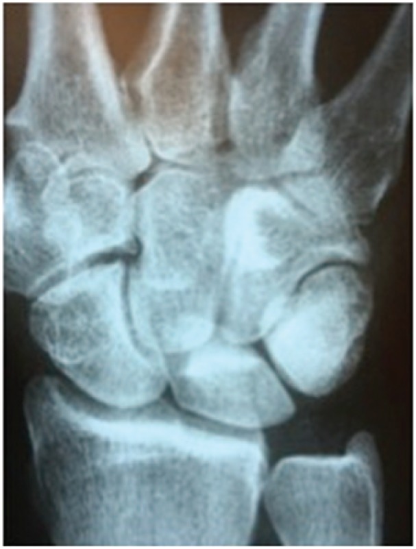
A 23-year-old right-handed professional baseball player presented with a 7- to 8-month history of left wrist pain precipitated by a head-first slide into second base. Pain was worse with swinging of the bat, usually at impact. The PA radiograph view showed a loose body at the ulnar border of the TH joint (Figure 1). Tenderness was present over the TH joint and worse with passive dorsiflexion and ulnar deviation. Conservative treatment including NSAIDs, rest, and corticosteroid injections failed to improve his symptoms, though injection done under fluoroscopic guidance with the wrist in maximal radial deviation did give initial relief. He underwent arthroscopic synovectomy and removal of the loose body with complete return to preinjury baseball activities.
Figure 1.

Posteroanterior wrist reveals loose body at triquetrohamate joint.
Case 2
A 51-year-old right-handed low handicap golfer presents with activity-related pain in the left wrist worse at ball strike increasing in intensity over a 2- to 3-year period. On examination, there is swelling, cystic in nature, over the dorsal ulnar aspect of the TH joint along with tenderness and increased pain with forced dorsiflexion and ulnar deviation. Radiographs revealed hypertrophic osseous changes involving the ulnar aspect of the hamate (Figure 2). After unsuccessful conservative treatment, the patient underwent arthroscopic synovectomy coupled with a limited open arthrotomy to allow for partial excision of the hamate to relieve the impingement (Figure 3). He was immobilized for 6 weeks, begun on progressive range and motion, followed by strengthening, with full return to pain-free activities including golf at 6 to 9 months.
Figure 2.

Intraoperative fluoroscopy has probe identifying hypertrophic hamate bone causing the impingement.
Figure 3.

Intraoperative view after partial resection of hamate.
Discussion
Ulnar-sided wrist pain can present as a diagnostic dilemma, and if misdiagnosed, treatment can result in pronounced morbidity. Furthermore, often the cause may be multifactorial, and failure to address this may result in continued symptoms. Successful management therefore requires a thorough knowledge of the different etiologies. The literature is replete with excellent reviews to help guide the hand surgeon through this diagnostic task.4,5 Triangular fibrocartilage tears, ECU tenosynovitis with subsheath injury, lunotriquetral interosseous ligament injury, along with ulnocarpal impaction, constitute the majority of conditions treated by the hand surgeon. In 1996, Nakao et al introduced the term triquetrohamate impaction syndrome in a case report, but except for this publication and brief mention in a review article, this injury has received little attention.1,3 This review encompasses the largest series to date and allows a thorough description of its presentation, history, physical exam findings, ancillary workup, and treatment.
In our patients, pain was localized to the dorsal ulnar aspect of the wrist at a point 1 to 2 cm distal to the TFCC complex. Symptoms arising from 2-handed golf and bat sports were predominantly found in the lead nondominant hand and usually experienced at impact with the ball. In this series, 2-handed sports, such as golf, baseball, and softball, made up 11 of 18 patients (>60%). These patients were the only ones to experience nondominant hand pain and this should guide the clinician to pain ulnarly in the nondominant lead hand, especially at impact. One-handed racket sports such as tennis produced symptoms in the dominant hand, also at impact with the ball. Although our series primarily involved patients involved in athletic activities (89%), it is our premise that with further knowledge and awareness, THIS may be diagnosed in the workplace or other activities that involve forced ulnar deviation and dorsiflexion. Also, with further recognition, earlier diagnosis may be achieved as in this series there was an average 7.4-month delay in diagnosis. A recent ex–college football player and low handicap golfer was treated with degenerative arthritis of the TH joint after a 5-year history, with the referring surgeon failing to identity the changes on radiograph (Figure 4).
Figure 4.
Posteroanterior view of wrist demonstrating joint space narrowing at the triquetrohamate joint with sclerotic changes due to repetitive impact trauma.
Findings on physical exam included tenderness over the distal aspect of the TH joint, an area 1 to 2 cm distal to the TFCC and 1 to 2 cm proximal to the base of the fifth metacarpal base. Pain is usually worse with both active and passive ulnar deviation and dorsiflexion, often associated with crepitus. Detailed exam is necessary to rule out neighboring pathology such as the TFCC and the ECU, but also realizing that these conditions may coexist and need treatment.
Ancillary studies should include standard wrist radiographs including PA, lateral, obliques, as well as a clenched fist to rule out ulnocarpal impaction. We have also utilized a PA in radial and ulnar deviation to better show joint narrowing and the presence of loose bodies. The radial deviation view done under fluoroscopic guidance has allowed injection of xylocaine and diagnostic confirmation of the TH joint as the source of pain. The use of MRI can further identify synovitis, chondral damage, and bone edema, while CT can better define the osseous impingement. Studies done in neutral and in ulnar deviation may further define the narrowing at the TH joint (Figure 5).
Figure 5.

Computed tomography scan in ulnar deviation reveals narrowing of joint space peripheral triquetrohamate joint.
Treatment begins with conservative methods including a short trial of rest, splinting, NSAIDs, occupational therapy, and, if necessary, judicious use of a corticosteroid injection. As stated previously, we have found the injection easier to perform under fluoroscopic guidance with the wrist in radial deviation. Core strengthening and sport-specific technique training are important in returning these patients to preinjury level. Using this conservative program, 56% (5 of 9) of patients were successful in achieving these goals. The remaining 44% returned to preinjury status with some limitations, again with conservative treatment. The remaining 50% of the patients (9 of 18) underwent surgical repair. Table 1 reveals a variety of treatment options utilized from arthroscopic synovectomy alone and/or combined with loose body removal, microfracture, and partial excision of hypertrophic hamate or triquetrum. Open procedures were necessary when the size of the carpal bone removed precluded arthroscopic removal. Also an open approach was used in cases with significant chondral damage, and the use of a dermal collagen interposition was felt indicated. The different techniques make it hard to compare specific long-term follow-up; however, surgical intervention did allow 7 of 9 patients (78%) to return to all activities without pain.
Triquetrohamate impaction syndrome is a defined, underreported cause of ulnar-sided wrist pain. It may coexist with more established causes, so the clinician needs to be aware of this presentation. The etiology of this syndrome seems to be repetitive impact of the opposing articular surfaces of the triquetrum and the hamate. Wrist position in ulnar deviation and dorsiflexion accentuates this contact. Kinematic description of wrist motion helps to explain the helicoid nature of this joint, and though many theories differ concerning the wrist, all document that as the wrist moves from neutral to ulnar deviation, the triquetrum translates ulnarly and dorsally along the helicoid slope of the hamate.2 This normal motion, in the face of repetitive injury, further increases this contact leading to pathologic impaction. Salient points that support the diagnosis of THIS are as follows:
In 2-handed sports, that is, golf, baseball, the lead nondominant hand is involved, and usually, symptoms are at impact when the wrist is in dorsiflexion and ulnar deviation. Otherwise, in one-handed activities, symptoms are located to the dominant extremity.
On exam, pain is localized on the dorsoulnar aspect of the TH joint with tenderness located in an area midway between the base of the fifth metacarpal and the ulnar styloid. The discomfort is increased with forced dorsiflexion and ulnar deviation. Locating the distal medial aspect of the triquetrum in radial deviation allows more space to exert deep palpation of the joint and confirm the location of the pain.
Radiographs in the PA projection in both radial deviations may better reveal joint space narrowing, osteophytes, and loose bodies. Magnetic resonance imaging and CT may reveal intraosseous edema, chondral damage, synovitis, and osseous changes. Injecting 2 to 3 cc of lidocaine into the TH joint under fluoroscopic guidance with the wrist in radial deviation helps to confirm the diagnosis. It also allows for placement of corticosteroid injections for therapeutic purposes.
Treatment begins with confirmation of TH impaction (THIS) as the correct diagnosis realizing other conditions can coexist. Once established, conservative treatment is initiated including NSAIDs, splints, rest, along with modification of activities. Core strengthening of trunk musculature is important. Judicious use of 1 to 2 corticosteroid injections may be helpful. Conservative treatment was successful in this series 56% of the time allowing full release without restrictions. If conservative treatment failed, surgical reconstruction was implemented. A number of procedures were utilized to treat the underlying condition; however, all addressed the pain secondary to TH impaction (THIS) and achieved 7 of 9 (78%) success.
This series of THIS represents the largest series to date. The emphasis of this preliminary report is to reintroduce this syndrome, first described as a case report by Nakao et al3 in 1996. Triquetrohamate impaction syndrome is an important cause of ulnar-sided wrist pain that deserves recognition. We have highlighted its presentation, findings on exam, recommended workup, and treatment. Our findings would support that approximately 50% of patients diagnosed can be handled conservatively with acceptable success. If conservative treatment fails, surgical intervention, through a multipronged approach, offers pain relief and return to preinjury status.
This report has confirmed the existence of THIS as an important and often unrecognized cause of ulnar-sided wrist pain. With heightened awareness of its presentation, earlier diagnosis and treatment may prevent avoidable morbidity.
Footnotes
Ethical Approval: This study was approved by our institutional review board.
Statement of Human and Animal Rights: All procedures followed were in accordance with the ethical standards of the responsible committee on human experimentation (institutional and national) and with the Helsinki Declaration of 1975, as revised in 2008.
Statement of Informed Consent: Informed consent was obtained from all patients for being included in the study.
Declaration of Conflicting Interests: The author(s) declared no potential conflicts of interest with respect to the research, authorship, and/or publication of this article.
Funding: The author(s) received no financial support for the research, authorship, and/or publication of this article.
References
- 1. Ek ETH, Suh N, Weiland A. Hand and wrist injuries in golf. J Hand Surg Am. 2013;38:2029-2033. [DOI] [PubMed] [Google Scholar]
- 2. Kamal RN, Rainbow MJ, Akelman E, Crisco JJ. In vivo triquetrum-hamate kinematics through a simulated hammering task wrist motion. J Bone Joint Surg Am. 2012;94:e85. [DOI] [PMC free article] [PubMed] [Google Scholar]
- 3. Nakao E, Nakamura R, Tsunoda K. Triquetrohamate impaction syndrome: a case report. J Hand Surg Am. 1996;21:778-780. [DOI] [PubMed] [Google Scholar]
- 4. Sachar K. Ulnar-sided wrist pain: evaluation and treatment of triangular fibrocartilage complex tears, ulnocarpal impaction syndrome, and lunotriquetral ligament tears. J Hand Surg Am. 2012;37:1489-1500. [DOI] [PubMed] [Google Scholar]
- 5. Shin AY, Deitch MA, Sachar K, Boyer MI. Ulnar-sided wrist pain. Diagnosis and treatment. J Bone Joint Surg Am. 2004;86:1560-1574. [PubMed] [Google Scholar]