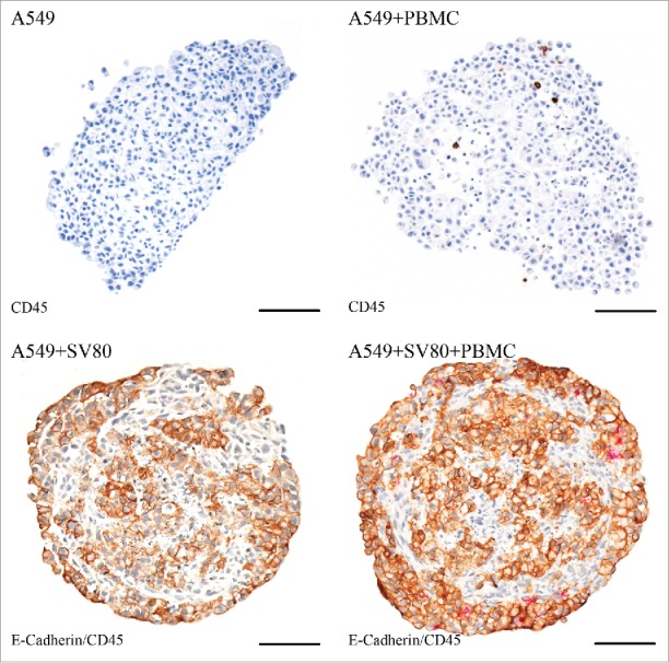
Figure 9.

Immunohistochemical staining – A549. Tissue sections were stained on CD45 (red) to visualize infiltrating CD45+ PBMCs in cancer cell and cancer cell/fibroblast microtissues. To discriminate between cancer cells and fibroblasts, a staining on E-cadherin (E-cad, brown) was used to visualize E-cad positive A549 cells (bar = 100 µm).
