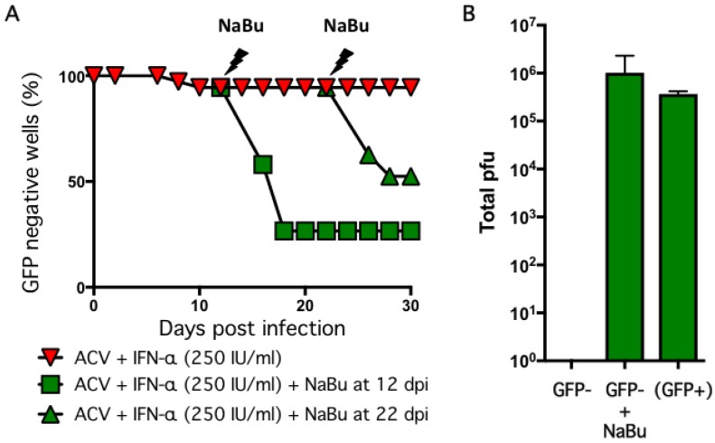
Figure 5.
Reactivation of infected cultures using sodium butyrate. (A) Neurons infected with HSV-1 GFP-Us11 at MOI = 0.01 in presence of 100 μM ACV and 250 IU/mL IFN-α were treated with 5 mM sodium butyrate (NaBu) and monitored for GFP fluorescence. The NaBu was added at either 12 or 22 dpi. The percentage of GFP negative wells is shown. (B) Supernatants were collected at 12 dpi from GFP negative wells [GFP−] or NaBu treated and maintained for a further six days [GFP+ + NaBu] or spontaneously reactivated wells collected at 8 dpi [(GFP+)] and assayed in parallel for infectious virus using Vero cells. Values represent total number of plaque-forming units (pfu) per well and graphed as the mean ± SEM.

