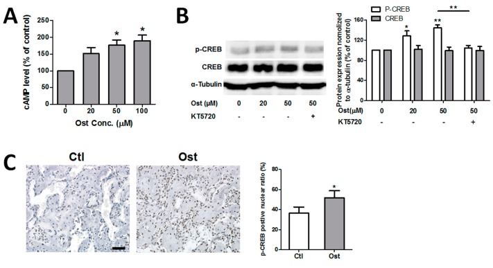
Figure 5.
Osthole activated cAMP/CREB pathway. (A) MC3T3-E1 cells were incubated with 0–100 μM osthole in growth medium for 2 h, and cellular cAMP levels were quantified with cAMP EIA kit (n = 3); (B) Cells were treated with 0, 20, or 50 μM osthole in osteogenic medium in the presence or absence of 4 μM KT5720 for 6 h. Proteins were separated by 10% SDS-PAGE and assessed with Western blotting, and the intensity of the bands was quantified (n = 5); (C) Callus sections at day 14 were blotted with p-CREB antibody and counterstained with hematoxylin. The p-CREB-positive nuclei percentage was counted (n = 4). One-way ANOVA, followed by Tukey’s test, was compared to the vehicle control; an unpaired Student t-test was compared between KT5720 ± groups, * p < 0.05, ** p < 0.01. Bar = 50 μm.

