Abstract
The development of ectothermic embryos is strongly affected by incubation temperature, and thermal imprinting of body growth and muscle phenotype has been reported in various teleost fishes. The complex epigenetic regulation of muscle development in vertebrates involves DNA methylation of the myogenin promoter. Body growth is a heritable and highly variable trait among fish populations that allows for local adaptations, but also for selective breeding. Here we studied the epigenetic effects of embryonic temperature and genetic background on body growth, muscle cellularity and myogenin expression in farmed Atlantic salmon (Salmo salar). Eggs from salmon families with either high or low estimated breeding values for body growth, referred to as Fast and Slow genotypes, were incubated at 8°C or 4°C until the embryonic ‘eyed-stage’ followed by rearing at the production temperature of 8°C. Rearing temperature strongly affected the growth rates, and the 8°C fish were about twice as heavy as the 4°C fish in the order Fast8>Slow8>Fast4>Slow4 prior to seawater transfer. Fast8 was the largest fish also at harvest despite strong growth compensation in the low temperature groups. Larval myogenin expression was approximately 4–6 fold higher in the Fast8 group than in the other groups and was associated with relative low DNA methylation levels, but was positively correlated with the expression levels of the DNA methyltransferase genes dnmt1, dnmt3a and dnmt3b. Juvenile Fast8 fish displayed thicker white muscle fibres than Fast4 fish, while Slow 8 and Slow 4 showed no difference in muscle cellularity. The impact of genetic background on the thermal imprinting of body growth and muscle development in Atlantic salmon suggests that epigenetic variation might play a significant role in the local adaptation to fluctuating temperatures over short evolutionary time.
Introduction
Ambient temperature controls and limits virtually all biochemical and physiological processes and behavioural activities in poikilothermic organisms. While heat stress during organogenesis may be teratogenic also to poikilothermic embryos, they show phenotypic plasticity to moderate changes in incubation temperature that strongly affect embryonic development and may have persistent effects on various phenotypic traits. Ducklings reared at relative low temperature showed slow growth and reduced thermoregulatory capacity after hatching [1], while embryonic temperature influenced growth rate and temperature choice in juvenile snapping turtles (Chelydra serpentine) [2] and swimming performance in wood frog tadpoles (Rana sylvatica) [3]. In zebrafish (Danio rerio), early temperature strongly affected metabolic enzymes in the skeletal muscle, swimming performance and thermal acclimation capacity of the adult fish [4,5]. In most teleosts, both hypertrophic and hyperplastic muscle growth continue after hatching [6–8], and the muscle phenotype at later stages has been shown to be programmed by embryonic temperature in various species [9–14]. The lasting effects of embryonic temperature on muscle have been demonstrated to involve temperature-dependent changes in the number of muscle precursor cells, which are responsible for postembryonic growth in teleosts [11,15,16].
Skeletal muscle development in vertebrates is regulated by the concerted action of the four myogenic regulatory factors (MRFs) MyoD, myf5, myogenin and MRF4. Mice knockout studies revealed that MyoD and myf5 are required to specify myoblasts, while myogenin and MRF4 act later to mediate differentiation of myoblasts into and fusion of myotubes [17–21]. Thermal effects on the embryonic expression of MyoD and myogenin have been reported in various teleosts, including Atlantic salmon and rainbow trout (Oncorhynchus mykiss) [22–24]. The upregulated myogenin expression and increased muscle development at elevated temperatures in Senegalese sole (Solea senegalensis) were associated with decreased methylation of the myogenin promoter and decreased expression of the DNA methyltransferases dnmt1 and dnmt3b [25]. Expression of myogenin was consistently downregulated at low temperatures in turkey embryos and in mouse skeletal muscle cells in vitro [26,27]. Further, demethylation of the mouse myogenin promoter activated the gene expression in somites during skeletal muscle differentiation [28,29], and inhibition of DNA methylation in mesenchymal and dental pulp stem cells up-regulated myogenin expression and induced myogenesis [30,31]. The complex epigenetic network regulating muscle development also involves histone modifications and myogenic microRNAs controlling the expression of key myogenic factors [32–36].
Studies linking genetic variation and epigenetic regulation have mainly been conducted in plants, yeast and Drosophila, but the inheritance of epigenetic alleles have also been reported in mice displaying different coat colour phenotypes, in niche-adapted Darwin finches, and in association with various human diseases [37–40]. Intriguingly, differences in adaptive phenotypic responses, including body size, to climate changes enabled by epigenetics were recently reported in two populations of winter skate (Leucoraja ocellata) [41]. Migratory salmonids have a tendency to spawn in their homing rivers differing in seasonal and regional thermal conditions, and evidence of local genetic adaptation in muscle cellularity and body growth have been reported in wild populations [11,42,43]. Substantial heritability in body growth exists in Atlantic salmon, and significant enhancements in growth rate and hence reduction in production time have been achieved in farmed Atlantic salmon through 11 generations of selective breeding since 1972 [44]. Here we examine the impact of genetic background and embryonic temperature on the epigenetic regulation of body growth and white muscle phenotype in farmed Atlantic salmon.
Material and methods
Experimental set up
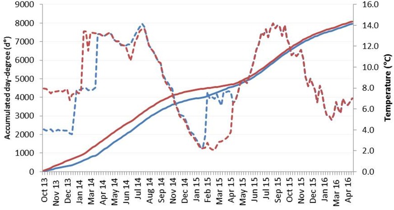
Families with fast and slow on-growth within the natural variation were selected from AquaGen’s breeding nucleus year class 2011 (11th generation with a heritability h2 of 0.3) by estimating breeding values (EBV) based on weight registrations at tagging, smolt weight, and harvest weight of the year class. Eight families with a mean EBV of 1.6 standard deviations (SD) higher than average and eight families with 1.6 SD lower than average were chosen and referred to as Fast and Slow genotypes, respectively. The families were created from eight different dams and fourteen different sires. The eight dams used to produce fast on-growth families originated from five families and the sires from seven families, while the eight dams used to produce slow on-growth families originated from three families and the sires from seven families. From October 2013, 600 fertilized eggs from each family were incubated at 4°C (3.9 ± 0.2°C) or 8°C (7.6 ± 0.5°C) until the eyed stage (360 day degrees, d° = number of days x temperature) that resulted in four experimental groups referred to as Fast4, Fast8, Slow4 and Slow8. Low-quality embryos were removed after physical shocking at 316 d° according to AquaGen’s standard protocol, and one of the Slow families was excluded due to high mortality at this stage. Dead and pin-eyed embryos were removed at the eyed stage, and the incubation temperature of the Fast4 and Slow4 groups was gradually increased to the standard production temperature of 8°C over three days. All groups were kept at this temperature until start feeding at which stage the temperature was further increased to 12.2 ± 1.1°C. Juveniles at the parr stage were RFID (radio frequency ID) tagged for individual identification at body weight of ~25 g (Fast8, Slow8) or ~8 g (Fast4, Slow4). From August 2014, a maximum of 200 individuals from each experimental group were kept for further on-growth at ambient temperatures varying from 2°C to 14°C in a flow-through system until sea transfer in April 2015 (Fig 1). For all groups to be transferred into seawater at similar total d° (calculated from fertilization to sea transfer), we increased the temperature in the Fast4 and Slow4 groups from ~2 to ~7°C for a period of 9 weeks prior to sea transfer (Fig 1, S1 Table). All groups were kept together in one sea cage (about 3000–4000 m3) until harvest in April 2016. Fish were fed by appetite with EWOS feed from start feeding until harvest.
Fig 1. Temperature and day-degrees profiles.
Temperature (°C, striped line) and accumulated day-degrees (d°, solid line) profiles between fertilization and harvest for the low (4°C, blue) and high (8°C, red) embryonic temperature groups. All fish were kept at the same temperature from the eyed-stage, except for the elevated temperature in the Fast4 and Slow4 groups prior to sea transfer. Details are given in Material and Methods.
Sampling
Body weight was measured in sub-samples at the start feeding, parr, and pre-smolt stages, while individual weight was recorded in smolts before sea transfer and in adults at harvest. All measurements were performed in fish anesthetized using metacain (Finquel vet., Scanvacc) in accordance to the supplier’s instructions. Prior to any sampling, fish were euthanized using an overdose of metacain. An overview of sampling dates and d° for each treatment group is provided in S1 Table. Muscle samples directly rostral of the dorsal fin were collected from start feeding larvae and parr of ~6g body weight, and fixated in RNA-later (Ambion) and in 96% ethanol. Samples in RNA-later were kept overnight at 4°C followed by storage at -20°C until further analysis, while samples in ethanol were stored at -20°C. For histology, a transversal section directly rostral of the dorsal fin was obtained from parr, fixed in 4% paraformaldehyde solution (PFA, Electron Microscopy Sciences), and were kept overnight at 4°C. Thereafter, samples were washed in phosphate buffered saline (PBS, Sigma-Aldrich) for 10 min, dehydrated in 50% ethanol (30 min) and 70% ethanol (2 x 1 hr), and stored at -20°C until further analysis.
Thermal growth coefficient (TGC; [45]) in freshwater and seawater was calculated by:
In the freshwater phase, TGC was calculated on mean weight (g) per treatment group from start feeding (BW2) to sea transfer (BW1), and d° using the temperature measured in tanks. In seawater phase, individual body weight (g) at sea transfer (BW2) and at harvest (BW1) was used, and d° using the temperature measured at 5m depth.
Standardized harvest weight (SHW) was used to standardize harvest weight independent of different smolt start weight at sea transfer [46]. SHW has been developed to standardize field trial weight data acquired in commercial Atlantic salmon farming where differences in production regimes can lead to uneven growth differences [47]. The more commonly used specific growth rate (SGR) is usually biased towards small fish when used to compare fish of uneven size. We used a smolt weight of 200 g, which was the lowest weight among the treated groups at transfer, and TGC combined with the sum of d° between sea transfer and harvest:
Difference in SHW (ΔSHW) for 4°C groups was calculated by:
DNA methylation analysis
Four families from each of the four treatment groups (n = 6 per family) were chosen for DNA methylation analysis. Total genomic DNA was isolated and purified from muscle tissue at the start feeding and parr stages using DNeasy Blood & Tissue Kit (Qiagen) according to the manufacturer’s protocol and DNA ethanol precipitation method, respectively, due to the differences in sample size. DNA quantity and quality were measured using a 1000-ND Nanodrop spectrophotometer (Nanodrop Technologies). DNA was bisulfite converted using the Epitect Fast Bisulfite Conversion Kit (Qiagen) according to the manufacturer’s protocol.
Six specific pyrosequencing assays (PyroMark Custom Assay, Qiagen) covering fourteen putative CpG sites identified in the Atlantic salmon myogenin promoter (NC_027321.1, S2 Fig) were designed using PyroMark Assay Design 2.0 (Qiagen) (S2 Table). Assay specific PCRs were performed using the PyroMark PCR Kit (Qiagen) according to manufacturer’s protocol. Singular PCR products were verified on a 1% agarose gel. Five of the fourteen CpG sites could be analyzed using pyrosequencing, as only two of the six primer sets (S2 Table) provided a singular PCR product. The PyroMark Q24 (Qiagen) was used in combination with the PyroMark 24 Advanced CpG Reagents (Qiagen) and Streptavidin Sepharose High Performance beads (GE Healthcare) to analyze the CpG methylation assays by pyrosequencing technology following the manufacturer’s PyroMark Q24 Advanced protocol using 25uL sequencing primer solution.
Gene expression analysis
Four families from each of the four treatment groups (n = 6–10 per family) were chosen for gene expression analysis in fast muscle tissue at the start feeding and parr stages. Total RNA was extracted and purified using the MagMax-96 Total RNA Isolation Kit (Applied Biosystems) with the MagMax-96 Magnetic Particle Processor (Applied Biosystems) and AllPrep DNA/RNA/miRNA Universal kit (Qiagen), respectively, following manufacturer’s instructions. Both methods were used due to differences in samples size. RNA extracted using the MagMax-96 kit was followed by a clean-up using the Turbo DNase Treatment Kit (Applied Biosciences). RNA quantity and quality were measured using a 1000-ND Nanodrop spectrophotometer. cDNA was synthesized using the Vilo Superscript Kit (Invitrogen) following the manufacturer’s instructions using 200ng of total RNA. The relative expression levels of myogenin, the DNA methyltransferase genes dnmt1 (XM_014193376), dnmt3a (XM_014136242.1), dnmt3b (XM_014146676.1) and the reference gene elongation factor 1α (ef1α, [48]) were determined using quantitative real-time PCR (qPCR), and primers were designed using Primer3 program (Applied Biosystems) (S1 Table). A two-fold standard dilution of muscle cDNA was set up for each primer set in order to determine the amplification efficiency. The qPCR was run in duplicates using the 7900HT Fast Real-Time PCR system (Applied Biosystems) with a total volume of 20μL containing 10μL Power SYBR Green PCR Master Mix (Applied Biosystems), 0.6μL 10μM forward and reverse primers, 8μL diluted cDNA. A cycling profile of 10 min at 95°C, followed by 40 cycles of 95°C for 15s, 60°C for 60s. Absence of genomic DNA was verified by running randomly chosen RNA samples. In order to rule out non-specific contamination a no template control was included and a melting curve analysis was performed to verify the measurement of a single specific product. SDS 2.3 software (Applied Biosystems) was used to collect all data that was thereafter analyzed using RQ manager 1.2 (Applied Biosystems). Relative gene expression was calculated based on the determined Ct values [49]. The treatment group with the lowest value was used as a calibrator and set to one (1).
Muscle cellularity
Samples were dehydrated in ethanol series (2 times 96% for 1 hr and 2 times 100% for 1 hr), followed by clearing in Histoclear (National Diagnostics) for 1 hr, Histoclear/paraffin (1:2; Histowax, Histolab Products AB) and embedding in paraffin using a Citadel 2000 Tissue processor (Thermo Scientific). Sections of 7 μm were cut using a microtome (Leica RM2255), and were mounted on microscope slides. After rehydration, the samples were stained with haematoxylin-eosin (Shandon Instant Haematoxylin and Shandon Instant Eosin (alcoholic), Thermo Scientific)). Maximum length and width of ~200–400 cells (n = 10–12 per group) were measured in an area of 1 mm2 using ImageJ (https://imagej.nih.gov/ij). The fast muscle cell diameter (μm) was estimated indirectly by regarding the cell as a circle [25] using the following formula: diameter = (maximum length * maximum width)1/2. The relative distribution was expressed as: (number of cells at a certain diameter / the total number cells) * 100.
Statistics
Data was tested for normal distribution using one-sample Kolmogorov-Smirnov statistic test. A general linear regression (GLM) was conducted to compare main effects of type of on-growth (fast, slow) and incubation temperature (4°C, 8°C) and the interaction between genotype and incubation temperature on body weight measured at the five developmental stages, and for TGC and SHW. Significant differences between experimental groups were analyzed by least square means (LS-means). As data for the expression and methylation levels was not normally distributed (K-S test p<0.05) a non-parametric Kruskall-Wallis test was performed following by Mann-WhitneyU test as posthoc test. Correlation analysis of the methylation sites and the expression data was performed using a non-parametric Spearman test. A student t-test was used to compare the average muscle fibre size between the groups. A two-sample Kolmogorov-Smirnov test was used to analyze differences in muscle fibre distributions. Data are presented as mean ± standard error and considered significantly different when p<0.05.
Results
Growth
Embryonic incubation at the low temperature of 4°C strongly delayed the developmental progress, but the 4°C and 8°C incubation groups reached the eyed stage and start feeding stage at quite similar number of d° (Fig 1, S3 Table). At start feeding the Fast8 and Slow8 fish were significantly heavier (0.19 g) than Fast4 and Slow4 (0.17 g), but the 4°C groups were heavier than the 8°C groups at the parr stage that was reached after 2085 d° and 1985 d°, respectively (Table 1). The body weight prior to seawater transfer was strongly influenced by both genotype and embryonic temperature, and the 8°C smolts were about twice as heavy as the 4°C smolts in the order Fast8>Fast4>Slow8>Slow4 after 4597 d° and 4313 d°, respectively. TGC in the freshwater phase was significantly higher in the high temperature groups (Fast8: 1.6, Slow8: 1.5) than in the low temperature groups (Fast4: 1.2, Slow4: 1.2), but the opposite was found in seawater (Fast8: 3.0 vs Fast4: 3.3, Slow8: 2.6 vs Slow4: 3.0) (Table 1). The catch-up growth of the 4°C fish in seawater reduced the weight difference between the 8°C and 4°C groups, and at harvest the Fast8 fish was 8% heavier than the Fast4 fish, while Slow8 was only 4% heavier than Slow4. SHW was significantly higher in both 4°C groups compared to 8°C groups that resulted in an estimated difference ΔSHW of 858 ± 45 g and 1019 ± 77 g for Fast4 and Slow4, respectively (Table 1).
Table 1. Biometrics.
| Fast 8 | Fast 4 | Slow 8 | Slow 4 | |||||
|---|---|---|---|---|---|---|---|---|
| Start-feed | 0.19±0.005a | (96) | 0.17±0.004b | (96) | 0.19±0.005a | (82) | 0.17±0.005b | (84) |
| Parr | 5.9±0.2bc | (79) | 7.0±0.2a | (80) | 5.5±0.2c | (70) | 6.5±0.2ab | (70) |
| Pre-smolt | 247.6±5.7b | (81) | 233.9±5.8b | (80) | 239.4±6.9b | (56) | 195.3±6.9a | (56) |
| Smolt | 446±2d | (1275) | 227±2b | (1339) | 417±3c | (613) | 200±2a | (950) |
| TGC FW | 1.6±0.02b | (8) | 1.2±0.02a | (8) | 1.5±0.02c | (7) | 1.2±0.02a | (7) |
| Harvest | 5458±36c | (905) | 5049±33b | (1061) | 4234±55a | (382) | 4084±38a | (800) |
| TGC SW | 3.0±0.0b | (899) | 3.3±0.0a | (1057) | 2.6±0.0c | (382) | 3.0±0.0b | (800) |
| SHW | 4032±30b | (899) | 4875±28a | (1057) | 3105±46c | (380) | 4123±32b | (798) |
| ΔSHW | 858±45a | (8) | 1019±77a | (7) |
Body weight (g) and growth estimates from start-feeding until harvest of Fast and Slow genotypes reared at 4°C or 8°C until the eyed stage given as mean ± SE (number of individuals).
Weight estimates are based on sub-samples of each family from start-feeding to pre-smolt, and on individual weight registrations from smolt until harvest.
Thermal growth coefficient (TGC) and standardized harvest weight (SHW) are estimated from individual weight registrations, while ΔSHW is estimated from mean SHW in each family.
Different letters indicate significant differences between groups (p<0.05).
DNA methylation of myogenin promoter
DNA methylation of the myogenin promoter was assessed in muscle tissue of start feeding larvae and parr juveniles by pyrosequencing five CpG sites upstream of the translation start site. At start feeding the Fast8 group showed significantly lower DNA methylation level at positions -610 and -598 than in the other groups, and also at position -258 when compared with Fast4 and Slow4 (Fig 2a). In addition, Fast8 showed significantly lower methylation level than Fast4 at position -234, while the methylation at position -255 was lower in Slow4 and Slow8 than in Fast4. The DNA methylation of the five sites was significantly lower in parr than in start feeding larvae (Fig 2b). The four treatment groups showed less variation at the parr stage, and only position -234 showed a significantly lower level in the Fast4 compared with the Slow4 group (p<0.05).
Fig 2. DNA methylation of myogenin promoter.
DNA methylation (%) of five CpG sites in the promoter region of the myogenin gene measured in start feeding larvae (a) and in parr (b) reared at 4°C or 8°C until the eyed stage. Fast and Slow denote the genetic background of the fish. Numbers 1–5 correspond to the CpG sites at position -610, -598, -258, -255 and -234, respectively. Different letters indicate significant differences between groups (p<0.05).
Gene expression patterns
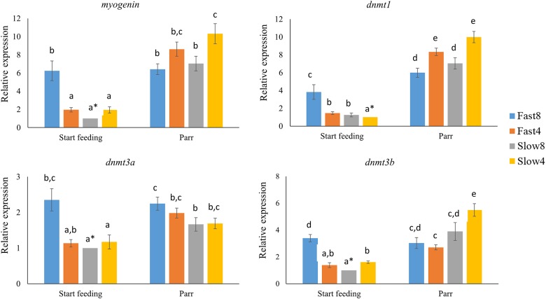
The relative expression levels of myogenin and the DNA methyltransferases dnmt1, dnmt3a and dnmt3b were determined in muscle tissue of start feeding larvae and parr using qPCR. At the larval stage, the mRNA levels of myogenin was approximately 4- to 6-fold higher in the Fast 8 group than in the other groups (p<0.05), in which no significant differences were found (Fig 3a). Similarly, Fast8 larvae expressed the dnmt1, dnmt3a and dnmt3b genes at significant higher levels than the other groups, except for the non-significant difference in dnmt3a mRNA levels between Fast8 and Fast4 (Fig 3b–3d). The four genes examined were expressed at higher levels at the parr stage than in the start-feeding larvae, except for the Fast8 parr displaying elevated levels of only dnmt1 (p<0.05; Fig 3a–3d). The Slow4 parr expressed myogenin, dnmt1 and dnmt3b at significantly higher levels than Fast 8 and Slow 8, while the dnmt3a expression was significantly higher in Fast8 compared Slow8.
Fig 3. Gene expression of myogenin and DNA methyltransferases.
Relative expression of myogenin and the DNA methyltransferases dnmt1, dnmt3a and dnmt3b in start feeding larvae and in parr from families with Fast and Slow genotypes after incubation at 4°C or 8°C until the eyed stage. Asterisks represent the calibrator group with the lowest expression value set to 1 for the gene expression analysis. Different letters indicate significant differences between groups and stages (p<0.05).
Independent of stage, growth rate and rearing temperature, positive correlations were found between the expression of myogenin and dnmt1 (ρ = 0.839; p<0.001), myogenin and dnmt3a (ρ = 0.762; p<0.001), myogenin and dnmt3b (ρ = 0.770; p<0.001) (S4 Table). Negative correlations were found between the methylation levels of the five studied sites and the expression of myogenin, dnmt1, dnmt3a and dnmt3b.
Muscle cellularity
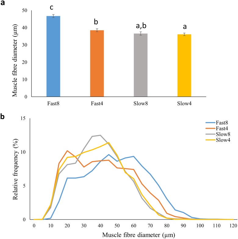
The average diameter of white muscle fibres was significantly larger in Fast8 compared to the other three groups at the parr stage, and also larger in Fast4 compared with Slow4 (p<0.05) (Fig 4a). The distribution of muscle fibre diameters (Fig 4b) showed that Fast8 possessed relatively more muscle fibres with a larger diameter than the other treatment groups (p<0.05; right-hand tail). Fast4 showed a distribution more towards the right-hand tail only compared to Slow4 (p<0.05).
Fig 4. White muscle cellularity in Atlantic salmon parr.
a) Average fibre diameter (μm), and b) size distribution (%) of fibre diameter in parr from families with Fast and Slow genotypes after incubation at 4°C or 8°C until the eyed-stage. Different letters present significant differences (p<0.05) between groups.
Discussion
This study demonstrated substantial effects of genetic background and embryonic temperature on muscle phenotype and body growth in farmed Atlantic salmon. While the temperature effects on body growth differed between the freshwater and seawater phases, fish from families of the Fast genotype were significantly heavier from the smolt stage until harvest than those with the Slow genotype. Independent of genotype, the high temperature fish were a two-fold heavier prior to seawater transfer than the low temperature fish, while the latter Fast4 and Slow4 fish grew significantly faster than Fast8 and Slow8 fish under the same conditions in seawater. The potential of compensatory growth experienced by the 4°C group was shown by using SHW as a model to standardize weight differences at sea transfer. The compensatory growth observed was probably resulting from different embryonic temperatures, although we cannot exclude possible effects of the temperature adjustments before seawater transfer and the timing of smoltification. Consistently, Atlantic salmon incubated at the low embryonic temperatures of 2°C or 5°C showed significant growth compensation after seawater transfer when compared to high temperature fish (8°C or 10°C) [14]. In comparison, salmon raised at embryonic temperature of 10°C prior to a decrease to 5°C grew faster than fish raised at 5°C during early stages, but the 5°C fish displayed higher foraging activity and a more sustained period of muscle growth [13]. Additionally, embryonic incubation at low temperature promoted hyperplasia in the white muscle of salmon larvae and juveniles [13,14,50], in agreement with the smaller fibre diameters in the Fast4 and Slow4 fish compared to Fast8 in the presents study. Intriguingly, Slow 8 and Slow4 showed no difference in muscle fibre thickness, suggesting that only the Fast genotype responded to elevated rearing temperature with hypertrophic growth. While large fibre size in fish skeletal muscle seems to be metabolically advantageous by reducing the cost of maintaining the membrane potential [51,52], a fast-growing strain of rainbow trout exhibited significantly smaller fibre diameter than a slow-growing strain from hatching to 24 cm body length that allowed for prolonged and greater muscle growth in the adult fish [53]. Consistently, pearlfish incubated at high embryonic temperature of 16°C showed reduced cell proliferation, but increased differentiation that gave rise to larger hatchlings, while their limited reserves of muscle precursor cells finally led to smaller adults than those incubated at 13 or 8.5°C [16].
Thermal imprinting of muscle cellularity involves modification of the proliferation and differentiation of the muscle precursor cells being regulated by conserved myogenic transcription factors [11,15,16]. Myogenin plays a crucial role in the differentiation of muscle precursor cells, and the ontogenic expression of myogenin in rainbow trout peaked in swim-up fry [54]. Correspondingly, the Fast8 fish displayed high myogenin expression at start-feeding, while the gene expression seemed to be delayed in the other treatment groups. Accordingly, low embryonic temperature delayed and prolonged expression of MyoD, myogenin and MyHC in rainbow trout that resulted in the recruitment of considerably more fibers compared to high temperature fish [22]. The elevated myogenin expression in the Fast8 fish coincided with the less methylated promoter region when compared to the other groups, in agreement with the inverse correlation between the myogenin expression and methylation levels in Senegalese sole at metamorphosis [25]. However, the expression of salmon myogenin was positively correlated with the gene expression of the DNA methyltransferases, in contrast to the negative correlation between the expression of myogenin and the levels of dnmt1 and dnmt3b in Senegalese sole [25]. The function of Dnmt1 is maintenance of DNA methylation levels after cell division, while Dnmt3a and 3b are directly involved in de novo methylation [55,56]. The conflicting results could be explained by the simultaneous processes of methylation of a variety of genes, and thereby the effects on individual genes may become less clear. The epigenetic regulation of myogenesis is further complicated by the involvement of histone modifications and microRNAs [34,57], and histone methylation of three pax7 genes was suggested to regulate myogenin expression in rainbow trout [35].
The thermal plasticity of muscle development and somatic growth in fish is thought to play a crucial role in the local adaptation to prevailing water temperature conditions, and evidence for genetic differences between populations has been provided in several species [11,12,58,59]. Eggs from two salmon populations spawning in a lowland or highland tributary of the Scottish River Dee System responded differently to the two temperature regimes when incubated together [11], while strong temperature and family effects on muscle cellularity were demonstrated in farmed families of Atlantic salmon originating from the Scottish River Shin [58]. The genetic gain for growth rate in Atlantic salmon has been estimated at 10–15% per generation [44], but no single SNP with a significant effect on growth has been identified across year-classes ([60], Moen et al. unpubl). The polygenic nature of this trait, together with the complex epigenetic mechanisms regulating skeletal muscle growth, makes it difficult to identify the mechanisms underlying the epigenetic variation found in Atlantic salmon.
Conclusions
This study demonstrates strong interactions between thermal phenotypic plasticity and genotypic diversity affecting body growth and muscle cellularity in Atlantic salmon. Epigenetic variation in skeletal muscle growth is for the first time documented by presenting differences in DNA methylation and expression of myogenin in farmed Atlantic salmon families with either high or low breeding values for on-growth. The persistent effects of embryonic temperature on body growth and muscle cellularity were consistently shown to differ between two genotypes. Thermal plasticity and epigenetic variation in body growth are probably prerequisites for the local adaptation of salmon populations to fluctuating environmental conditions, and may potentially make them more resilient to global warming.
Supporting information
Fourteen potential DNA methylation sites (bold CG) were identified in the proximal promoter region of Atlantic salmon myogenin. Five CG sites (green coloured) were assessed by pyrosequencing. Underlined sites indicate two E-boxes (CAnnTG) potentially binding myogenic regulatory factors. The promoter sequence ends with the ATG translational start site.
(TIF)
Overview of date and day-degree (d°) for tissue sampling and weight registrations for the two temperature groups of 4°C and 8°C.
(DOCX)
Overview of primers used for qPCR and assays used for DNA methylation analysis by pyrosequencing. Pyrosequencing assays 1 and 5 provided a singular PCR product.
(DOCX)
Main effects of type of on-growth (fast, slow) and incubation temperature (4°C, 8°C) and the interaction between genotype and incubation temperature on body weight measured at the five developmental stages, and for TGC and SHW.
(DOCX)
Spearman correlation analysis between gene expression of myogenin, dnmt1, dnmt3a and dnmt3b, and the methylation levels of the five studied methylation sites (CpG). CpG1-5 refer to the putative CpG sites in the myogenin promoter located at -610, -598, -258, -255 and -234, respectively. P<0.05 are considered significantly different.
(DOCX)
Acknowledgments
We would like to thank Hege Munck, Katrine Kirste, Audny Johansen and Tina Thesslund (Nofima AS), and Fride Tonning (University of Tromsø) for their technical assistance. We are also very grateful to Ingun Næve, Sven Arild Korsvoll and Marit Prestrud (AquaGen AS) for their assistance during sampling.
Data Availability
All relevant data are within the paper and its Supporting Information files.
Funding Statement
The authors received funding from Norwegian Research Council Project No. 221734/O30. Nofima AS provided support in the form of salaries for authors [EB, HJ, OA] and AquaGen AS provided support in the form of salaries for authors [MM, AA, NS] but did not have any additional role in the study design, data collection and analysis, decision to publish, or preparation of the manuscript. The specific roles of these authors are articulated in the ‘author contributions’ section.
References
- 1.DuRant SE, Hopkins WA, Wilson A, Hepp GR. Incubation temperature affects the metabolic cost of thermoregulation in a young precocial bird. Funct Ecol. 2012; 26: 416–422. [Google Scholar]
- 2.O’Steen S. Embryonic temperature influences juvenile temperature choice and growth rate in snapping turtles Chelydra serpentine. J Exp Biol. 1998; 201: 439–449. [PubMed] [Google Scholar]
- 3.Watkins TB, Vraspir J. Both incubation temperature and posthatching temperature affect swimming performance and morphology of wood frog tadpoles (Rana sylvatica). Physiol Biochem Zool. 2006; 79: 140–149. doi: 10.1086/498182 [DOI] [PubMed] [Google Scholar]
- 4.Scott GR, Johnston IA. Temperature during embryonic development has persistent effects on thermal acclimation capacity in zebrafish. Proc Natl Acad Sci USA. 2012; 109: 14247–14252. doi: 10.1073/pnas.1205012109 [DOI] [PMC free article] [PubMed] [Google Scholar]
- 5.Schnurr ME, Yin Y, Scott GR. Temperature during embryonic development has persistent effects on metabolic enzymes in the muscle of zebrafish. J Exp Biol. 2014; 217: 1370–1380. doi: 10.1242/jeb.094037 [DOI] [PubMed] [Google Scholar]
- 6.Weatherley AH, Gill HS, Lobo AF. Recruitment and maximal diameter of axial muscle fibres in teleosts and their relationship to somatic growth and ultimate size. J Fish Biol. 1988; 33: 851–859. [Google Scholar]
- 7.Veggetti A, Mascarello F, Scapolo PA, Rowlerson A, Carnevali MC. Muscle growth and myosin isoform transitions during development of a small teleost fish, Poecilia reticulata (Peters) (Atheriniformes, Poeciliidae): a histochemical, immunohistochemical, ultrastructural and morphometric study. Anat Embryol. 1993; 187: 353–361. [DOI] [PubMed] [Google Scholar]
- 8.Steinbacher P, Haslett JR, Six M, Gollmann HP, Sänger AM, Stoiber W. Phases of myogenic cell activation and possible role of dermomyotome cells in teleost muscle formation. Dev Dyn. 2006; 235: 3132–3143. doi: 10.1002/dvdy.20950 [DOI] [PubMed] [Google Scholar]
- 9.Stickland NC, White RN, Mescall PE, Crook AR, Thorpe JE. The effect of temperature on myogenesis in embryonic development of the Atlantic salmon (Salmo salar L.). Anat Embryol. 1988; 178: 253–257 [DOI] [PubMed] [Google Scholar]
- 10.Usher ML, Stickland NC, Thorpe JE (1994). Muscle development in Atlantic salmon (Salmo salar) embryos and the effect of temperature on muscle cellularity. J Fish Biol. 1994; 44: 953–964. [Google Scholar]
- 11.Johnston IA, McLay HA, Abercromby M, Robins D. Phenotypic plasticity of early myogenesis and satellite cell numbers in Atlantic salmon spawning in upland and lowland tributaries of a river system. J Exp Biol. 2000; 203: 2539–2552. [DOI] [PubMed] [Google Scholar]
- 12.Johnston IA, Vieira VLA, Temple GK. Functional consequences and population differences in the developmental plasticity of muscle to temperature in Atlantic herring. Mar Ecol Prog Ser. 2001; 213: 285–300. [Google Scholar]
- 13.Albokhadaim I, Hammond CL, Ashton C, Simbi BH, Bayol S, Farrington S, Stickland N. Larval programming of post-hatch muscle growth and activity in Atlantic salmon (Salmo salar). J Exp Biol. 2007; 210: 1735–1741. doi: 10.1242/jeb.003194 [DOI] [PubMed] [Google Scholar]
- 14.Macqueen DJ, Robb DHF, Olsen T, Melstveit L, Paxton CGM, Johnston IA. Temperature until the ‘eyed-stage’ of embryogenesis programmes the growth trajectory and muscle phenotype of adult Atlantic salmon. Biol Lett. 2008; 4: 294–298 doi: 10.1098/rsbl.2007.0620 [DOI] [PMC free article] [PubMed] [Google Scholar]
- 15.Matschak TW, Stickland NC. The growth of Atlantic salsmon (Salmo salar L.) myosatellite cells in culture at two different temperatures. Experientia. 1995; 51: 260–266. [DOI] [PubMed] [Google Scholar]
- 16.Steinbacher P, Marschallinger J, Obermayer A, Neuhofer A, Sänger AM, Stoiber W. Temperature-dependent modification of muscle precursor cell behaviour is an underlying reason for lasting effects on muscle cellularity and body growth of teleost fish. J Exp Biol. 2011; 214: 1791–1801. doi: 10.1242/jeb.050096 [DOI] [PMC free article] [PubMed] [Google Scholar]
- 17.Hasty P, Bradley A, Morris JH, Edmondson DG, Venuti JM, Olson EN et al. Muscle deficiency and neonatal death in mice with a targeted mutation in the myogenin gene. Nature. 1993; 364: 501–506 doi: 10.1038/364501a0 [DOI] [PubMed] [Google Scholar]
- 18.Nabeshima Y, Hanaoka K, Hayasaka M, Esumi E, Li S, Nonaka I. Myogenin gene disruption results in perinatal lethality because of severe muscle defect. Nature. 1993; 364: 532–535. doi: 10.1038/364532a0 [DOI] [PubMed] [Google Scholar]
- 19.Bergstrom DA, Tapscott SJ. Molecular distinction between specification and differentiation in the myogenic basic helix-loop-helix transcription factor family. Mol Cell Biol. 2001; 21: 404–412. [DOI] [PMC free article] [PubMed] [Google Scholar]
- 20.Rescan PY. Muscle growth patterns and regulation during fish ontogeny. Gen Comp Endocrinol. 2005; 142: 111–116. doi: 10.1016/j.ygcen.2004.12.016 [DOI] [PubMed] [Google Scholar]
- 21.Buckingham M. Gene regulatory networks and transcriptional mechanisms that control myogenesis. Dev Cell. 2014; 28: 225–238 doi: 10.1016/j.devcel.2013.12.020 [DOI] [PubMed] [Google Scholar]
- 22.Xie SQ, Mason PS, Wilkes D, Goldspink G, Fauconneau B, Stickland NC. Lower environmental temperature delays and prolongs myogenic factor expression and muscle differentiation in rainbow trout (Onchrhynchus mykiss) embryos. Differentiation. 2001; 68: 106–114 doi: 10.1046/j.1432-0436.2001.680204.x [DOI] [PubMed] [Google Scholar]
- 23.Wilkes D, Xie SQ, Stickland NC, Alami-Durante H, Kentouri M, Sterioti A et al. Temperature and myogenic factor transcript levels during early development determines muscle growth potential in rainbow trout (Oncorhynchus mykiss) and sea bass (Dicentrarchus labrax). J Exp Biol. 2001; 204: 2763–2771. [DOI] [PubMed] [Google Scholar]
- 24.Macqueen DJ, Robb D, Johnston IA. Temperature influences the coordinated expression of myogenic regulatory factors during embryonic myogenesis in Atlantic salmon (Salmo salar L.). J Exp Biol. 2007; 210: 2781–2794 doi: 10.1242/jeb.006981 [DOI] [PubMed] [Google Scholar]
- 25.Campos C, Valente LMP, Conceicao LEC, Engrola S, Fernandes JMO. Temperature affects methylation of the myogenin putative promoter, its expression and muscle cellularity in Senegalese sole larvae. Epigenetics. 2013; 8: 389–397 doi: 10.4161/epi.24178 [DOI] [PMC free article] [PubMed] [Google Scholar]
- 26.Maltby V, Somaiya A, French NA, Stickland NC. In ovo temperature manipulation influences post-hatch muscle growth in the turkey. Br Poul Sci. 2004; 45: 491–498. [DOI] [PubMed] [Google Scholar]
- 27.Shima A, Matsuda R. The expression of myogenin, but not of MyoD, is temperature-sensitive in mouse skeletal muscle cells. Zool Sci. 2008; 25: 1066–1074 doi: 10.2108/zsj.25.1066 [DOI] [PubMed] [Google Scholar]
- 28.Fuso A, Ferraguti G, Grandoni F, Ruggeri R, Scarpa S, Strom R et al. Early demethylation of non-CpG, CpC-rich, elements in the myogenin 5’-flanking region: A priming effect on the spreading of active demethylation? Cell Cycle. 2010; 9: 3965–3976. doi: 10.4161/cc.9.19.13193 [DOI] [PMC free article] [PubMed] [Google Scholar]
- 29.Palacios D, Mozzetta C, Consalvi S, Caretti G, Saccone V, Proserpio V et al. TNF/p38α/polycomb signaling to Pax7 locus in satellite cells links inflammation to the epigenetic control of muscle regeneration. Cell Stem Cell. 2010; 7: 455–469. doi: 10.1016/j.stem.2010.08.013 [DOI] [PMC free article] [PubMed] [Google Scholar]
- 30.Nakatsuka R., Nozaki T., Uemura Y., Matsuoka Y., Sasaki Y., Shinohara M et al. 5-Aza-2′-deoxycytidine treatment induces skeletal myogenic differentiation of mouse dental pulp stem cells. Arch Oral Biol. 2010; 55: 350–357. doi: 10.1016/j.archoralbio.2010.03.003 [DOI] [PubMed] [Google Scholar]
- 31.Hupkes M, Jonsson MK, Scheenen WJ, van Rotterdam W, Sotoca AM, van Someren EP et al. 2011. Epigenetics: DNA demethylation promotes skeletal myotube maturation. FASEB J. 2011; 25: 3861–3872. doi: 10.1096/fj.11-186122 [DOI] [PubMed] [Google Scholar]
- 32.Brand-Saberi B. Genetic and epigenetic control of skeletal muscle development.Ann Anat. 2005; 187: 199–207. doi: 10.1016/j.aanat.2004.12.018 [DOI] [PubMed] [Google Scholar]
- 33.Yang CC, Liu H, Chen SL, Wang TH, Hsieh CL, Huang Y et al. Epigenetic silencing of myogenic gene program by Myb-binding protein 1a suppresses myogenesis. EMBO J. 2012; 31:1739–1751. doi: 10.1038/emboj.2012.24 [DOI] [PMC free article] [PubMed] [Google Scholar]
- 34.Moresi V, Marroncelli N, Coletti D, Adamo S. Regulation of skeletal muscle development and homeostasis by gene imprinting, histone acetylation and microRNA. Biochim Biophys Acta. 2015; 1849: 309–316. doi: 10.1016/j.bbagrm.2015.01.002 [DOI] [PubMed] [Google Scholar]
- 35.Seiliez I, Froehlich JM, Marandel L, Gabillard JC, Biga PR. Evolutionary history and epigenetic regulation of the three paralogous pax7 genes in rainbow trout. Cell Tissue Res. 2015; 359: 715–727. doi: 10.1007/s00441-014-2060-0 [DOI] [PMC free article] [PubMed] [Google Scholar]
- 36.Laker RC, Ryall JG. DNA Methylation in Skeletal Muscle Stem Cell Specification, Proliferation, and Differentiation. Stem Cells Int. 2016; Article ID 5725927 [DOI] [PMC free article] [PubMed] [Google Scholar]
- 37.Morgan HD, Sutherland HG, Martin DI, Whitelaw E. Epigenetic inheritance at the agouti locus in the mouse. Nat Genet. 1999; 23: 314–318. doi: 10.1038/15490 [DOI] [PubMed] [Google Scholar]
- 38.Terry MB, Ferris JS, Pilsner R, Flom JD, Tehranifar P, Santella RM et al. Genomic DNA methylation among women in a multiethnic New York City birth cohort. Cancer Epidemiol Biomarkers Prev. 2008; 17: 2306–2310. doi: 10.1158/1055-9965.EPI-08-0312 [DOI] [PMC free article] [PubMed] [Google Scholar]
- 39.Feil R, Fraga MF. Epigenetics and the environment: emerging patterns and implications. Nat Rev Genet. 2012; 13:97–109 doi: 10.1038/nrg3142 [DOI] [PubMed] [Google Scholar]
- 40.Skinner MK, Gurerrero-Bosagna C, Haque MM, Nilsson EE, Koop JA, Knutie SA et al. Epigenetics and the evolution of Darwin’s finches. Genome Biol Evol. 2014; 6:1972–1989. doi: 10.1093/gbe/evu158 [DOI] [PMC free article] [PubMed] [Google Scholar]
- 41.Lighten J, Incarnato D, Ward BJ, van Oosterhout C, Bradbury I, Hanson M et al. Adaptive phenotypic response to climate enabled by epigenetics in a K-strategy species, the fish Leucoraja ocellata (Rajidae). R Soc Open Sci. 2016; 3:160299 doi: 10.1098/rsos.160299 [DOI] [PMC free article] [PubMed] [Google Scholar]
- 42.Sundström LF, Löhmus M, Devlin RH. Selection on increased intrinsic growth rates in coho salmon, Oncorhynchus kisutch. Evolution. 2005; 59: 1560–1569. [PubMed] [Google Scholar]
- 43.Harvey AC, Glover KA, Taylor MI, Creer S, Carvalho GR. A common garden design reveals population-specific variability in potential impacts of hybridization between populations of farmed and wild Atlantic salmon, Salmo salar L. Evol Appl. 2016; 9: 435–449. doi: 10.1111/eva.12346 [DOI] [PMC free article] [PubMed] [Google Scholar]
- 44.Gjedrem T. Genetic improvement of cold-water fish species. Aquac Res. 2000; 31:25–31 [Google Scholar]
- 45.Cho CY. Feeding systems for rainbow trout and other salmonids with reference to current estimates of energy and protein-requirements. Aquaculture. 1992; 100: 107–123. [Google Scholar]
- 46.Aunsmo A, Bjerke Larssen R, Valle PS, Sandberg M, Evensen Ø, Midtlyng PI, Østvik A, Skjerve E. Improved field trial methodology for quantifying vaccination side-effects in farmed Atlantic salmon (Salmo salar L.). Aquaculture. 2008; 284: 19–24. [Google Scholar]
- 47.Aunsmo A, Krontveit R, Valle PS, Bohlin J. Field validation of growth models used in Atlantic salmon farming. Aquaculture 2014; 249–257 [Google Scholar]
- 48.Julin K, Johansen LH, Sommer AI. Reference genes evaluated for use in infectious pancreatic necrosis virus real-time RT-qPCR assay applied during different stages of an infection. J Virol Methods. 2009; 162: 30–39. doi: 10.1016/j.jviromet.2009.07.003 [DOI] [PubMed] [Google Scholar]
- 49.Pfaffl MW. A new mathematical model for relative quantification in real-time RT–PCR. Nucleic Acids Res. 2001; 29: e45–e45. [DOI] [PMC free article] [PubMed] [Google Scholar]
- 50.Nathanailides C, Lopez-Albors O, Stickland NC. Influence of prehatch temperature on the development of muscle cellularity in posthatch Atlantic salmon (Salmo salar). Can. J. Fish. Aquat.Sci. 1995; 52:675–680. [Google Scholar]
- 51.Johnston IA, Kristjánsson BK, Paxton CG, Vieira VLA, Macqueen DJ, Bell MA. Universal scaling rules predict evolutionary patterns of myogenesis in species with intermediate growth. Proc R Soc Lond B Biol Sci. 2012; 279: 2255–2261 [DOI] [PMC free article] [PubMed] [Google Scholar]
- 52.Jimenez AG, Dillaman RM, Kinsey ST. Large fibre size in skeletal muscle is metabolically advantageous. Nat Commun. 2013; 4: 2150 doi: 10.1038/ncomms3150 [DOI] [PMC free article] [PubMed] [Google Scholar]
- 53.Valente LMP, Rocha E, Gomes EFS, Silva MW, Oliveira MH, Monteiro RAF et al. Growth dynamics of white and red muscle fibres in fast-and slow-growing strains of rainbow trout. J Fish Biol. 1999; 55: 675–691. [Google Scholar]
- 54.Johansen KA, Overturf K. Quantitative expression analysis of genes affecting muscle growth during development of rainbow trout (Oncorhynchus mykiss). Mar Biotechnol. 2005; 7: 576–587. doi: 10.1007/s10126-004-5133-3 [DOI] [PubMed] [Google Scholar]
- 55.Bird A. DNA methylation patterns and epigenetic memory. Genes Develop. 2002; 16: 6–21. doi: 10.1101/gad.947102 [DOI] [PubMed] [Google Scholar]
- 56.Suzuki MM, Bird A. DNA methylation landscapes: provocative insights from epigenomics. Nat Rev Genet. 2008; 9, 465–476. doi: 10.1038/nrg2341 [DOI] [PubMed] [Google Scholar]
- 57.Bizuayehu TT, Johansen SD, Puvanendran V, Toften H, Babiak I. Temperature during early development has long-term effects on microRNA expression in Atlantic cod. BMC Genomics. 2015; 16: 305 doi: 10.1186/s12864-015-1503-7 [DOI] [PMC free article] [PubMed] [Google Scholar]
- 58.Johnston IA, McLay HA. Temperature and family effects on muscle cellularity at hatch and first feeding in Atlantic salmon (Salmo salar L.). Can J Zool. 1997; 75: 64–74. [Google Scholar]
- 59.Johnston IA, Abercromby M, Vieira VL, Sigursteindóttir RJ, Kristjánsson BK, Sibthorpe D et al. Rapid evolution of muscle fibre number in post-glacial populations of Arctic charr Salvelinus alpinus. J Exp Biol. 2004; 207: 4343–4360. doi: 10.1242/jeb.01292 [DOI] [PubMed] [Google Scholar]
- 60.Tsai HY, Hamilton A, Guy DR, Tinch AE, Bishop SC, Houston RD. The genetic architecture of growth and fillet traits in farmed Atlantic salmon (Salmo salar). BMC Genetics. 2015; 16:51 doi: 10.1186/s12863-015-0215-y [DOI] [PMC free article] [PubMed] [Google Scholar]
Associated Data
This section collects any data citations, data availability statements, or supplementary materials included in this article.
Supplementary Materials
Fourteen potential DNA methylation sites (bold CG) were identified in the proximal promoter region of Atlantic salmon myogenin. Five CG sites (green coloured) were assessed by pyrosequencing. Underlined sites indicate two E-boxes (CAnnTG) potentially binding myogenic regulatory factors. The promoter sequence ends with the ATG translational start site.
(TIF)
Overview of date and day-degree (d°) for tissue sampling and weight registrations for the two temperature groups of 4°C and 8°C.
(DOCX)
Overview of primers used for qPCR and assays used for DNA methylation analysis by pyrosequencing. Pyrosequencing assays 1 and 5 provided a singular PCR product.
(DOCX)
Main effects of type of on-growth (fast, slow) and incubation temperature (4°C, 8°C) and the interaction between genotype and incubation temperature on body weight measured at the five developmental stages, and for TGC and SHW.
(DOCX)
Spearman correlation analysis between gene expression of myogenin, dnmt1, dnmt3a and dnmt3b, and the methylation levels of the five studied methylation sites (CpG). CpG1-5 refer to the putative CpG sites in the myogenin promoter located at -610, -598, -258, -255 and -234, respectively. P<0.05 are considered significantly different.
(DOCX)
Data Availability Statement
All relevant data are within the paper and its Supporting Information files.


