

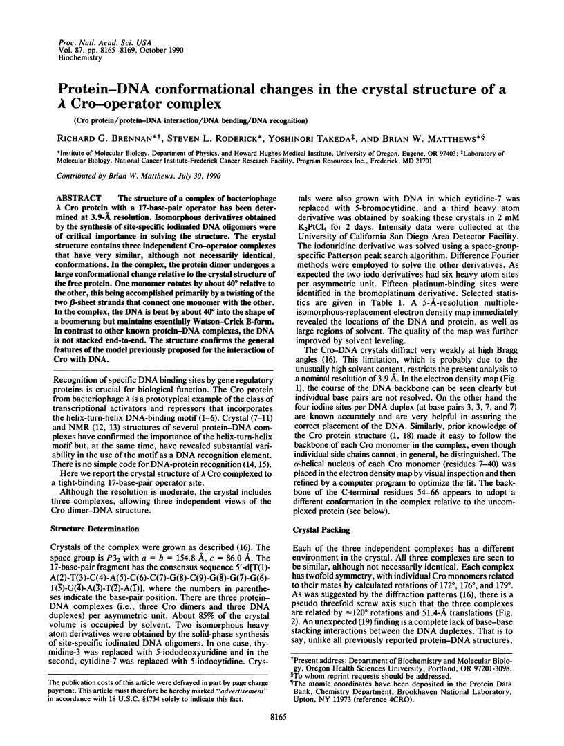
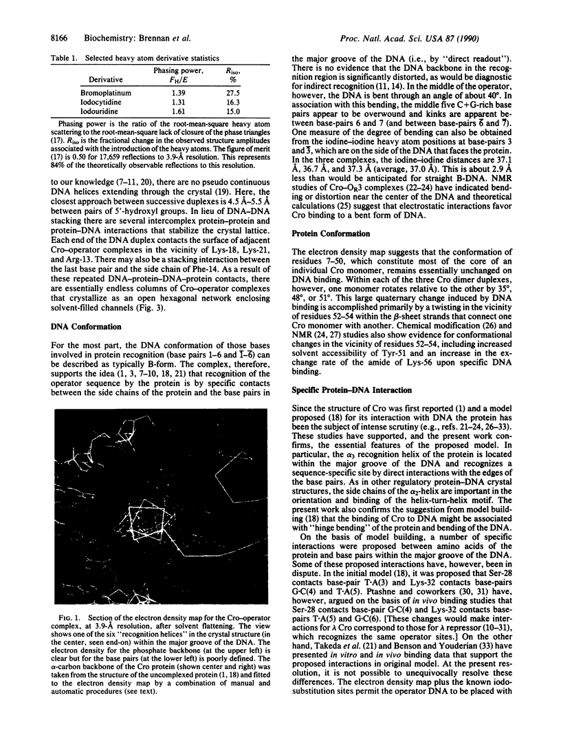
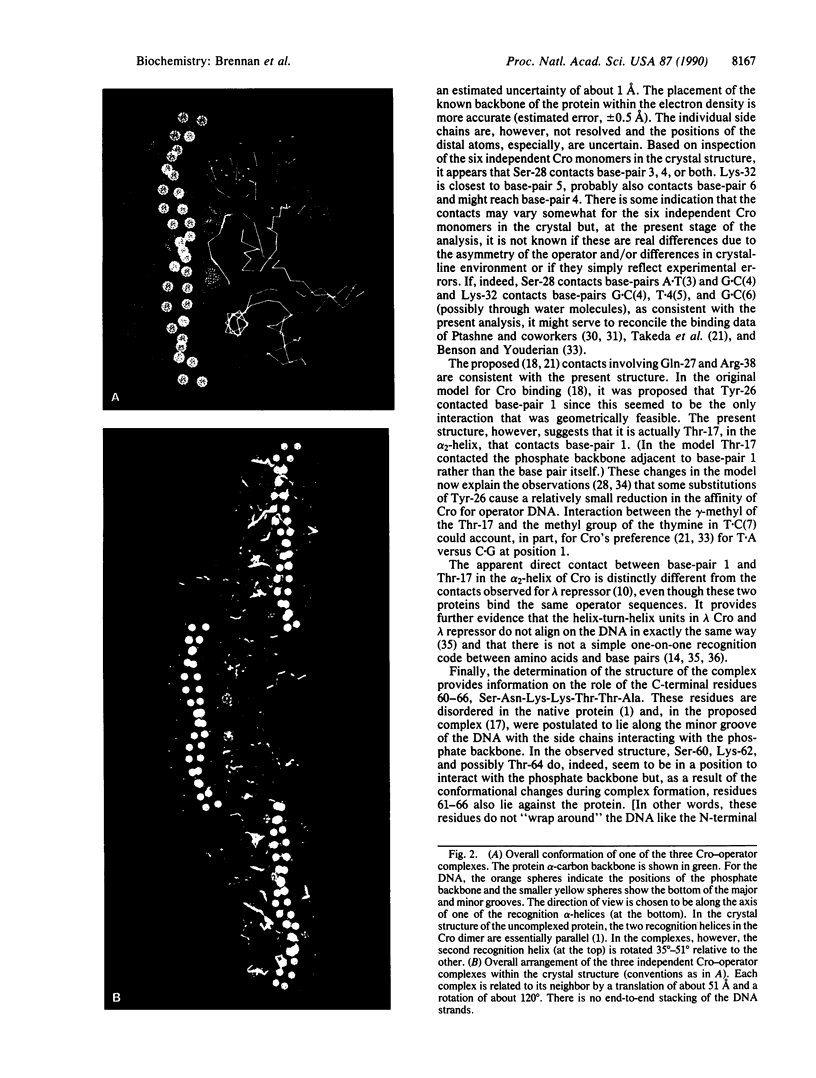
Abstract
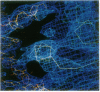
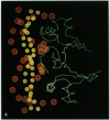
The structure of a complex of bacteriophage lambda Cro protein with a 17-base-pair operator has been determined at 3.9-A resolution. Isomorphous derivatives obtained by the synthesis of site-specific iodinated DNA oligomers were of critical importance in solving the structure. The crystal structure contains three independent Cro-operator complexes that have very similar, although not necessarily identical, conformations. In the complex, the protein dimer undergoes a large conformational change relative to the crystal structure of the free protein. One monomer rotates by about 40 degrees relative to the other, this being accomplished primarily by a twisting of the two beta-sheet strands that connect one monomer with the other. In the complex, the DNA is bent by about 40 degrees into the shape of a boomerang but maintains essentially Watson-Crick B-form. In contrast to other known protein-DNA complexes, the DNA is not stacked end-to-end. The structure confirms the general features of the model previously proposed for the interaction of Cro with DNA.
Full text
PDF
Images in this article
Selected References
These references are in PubMed. This may not be the complete list of references from this article.
- Aggarwal A. K., Rodgers D. W., Drottar M., Ptashne M., Harrison S. C. Recognition of a DNA operator by the repressor of phage 434: a view at high resolution. Science. 1988 Nov 11;242(4880):899–907. doi: 10.1126/science.3187531. [DOI] [PubMed] [Google Scholar]
- Anderson J. E., Ptashne M., Harrison S. C. Structure of the repressor-operator complex of bacteriophage 434. 1987 Apr 30-May 6Nature. 326(6116):846–852. doi: 10.1038/326846a0. [DOI] [PubMed] [Google Scholar]
- Anderson J., Ptashne M., Harrison S. C. Cocrystals of the DNA-binding domain of phage 434 repressor and a synthetic phage 434 operator. Proc Natl Acad Sci U S A. 1984 Mar;81(5):1307–1311. doi: 10.1073/pnas.81.5.1307. [DOI] [PMC free article] [PubMed] [Google Scholar]
- Anderson W. F., Ohlendorf D. H., Takeda Y., Matthews B. W. Structure of the cro repressor from bacteriophage lambda and its interaction with DNA. Nature. 1981 Apr 30;290(5809):754–758. doi: 10.1038/290754a0. [DOI] [PubMed] [Google Scholar]
- Anderson W. F. Proposed alpha-helical super-secondary structure associated with protein-dna recognition. J Mol Biol. 1982 Aug 25;159(4):745–751. doi: 10.1016/0022-2836(82)90111-5. [DOI] [PubMed] [Google Scholar]
- Benson N., Sugiono P., Youderian P. DNA sequence determinants of lambda repressor binding in vivo. Genetics. 1988 Jan;118(1):21–29. doi: 10.1093/genetics/118.1.21. [DOI] [PMC free article] [PubMed] [Google Scholar]
- Benson N., Youderian P. Phage lambda Cro protein and cI repressor use two different patterns of specific protein-DNA interactions to achieve sequence specificity in vivo. Genetics. 1989 Jan;121(1):5–12. doi: 10.1093/genetics/121.1.5. [DOI] [PMC free article] [PubMed] [Google Scholar]
- Boelens R., Scheek R. M., van Boom J. H., Kaptein R. Complex of lac repressor headpiece with a 14 base-pair lac operator fragment studied by two-dimensional nuclear magnetic resonance. J Mol Biol. 1987 Jan 5;193(1):213–216. doi: 10.1016/0022-2836(87)90638-3. [DOI] [PubMed] [Google Scholar]
- Brennan R. G., Matthews B. W. Structural basis of DNA-protein recognition. Trends Biochem Sci. 1989 Jul;14(7):286–290. doi: 10.1016/0968-0004(89)90066-2. [DOI] [PubMed] [Google Scholar]
- Brennan R. G., Matthews B. W. The helix-turn-helix DNA binding motif. J Biol Chem. 1989 Feb 5;264(4):1903–1906. [PubMed] [Google Scholar]
- Brennan R. G., Takeda Y., Kim J., Anderson W. F., Matthews B. W. Crystallization of a complex of cro repressor with a 17 base-pair operator. J Mol Biol. 1986 Mar 5;188(1):115–118. doi: 10.1016/0022-2836(86)90488-2. [DOI] [PubMed] [Google Scholar]
- Eisenbeis S. J., Nasoff M. S., Noble S. A., Bracco L. P., Dodds D. R., Caruthers M. H. Altered Cro repressors from engineered mutagenesis of a synthetic cro gene. Proc Natl Acad Sci U S A. 1985 Feb;82(4):1084–1088. doi: 10.1073/pnas.82.4.1084. [DOI] [PMC free article] [PubMed] [Google Scholar]
- Hochschild A., Douhan J., 3rd, Ptashne M. How lambda repressor and lambda Cro distinguish between OR1 and OR3. Cell. 1986 Dec 5;47(5):807–816. doi: 10.1016/0092-8674(86)90523-4. [DOI] [PubMed] [Google Scholar]
- Hochschild A., Ptashne M. Homologous interactions of lambda repressor and lambda Cro with the lambda operator. Cell. 1986 Mar 28;44(6):925–933. doi: 10.1016/0092-8674(86)90015-2. [DOI] [PubMed] [Google Scholar]
- Jordan S. R., Pabo C. O. Structure of the lambda complex at 2.5 A resolution: details of the repressor-operator interactions. Science. 1988 Nov 11;242(4880):893–899. doi: 10.1126/science.3187530. [DOI] [PubMed] [Google Scholar]
- Kirpichnikov M. P., Hahn K. D., Buck F., Rüterjans H., Chernov B. K., Kurochkin A. V., Skryabin K. G., Bayev A. A. 1H NMR study of the interaction of bacteriophage lambda Cro protein with the OR3 operator. Evidence for a change of the conformation of the OR3 operator on binding. Nucleic Acids Res. 1984 Apr 25;12(8):3551–3561. doi: 10.1093/nar/12.8.3551. [DOI] [PMC free article] [PubMed] [Google Scholar]
- Lamerichs R. M., Boelens R., van der Marel G. A., van Boom J. H., Kaptein R., Buck F., Fera B., Rüterjans H. H NMR study of a complex between the lac repressor headpiece and a 22 base pair symmetric lac operator. Biochemistry. 1989 Apr 4;28(7):2985–2991. doi: 10.1021/bi00433a037. [DOI] [PubMed] [Google Scholar]
- Lee S. J., Shirakawa M., Akutsu H., Kyogoku Y., Shiraishi M., Kitano K., Shin M., Ohtsuka E., Ikehara M. Base sequence-specific interactions of operator DNA fragments with the lambda-cro repressor coupled with changes in their conformations. EMBO J. 1987 Apr;6(4):1129–1135. doi: 10.1002/j.1460-2075.1987.tb04868.x. [DOI] [PMC free article] [PubMed] [Google Scholar]
- Leighton P., Lu P. Lambda cro repressor complex with OR3 DNA: 15N NMR observations. Biochemistry. 1987 Nov 17;26(23):7262–7271. doi: 10.1021/bi00397a011. [DOI] [PubMed] [Google Scholar]
- Matthew J. B., Ohlendorf D. H. Electrostatic deformation of DNA by a DNA-binding protein. J Biol Chem. 1985 May 25;260(10):5860–5862. [PubMed] [Google Scholar]
- Matthews B. W. Protein-DNA interaction. No code for recognition. Nature. 1988 Sep 22;335(6188):294–295. doi: 10.1038/335294a0. [DOI] [PubMed] [Google Scholar]
- McClarin J. A., Frederick C. A., Wang B. C., Greene P., Boyer H. W., Grable J., Rosenberg J. M. Structure of the DNA-Eco RI endonuclease recognition complex at 3 A resolution. Science. 1986 Dec 19;234(4783):1526–1541. doi: 10.1126/science.3024321. [DOI] [PubMed] [Google Scholar]
- McKay D. B., Steitz T. A. Structure of catabolite gene activator protein at 2.9 A resolution suggests binding to left-handed B-DNA. Nature. 1981 Apr 30;290(5809):744–749. doi: 10.1038/290744a0. [DOI] [PubMed] [Google Scholar]
- Metzler W. J., Lu P. Lambda cro repressor complex with OR3 operator DNA. 19F nuclear magnetic resonance observations. J Mol Biol. 1989 Jan 5;205(1):149–164. doi: 10.1016/0022-2836(89)90372-0. [DOI] [PubMed] [Google Scholar]
- Ohlendorf D. H., Anderson W. F., Fisher R. G., Takeda Y., Matthews B. W. The molecular basis of DNA-protein recognition inferred from the structure of cro repressor. Nature. 1982 Aug 19;298(5876):718–723. doi: 10.1038/298718a0. [DOI] [PubMed] [Google Scholar]
- Ohlendorf D. H., Anderson W. F., Lewis M., Pabo C. O., Matthews B. W. Comparison of the structures of cro and lambda repressor proteins from bacteriophage lambda. J Mol Biol. 1983 Sep 25;169(3):757–769. doi: 10.1016/s0022-2836(83)80169-7. [DOI] [PubMed] [Google Scholar]
- Otwinowski Z., Schevitz R. W., Zhang R. G., Lawson C. L., Joachimiak A., Marmorstein R. Q., Luisi B. F., Sigler P. B. Crystal structure of trp repressor/operator complex at atomic resolution. Nature. 1988 Sep 22;335(6188):321–329. doi: 10.1038/335321a0. [DOI] [PubMed] [Google Scholar]
- Pabo C. O., Lewis M. The operator-binding domain of lambda repressor: structure and DNA recognition. Nature. 1982 Jul 29;298(5873):443–447. doi: 10.1038/298443a0. [DOI] [PubMed] [Google Scholar]
- Pakula A. A., Sauer R. T. Amino acid substitutions that increase the thermal stability of the lambda Cro protein. Proteins. 1989;5(3):202–210. doi: 10.1002/prot.340050303. [DOI] [PubMed] [Google Scholar]
- Pakula A. A., Young V. B., Sauer R. T. Bacteriophage lambda cro mutations: effects on activity and intracellular degradation. Proc Natl Acad Sci U S A. 1986 Dec;83(23):8829–8833. doi: 10.1073/pnas.83.23.8829. [DOI] [PMC free article] [PubMed] [Google Scholar]
- Sarai A., Takeda Y. Lambda repressor recognizes the approximately 2-fold symmetric half-operator sequences asymmetrically. Proc Natl Acad Sci U S A. 1989 Sep;86(17):6513–6517. doi: 10.1073/pnas.86.17.6513. [DOI] [PMC free article] [PubMed] [Google Scholar]
- Schevitz R. W., Otwinowski Z., Joachimiak A., Lawson C. L., Sigler P. B. The three-dimensional structure of trp repressor. 1985 Oct 31-Nov 6Nature. 317(6040):782–786. doi: 10.1038/317782a0. [DOI] [PubMed] [Google Scholar]
- Takeda Y., Kim J. G., Caday C. G., Steers E., Jr, Ohlendorf D. H., Anderson W. F., Matthews B. W. Different interactions used by Cro repressor in specific and nonspecific DNA binding. J Biol Chem. 1986 Jul 5;261(19):8608–8616. [PubMed] [Google Scholar]
- Takeda Y., Sarai A., Rivera V. M. Analysis of the sequence-specific interactions between Cro repressor and operator DNA by systematic base substitution experiments. Proc Natl Acad Sci U S A. 1989 Jan;86(2):439–443. doi: 10.1073/pnas.86.2.439. [DOI] [PMC free article] [PubMed] [Google Scholar]
- Wolberger C., Dong Y. C., Ptashne M., Harrison S. C. Structure of a phage 434 Cro/DNA complex. Nature. 1988 Oct 27;335(6193):789–795. doi: 10.1038/335789a0. [DOI] [PubMed] [Google Scholar]