Abstract
Tc40 is an immunodominant antigen present in natural Trypanosoma cruzi infections. This immunogen was thoroughly mapped by using overlapping amino acid sequences identified by gene cloning and chemical peptide synthesis. To map continuous epitopes of the Tc40 antigen, an epitope expression library was constructed and screened with sera from human chagasic patients. A major, linear B-cell epitope spanning residues 403 to 426 (PAKAAAPPAA) was identified in the central domain of Tc40. A synthetic peptide spanning this region reacted strongly with 89.8% of the serum samples from T. cruzi-infected individuals. This indicates that the main antigenic site is defined by the linear sequence of the peptide rather than a conformation-dependent structure. The major B-cell epitope of Tc40 shares a high degree of sequence identity with T. cruzi ribosomal and RNA binding proteins, suggesting the existence of cross-reactivity among these molecules.
Trypanosoma cruzi is a parasitic protozoan that causes Chagas' disease, an ailment without effective drug treatment that affects 16 million to 18 million people in South and Central America. Chagas' disease is routinely diagnosed by serological methods based on crude or semipurified T. cruzi antigens, but there is considerable variation in the reproducibility and reliability of the results (3, 21). In order to obtain better targets for more sensitive and specific diagnostic tests, T. cruzi antigens have been isolated by immunoscreening of DNA expression libraries with sera from human chagasic patients and infected animals (5, 6). Most of the T. cruzi antigens identified by this procedure contain extensive arrays of tandemly repeated amino acid sequences (5, 6). Natural humoral responses to many T. cruzi antigens appear to be directed to epitopes encoded by these repeat units. It has been suggested that the immune response to parasite repetitive antigens does not protect the host because it masks the host's immune response to more critical epitopes on different molecules (1, 2, 6, 10, 14).
The chronic phase of Chagas' disease has a wide variety of clinical manifestations, ranging from severe cardiomyopathy and massive damage in segments of the digestive tract (megacolon and/or megaesophagus) to the absence of relevant clinical symptoms (indeterminate form) (21). Despite the obvious clinical importance of Chagas' disease, the pathogenesis of the chronic phase of Chagas' disease is still poorly understood. It is not clear what role the parasite antigens may have in the development and ultimate appearance of chronic manifestations. Correct identification of epitopes in T. cruzi immunodominant antigens will greatly help in the diagnosis and prognosis of Chagas' disease and the characterization of targets for immunization-vaccination and immunosuppression.
In a previous study, we isolated a DNA recombinant clone (Tc40) encoding a T. cruzi immunodominant antigen recognized by a large number of serum samples from patients with chronic Chagas' disease (11). In contrast to most T. cruzi antigens, Tc40 does not contain amino acid tandem repeats. Information is available for only a limited number of the identified, nonrepetitive T. cruzi antigens (7, 12, 13, 16, 18, 19). In the present study we mapped the B-cell epitopes of Tc40 using an epitope DNA expression library and overlapping amino acid sequences generated by gene cloning and chemical peptide synthesis. We found that the immunodominant, B-cell epitope of Tc40 shares a high degree of sequence identity with different T. cruzi antigenic ribonucleoproteins such as ribosomal and RNA binding proteins, suggesting the existence of intermolecular cross-reactions among parasite antigens.
MATERIALS AND METHODS
Human sera and guinea pig antisera.
Serum samples (n = 166) from patients with chronic Chagas' disease were collected in different Latin American countries (Argentina, Honduras), mainly Brazil. Diagnosis of the disease was made according to clinical symptoms and serological analysis (ImmunoCruzi and BioELISAcruzi; Biolab-Mérieux, Rio de Janeiro, Brazil). Nonchagasic sera were also tested by enzyme-linked immunosorbent assay (ELISA; bioELISAcruzi) to confirm the lack of anti-T. cruzi reactivity. The 116 samples from nonchagasic individuals included (i) 62 samples from healthy blood donors and (ii) 54 samples from patients with unrelated diseases, as defined by clinical, epidemiological, and serological diagnosis of the respective diseases, as follows: 7 patients with toxoplasmosis, 26 patients with kala azar, 5 patients with mononucleosis, 8 patients with autoimmune diseases, and 8 patients with filariasis.
Guinea pig antiserum to Schistosoma japonicum gluthatione-S-transferase (GST) and GST-Tc40 fusion proteins were obtained as reported previously (11). Monoclonal antibody against protein 10 of phage T7 was provided by Novagen, Le Perray en Yvelines, France.
Expression of recombinant proteins in the pGEX vector.
DNA fragments corresponding to the 5′, central, and 3′ regions of the Tc40 gene were generated as described elsewhere (11). The three inserts were subcloned into the pGEX plasmid (Pharmacia Biotech, Les Ulis, France) to express the amino-terminal, carboxy-terminal, and central domains of the Tc40 protein fused to GST. Fusion proteins were expressed in the Escherichia coli DH5α strain (Gibco BRL, Cergy Pontoise, France), after induction with isopropyl-β-d-thiogalactopyranoside. Sodium dodecyl sulfate-polyacrylamide gel electrophoresis (SDS-PAGE) and immunoblotting were performed as described previously (11).
Construction and screening of the Tc40 epitope library.
The Tc40 epitope library was constructed with the pTOPE plasmid, according to the instructions of the manufacturer (Novagen). Briefly, the λgt11 Tc40 insert, which represents the central domain of the Tc40 gene, was partially cleaved with DNase I. The random DNA fragments were fractionated by electrophoresis in a 2% Mermaid gel (Bio-Rad, Ivry sur Seine, France), and fragments averaging 50 to 150 bp in size were eluted from the gel. DNA fragments were successively treated with T4 DNA polymerase to generate blunt ends and with Tth DNA polymerase to add a dA residue to each 3′ end. The fragments were then ligated into the pTOPE plasmid, which contains single dT overhangs. The library was grown in the E. coli Novablue (DE3) strain (Novagen). Immunoscreening of the Tc40 epitope library was carried out with a pool of human chagasic sera as well as anti-GST-Tc40 serum raised in a guinea pig.
Nucleotide sequencing of the selected clones was performed by the dideoxynucleotide chain-termination method by using Taq dye terminator cycle sequencing chemistry in an ABI PRISM 377 DNA sequencer. The sequences were analyzed and compared by using the PCGENE and DNASTAR programs. The sequences were analyzed for similarities to sequences in the GenBank database by using the BLAST algorithm at the National Center for Biotechnology Information Internet site.
Peptide synthesis.
Peptides S23G and BIOS23G were synthesized at bioMérieux (Marcy l'Etoile, France) facilities by using 9-fluorenylmethoxy carbonyl chemistry with 1-hydroxy-benzotriazole and 2-(1H-benzotriazol-1-yl)-1,1,3,3-tetramethyluronium hexafluorophosphate as coupling agents. Standard solid-phase synthesis procedures were used, and the peptides were purified by high-performance liquid chromatography. Analysis by mass spectrometry demonstrated that the purities of the peptides were more than 90%.
ELISA.
Evaluation of peptide BIO-S23G was performed by indirect ELISA. Each well of Maxisorb, 96-well microtiter plates (Nunc, Roskilde, Denmark) was coated with 1 μg of streptavidin overnight at 4°C. The plates were then washed three times in phosphate-buffered saline (PBS) containing 0.05% Tween 20 (PBST), and each well was sensitized with 1 μg of peptide BIOS23G for 2 h at 37°C. The plates were washed as described above and blocked with PBST containing 10% horse serum for 2 h at 37°C. After a further washing cycle in PBST, human sera diluted 1/100 in the blocking solution were added to the wells and the plates were incubated for 2 h at 37°C. This was followed by three washes in PBST and incubation with goat, anti-human immunoglobulin G peroxidase conjugate (diluted 1/30,000; Jackson) for a further 90 min. The plates were washed and incubated with a mixture of ortho-phenylenediamine and H2O2 (bioMérieux) for 10 min. The reaction was stopped by addition of 1 N sulfuric acid, and the absorbances of the plates at 492 nm were read on an AXIA microreader (bioMérieux). The cutoff values were calculated as the mean optical density at 492 nm (OD492) of sera from 20 healthy individuals plus 3 standard deviations. These individuals were from an area in Brazil where Chagas' disease is not endemic, and they were not included in the panel of 62 samples from healthy blood donors described above.
RESULTS
Characterization of a major human B-cell epitope in Tc40.
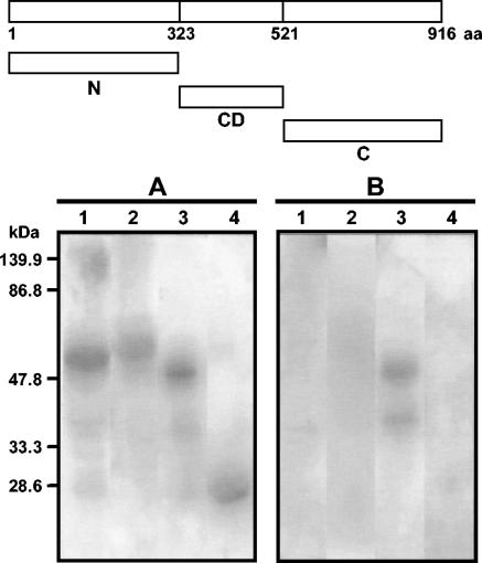
Previous work (11) has indicated that epitopes for human chagasic antibodies are located in the central domain of the Tc40 molecule. To confirm this result, constructs encoding the N-terminal, C-terminal, and central domains of Tc40 were expressed as GST fusion proteins in E. coli. All three fusion proteins were recognized by an antiserum raised against GST, as indicated by the presence of bands with the expected molecular mass (Fig. 1A). In contrast, only the fusion protein containing the Tc40 central domain reacted with human chagasic serum (Fig. 1B), indicating the presence of epitopes in this region reactive with human serum.
FIG. 1.
Identification of antigenic regions of Tc40 by immunoblot analysis. A schematic representation of the Tc40 protein (916 amino acids) is shown at the top. Regions encoding the amino-terminal domain (N), the central domain (CD), or the carboxy-terminal domain (C) of Tc40 were expressed in E. coli as GST fusion proteins. Fusion proteins were separated by SDS-PAGE, transferred onto nitrocellulose membranes, and incubated with rabbit antiserum against GST (A) or a pool of human chronic chagasic sera (B). Lanes: 1, N-terminal domain of Tc40; 2, C-terminal domain of Tc40; 3, central domain of Tc40; 4, GST without fusion.
The DNA fragment encoding the central domain of Tc40 (amino acids 323 to 520) was digested with DNase I, and the resulting DNA fragments (50 to 150 bp) were subcloned into the pTOPE plasmid. Sequencing of randomly picked, shotgun clones (which can contain inserts in either the correct or the inverse orientation for Tc40 expression and in one of three open reading frames) showed that the clones represented random fragments from the original insert. Approximately 3,000 phagemids were screened, and two clones expressing strongly reactive epitopes were detected and purified. Both fusion proteins were recognized by guinea pig antibodies against Tc40 (Fig. 2B), but only one reacted with human chagasic serum (Fig. 2C). No reaction was observed with normal human serum (Fig. 2D). These results indicate the existence of a major epitope for human chagasic antibodies in Tc40.
FIG. 2.
Characterization of clones selected from a Tc40 epitope library constructed in the pTOPE vector. The DNA fragment encoding the central domain of Tc40 (amino acids 323 to 520) was digested with DNase I, and the resulting fragments were subcloned into the pTOPE plasmid. Protein extracts from E. coli expressing only the T7 protein 10 gene (lane 1) or fused with Tc40 epitopes (lanes 2 and 3) were separated by SDS-PAGE and transferred onto nitrocellulose membranes. Blots were incubated with a monoclonal antibody against the T7 tag (A), guinea pig antiserum against Tc40 (B), a pool of sera from individuals with chronic Chagas' disease (C), and a pool of sera from healthy individuals (D).
The epitope recognized by human chagasic antibodies is composed of 24 amino acids (residues 403 to 426 in Tc40) (Fig. 3). In order to confirm whether anti-Tc40 antibodies from chronic chagasic patients were directed mainly to this epitope, a peptide (S23G) encompassing residues 402 to 426 of Tc40 was synthesized and tested for its ability to inhibit binding of human chagasic antibodies to the Tc40 central domain GST fusion protein by competitive ELISA. The S23G peptide inhibited binding of human antibodies to Tc40 in a concentration-dependent manner (data not shown). The S23G peptide and Tc40 fusion protein displayed similar levels of inhibition of binding, indicating that the S23G peptide contains the major human B-cell epitope in Tc40.
FIG. 3.
Localization of the major B-cell epitopes of Tc40. The predicted amino acid sequence of the central domain of Tc40 is presented. The epitope recognized by both guinea pig antiserum against Tc40 and human chagasic antibodies is underlined, and the epitope recognized only by guinea pig antibodies is double underlined.
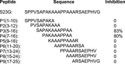
To define the core of the immunodominant peptide, nine different overlapping peptides encompassing the S23G sequence were synthesized and tested for their ability to inhibit the binding of human antibodies to Tc40-GST (Fig. 4). One sequence (PAKAAAPPAA) was deemed the core epitope, based on its ability to inhibit the binding of human antibodies to Tc40-GST by 80%. These results suggest that the main antigenic site of Tc40 is defined by the linear sequence of the peptide rather than a conformation-dependent structure. The existence of preferential conformational properties in the major Tc40 epitope was also investigated by using circular dichroism. The results indicate that the S23G peptide preferentially adopts an extended conformation, supporting the existence of a linear epitope (data not shown).
FIG. 4.
Identification of the human immunodominant epitope in Tc40. Nine different overlapping peptides (peptides P1 to P9) encompassing the S23G peptide were synthesized and used to inhibit the binding of human chagasic antibodies to GST-Tc40. The numbers in parentheses indicate the amino acid positions in the S23G peptide.
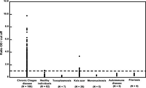
The S23G peptide was tested by ELISA with sera from the 166 chronic chagasic patients which had previously been shown to react with the Tc40-GTS fusion protein. To improve the binding of the S23G peptide to the microtiter plates, biotin was coupled to the N terminus, creating the Bio-S23G peptide. The Bio-S23G peptide reacted with sera from 149 chronic chagasic patients (sensitivity, 89.8%), assuming a cutoff absorbance value equal to the mean OD492 of sera from 20 healthy individuals plus 3 standard deviations (Fig. 5). In contrast, the values for all 62 serum samples from healthy individuals were below the designated cutoff, giving a specificity of 100%. The specificity of the Bio-SG23 peptide was analyzed with sera from 116 nonchagasic individuals, including healthy blood donors (n = 62), patients with other pathologies (n = 28), and patients with leishmaniasis (n = 26). Bio-SG23 presented a specificity of 100% with sera from 90 nonchagasic individuals, excluding those with leishmaniasis (n = 26). However, when sera from the 26 leishmaniasis patients were included in the study, Bio-SG23 presented a specificity of 97.6%.
FIG. 5.
A492 obtained by Bio-SG23 ELISA with sera from chagasic and nonchagasic patients. To improve the binding of the S23G peptide to the microtiter plates, biotin was coupled to the N terminus, creating the Bio-S23G peptide. The horizontal dashed line represents the cutoff region.
The Tc40 immunodominant epitope is distributed over different antigenic ribonucleoproteins of T. cruzi.
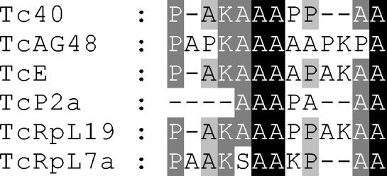
The Tc40 gene encodes a polypeptide of ∼100 kDa that is highly conserved (98% identity at the amino acid level) in T. cruzi strains G and CL. Antibodies against Tc40 reacted with a protein with the expected molecular mass of 100 kDa and with two additional proteins of 41 and 38 kDa (11), suggesting the existence of cross-reactive epitopes in these molecules. To further clarify this point, the amino acid sequence of the Tc40 epitope (PAKAAAPPAA) was compared with published T. cruzi sequences. The Tc40 epitope is quite similar to a conserved motif found in several T. cruzi ribosomal proteins (TcP2a, TcRpL19, TcRpL7, TcE-L19E) and in a T. cruzi RNA binding protein (TcRB48) (4, 8, 9, 12) (Fig. 6). Like Tc40, these ribonucleoproteins strongly reacted with human chagasic sera and have been used for the serological diagnosis of Chagas' disease (4, 8, 9, 12). The numbers and location of this motif can vary between different T. cruzi ribonucleoproteins (Fig. 6). The Tc40 epitope has ∼83% identity with a sequence repeated nine times at the C terminus of ribosomal protein TcRpL19, while at the N terminus of TcRpL7a there are three repeats with partial homology to Tc40. It is noteworthy that ribosomal proteins have molecular masses of 35 to 38 kDa, similar to the molecular mass of the 41-kDa protein recognized by the anti-Tc40 antibodies. We have compared Tc40 with other parasite proteins deposited in GenBank. Leishmania major contains a Tc40-like protein (GenBank accession no. AL356246) that displays 31% identity with the T. cruzi Tc40 at the amino acid level. However, only three residues (AAP) of the Tc40 epitope in L. major (TTAGAAPVR) are conserved.
FIG. 6.
Comparison of amino acid sequences of the major human Tc40 epitope and repetitive motifs of T. cruzi ribonucleoproteins. Alignments were done by using the Clustal W program of MegAlign software (DNASTAR Inc.). Conserved residues are shaded in black (100% conservation), dark gray (83% conservation), and light gray (67% conservation). Sequences are as follows: Tc40, Tc40 human epitope; TcAG48, T. cruzi RNA binding protein-like protein (GenBank accession no. AF316151); TcE, T. cruzi ribosomal L19E homologue protein; TcP2a, T. cruzi ribosomal protein, JL5 variant (GenBank accession no. X69509); TcRpL19, T. cruzi ribosomal L19-like protein (GenBank accession no. AF316149); TcRpL7a, T. cruzi ribosomal L7a-like protein (GenBank accession no. AF316150).
DISCUSSION
An expression library was constructed to map continuous epitopes of the T. cruzi Tc40 antigen. Screening of the library with sera from human chagasic patients resulted in the identification of a linear B-cell epitope spanning residues 403 to 426 (PAKAAAPPAA) in the central domain of Tc40. A synthetic peptide spanning this region strongly reacted with 89.8% of the serum samples from T. cruzi-infected individuals, indicating that the main antigenic site was indeed defined by the linear sequence of the peptide rather than a conformation-dependent structure. The fact that sera from 10% of the chagasic patients tested did not react with this epitope could imply the existence of weak linear epitopes and/or conformation-dependent epitopes. The sensitivity (∼90%) of the Tc40 epitope for the serodiagnosis of Chagas' disease is comparable to that of other T. cruzi antigens used for serodiagnosis of the disease (5). It has previously been demonstrated (5, 20) that the immune response against a single protein is not universally present in the infected population, and therefore, no single recombinant antigen or synthetic peptide is sensitive enough to prevent the risk of T. cruzi transmission by transfusion. To overcome this problem, a multiepitope, synthetic peptide carrying four T. cruzi repeating B-cell epitopes (PEP-2, TcD, TcE, and TcLo1.2) has recently been evaluated with sera from chagasic and nonchagasic patients. The sensitivity and specificity of this peptide were 99.6 and 99.3%, respectively (8, 9). It is noteworthy that the Tc40 epitope shares strong homology with TcE (KAAIAPAKAAAAPAKAATAPA) (Fig. 6), which is located in the 35-kDa L19E ribosomal sequence (8).
We found that the major B-cell epitope of Tc40 contains a high degree of sequence identity with a repeated motif found at the amino or carboxy terminus of T. cruzi ribosomal and RNA binding proteins, suggesting the existence of cross-reactivity between these molecules. It is not known why ribonucleoproteins have these repeated motifs. The presence of a common motif among different proteins may simply reflect a common ancestral relationship between their sequences and does not necessarily indicate a shared interactive function.
Ribonucleoproteins are highly immunogenic in vertebrate hosts and have been used for the serodiagnosis of Chagas' disease (4, 8, 9, 12). Requena et al. (15) suggested that the dominant immune response against these proteins could be due to the abundance and stability of nucleoprotein particles and their capacity to be phagocytosed and processed by antigen-presenting cells. It has been suggested that the repetitive epitopes found in many parasite proteins do not elicit protective immune responses but instead have evolved as an immune evasion mechanism by using their ability to induce thymus-independent B-cell activation (17).
Acknowledgments
We thank François Penin, UMR5086 CNRS-Université Claude Bernard, Laboratoire de Bioinformatique et RMN Structurales, IFR 128 Biosciences Lyon Gerland, Institut de Biologie et Chimie des Protéines, Lyon, France, for helpful discussions.
Mylène Lesénéchal was a doctoral fellow from the MRE, French Government. This work was supported by bioMérieux SA grants to Glaucia Paranhos-Bacccalà and by FAPESP and CNPq (Brazil) grants to J. Franco da Silveira.
REFERENCES
- 1.Alvarez, P., M. S. Leguizamón, C. A. Buscaglia, T. A. Pitcovsky, and O. Campetella. 2001. Multiple overlapping epitopes in the repetitive unit of the shed acute-phase antigen from Trypanosoma cruzi enhance its immunogenic properties. Infect. Immun. 69:7946-7949. [DOI] [PMC free article] [PubMed] [Google Scholar]
- 2.Buscaglia, C. A., O. Campetella, M. S. Leguizamón, and A. C. Frasch. 1998. The repetitive domain of Trypanosoma cruzi trans-sialidase enhances the immune response against the catalytic domain. J. Infect. Dis. 177:431-436. [DOI] [PubMed] [Google Scholar]
- 3.Camargo, M. E., E. L. Segura, I. G. Kagan, J. M. P. Souza, J. R. Carvalheiro, J. F. Yanovsky, and M. C. S. Guimarães. 1986. Collaboration on the standardization of Chagas' disease in the Americas: an appraisal. PAHO Bull. 20:233-244. [PubMed] [Google Scholar]
- 4.DaRocha, W. D., D. C. Bartholomeu, C. D. S. Macedo, F. H. Horta, D. Cunha-Neto, J. E. Donelson, and S. M. R. Teixeira. 2002. Characterization of cDNA clones encoding ribonucleoprotein antigens expressed in Trypanosoma cruzi amastigotes. Parasitol. Res. 88:292-300. [DOI] [PubMed] [Google Scholar]
- 5.Franco da Silveira, J., E. S. Umezawa, and A. O. Luquetti. 2001. Chagas' disease: recombinant Trypanosoma cruzi antigens for serological diagnosis. Trends Parasitol. 17:286-291. [DOI] [PubMed] [Google Scholar]
- 6.Frasch, A. C. C., J. J. Cazzulo, L. Aslund, and U. Pettersson. 1991. Comparison of genes encoding Trypanosoma cruzi antigens. Parasitol. Today 7:148-151. [DOI] [PubMed] [Google Scholar]
- 7.Godsel, L. M., R. S. Tibbetts, C. L Olson, B. M. Chaudoir, and D. M. Engman. 1995. Utility of recombinant flagelar calcium-binding protein for serodiagnosis of Trypanosoma cruzi infection. J. Clin. Microbiol. 33:2082-2085. [DOI] [PMC free article] [PubMed] [Google Scholar]
- 8.Houghton, R. L., D. R. Benson, L. D. Reynolds, P. D. McNeill, P. R. Sleath, M. J. Lodes, D. A. Leiby, R. Badaro, and S. G. Reed. 1999. A multi-epitope synthetic peptide and recombinant protein for the detection of antibodies to Trypanosoma cruzi in radioimmunoprecipitation-confirmed and consensus-positive sera. J. Infect. Dis. 179:1226-1234. [DOI] [PubMed] [Google Scholar]
- 9.Houghton, R. L., D. R. L. Benson, L. Reynolds, P. McNeill, P. Sleath, M. Lodes, Y. A. Skeiky, R. Badaro, A. U. Krettli, and S. G. Reed. 2000. Multiepitope synthetic peptide and recombinant protein for the detection of antibodies to Trypanosoma cruzi in patients with treated or untreated Chagas' disease. J. Infect. Dis. 181:325-330. [DOI] [PubMed] [Google Scholar]
- 10.Kemp, D. J., R. L. Coppel, and R. F. Anders. 1987. Repetitive proteins and genes of malaria. Annu. Rev. Microbiol. 141:181-208. [DOI] [PubMed] [Google Scholar]
- 11.Lesenechal, M., R. A. Mortara, L. Duret, M. Jolivet, M. E. Camargo, J. Franco da Silveira, and G. Paranhos-Baccala. 1997. Cloning and characterization of a gene encoding a novel antigen of Trypanosoma cruzi. Mol. Biochem. Parasitol. 87:193-204. [DOI] [PubMed] [Google Scholar]
- 12.Levin, M. J., M. Vazquez, D. Kaplan, and A. G. Schijman. 1993. The Trypanosoma cruzi ribosomal P protein family: classification and antigenicity. Parasitol. Today 9:381-384. [DOI] [PubMed] [Google Scholar]
- 13.Levitus, G., M. Hontebeyrie-Joskowicz, M. H. Van Regenmortel, and M. J. Levin. 1991. Humoral autoimmune response to ribosomal P proteins in chronic Chagas heart disease. Clin. Exp. Immunol. 85:413-417. [DOI] [PMC free article] [PubMed] [Google Scholar]
- 14.Pitcovsky, T. A., J. Mucci, P. Alvarez, M. S. Leguizamon, O. Burrone, P. M. Alzari, and O. Campetella. 2001. Epitope mapping of trans-sialidase from Trypanosoma cruzi reveals the presence of several cross-reactive determinants. Infect. Immun. 69:1869-1875. [DOI] [PMC free article] [PubMed] [Google Scholar]
- 15.Requena, J. M., C. Alonso, and M. Soto. 2000. Evolutionary conserved proteins as prominent immunogens during Leishmania infections. Parasitol. Today 16:246-250. [DOI] [PubMed] [Google Scholar]
- 16.Schijman, A. G., G. Levitus, and M. J. Levin. 1992. Characterization of the C-terminal region of a Trypanosoma cruzi 38-kDa ribosomal P0 protein that does not react with lupus anti-P autoantibodies. Immunol. Lett. 33:15-20. [DOI] [PubMed] [Google Scholar]
- 17.Schofield, L. 1991. On the function of repetitive domains in protein antigens of Plasmodium and other eukaryotic parasites. Parasitol. Today 7:99-105. [DOI] [PubMed] [Google Scholar]
- 18.Skeiky, Y. A., D. R. Benson, M. Parsons, K. B. Elkon, and S. G. Reed. 1992. Cloning and expression of Trypanosoma cruzi ribosomal protein P0 and epitope analysis of anti-P0 autoantibodies in Chagas' disease patients. J. Exp. Med. 176:201-211. [DOI] [PMC free article] [PubMed] [Google Scholar]
- 19.Skeiky, Y. A., D. R. Benson, J. A. Guderian, P. R. Sleath, M. Parsons, and S. G. Reed. 1993. Trypanosoma cruzi acidic ribosomal P protein gene family. Novel P proteins encoding unusual cross-reactive epitopes. J. Immunol. 151:5504-5515. [PubMed] [Google Scholar]
- 20.Umezawa, E. S., S. F. Bastos, M. E. Camargo, L. M. Yamauchi, M. R. Santos, A. Gonzalez, B. Zingales, M. J. Levin, O. Souza, R. Rangel-Aldao, and J. Franco da Silveira. 1999. Evaluation of recombinant antigens for Chagas' disease serodiagnosis in South and Central America. J. Clin. Microbiol. 37:1554-1560. [DOI] [PMC free article] [PubMed] [Google Scholar]
- 21. World Health Organization. 2002. Control of Chagas disease. Second report of a WHO expert committee. W.H.O. Tech. Rep. Ser. 905:1-28. [Google Scholar]


