Abstract
We report a novel approach that allows for the rapid identification of proteins mediating phosphorylation in signaling cascades after specific stimulation. As a proof of concept, we used the interferon- γ (IFN-γ)-induced phosphorylation of signal transducer and activator of transcription-1 (Stat1) in a human promonocytic cell line, which was previously shown to be deficient in this signaling pathway. By using retroviral cDNA expression libraries, transduced selector cells expressing single cDNAs were stimulated with IFN-γ, then fixed, permeabilized and stained intracellularly for phospho-Stat1 levels. Cells responding to the stimulation, which showed increased levels of phosphorylated Stat1, were enriched using fluorescence activated cell sorting (FACS). Genomic DNA was isolated from the enriched cell population and served as a template for cDNA amplification using PCR. After only one round of selection, a cDNA encoding the β-chain of the IFN-γ receptor (IFNGR2) was obtained and demonstrated to restore the selected phenotype. The approach now allows one to use phospho-events as reporters, alone or in tandem, for screening of signaling network states, overcoming a prior need to rely on the reporter genes that are often only indirect measures of phenotypes desired in a screen.
INTRODUCTION
Expression cloning is a powerful tool utilized for the identification of cDNAs accounting for a certain phenotype of interest. However, the first approaches to screening cDNA libraries were based on the transfection of plasmid libraries into suitable and easily transfectable selector cells followed by screening for a transiently appearing rather than stable phenotype (1). Furthermore, by this approach multiple cDNAs were often expressed in single cells leading to difficulties in isolating the correct cDNA. In contrast, retroviral cDNA libraries are readily transduced into a wide range of cells at high efficiency and can incorporate a single gene expression cassette into the host cell genome. This allows for genetic screens on a single cell level owing to the stable and sustained expression of the integrated gene cassette. Such retroviral cDNA libraries developed in our laboratory have been successfully applied to the identification of protein–protein interactions and resultant phenotypes (2). Among others, genetic screens of such libraries facilitated the characterization of viral receptors (3–5), cellular factors restricting viral replication (6) and proteins involved in the regulation of apoptosis (7,8).
In this study, we considered a different application for retroviral cDNA libraries. Recently, we have developed methods to measure the phosphorylation state of proteins involved in specific signal transduction pathways on a single cell level using flow cytometry (9–14). However, the identification of genes mediating the phosphorylation of any given target protein relies on the availability of specific pharmacological inhibitors directed against known kinases or receptors expected to play a pivotal role in upstream events in the signaling cascade studied. Thus, we sought to establish novel methods that would facilitate the identification of cellular factors crucial for the phosphorylation of cytoplasmic proteins. Therefore, we combined retroviral cDNA library technology and phospho-specific flow cytometry in an attempt to develop a method that could identify players in signaling pathways on a genome-wide level. As a proof of concept, we established a subclone of a human myeloid cell line that did not phosphorylate signal transducer and activator of transcription-1 (Stat1) in response to interferon-γ (IFN-γ) (15,16). This cell line served as the selector cell in the screening of a retroviral cDNA library for proteins reconstituting responsiveness to IFN-γ by phosphorylating Stat1.
A retroviral cDNA library derived from human peripheral blood lymphocytes was transduced into the selector cells and subjected to a genetic screen by intracellular phospho-protein staining using antibodies raised against phophorylated Stat1 (pStat1) and subsequent enrichment of responsive cells by cell sorting. After only one round of screening, a single cDNA encoding the β-chain of the IFN-γ receptor [IFNGR2; (17)] was isolated from genomic DNA of selected cells and shown to restore the responsiveness of selector cells upon expression. This novel approach should be a powerful tool in the efforts to identify and characterize yet unknown components of phospho-protein networks.
MATERIALS AND METHODS
cDNA library, plasmids and bacteria strains
The retroviral cDNA library derived from human peripheral blood lymphocytes of 52 healthy human donors (pLib PBL cDNA; Clontech, Heidelberg, Germany) and the plasmids, BMN-I-GFP (http://www.stanford.edu/group/nolan/plasmid_maps/pmaps.html) and BMN-I-GFP-cDNA1.2, were transformed into and expanded in the Escherichia coli strain XL1-blue in Luria–Bertani (LB) medium containing ampicillin (100 μg/ml). For the expansion of bacteria transformed using pCR-Blunt II TOPO-based constructs, LB supplemented with 50 μg/ml kanamycin was used. Purification of plasmid DNA was performed using QIAprep Spin Miniprep kit and QIAfilter Plasmid Maxi kit (Qiagen).
Cells and transduction protocol
Wild-type (wt) human promonocytic U937 cells were purchased from ATCC (CRL-1593). The U937 cell clone #10 (plus) was kindly provided by G. Poli (Turin, Italy) and has been previously shown to support human immunodeficiency virus type 1 (HIV-1) replication in the presence of IFN-γ (16). U937 cell clone #10.13 was obtained by biological cloning using limiting dilution of cell clone #10 and subsequent testing for IFN-γ responsiveness using fluorescence activated cell sorting (FACS) analysis. MLV(VSV-G) pseudotype vector particles were generated by transient transfection of Phoenix gp cells (http://www.stanford.edu/group/nolan/retroviral_systems/retsys.html) with VSV-G-encoding construct pCR-VSV and respective transfer vectors using FuGENE (Roche) in accordance with the manufacturer's instructions. Cell-free supernatants were harvested 2 days post-transfection and subsequently used to transduce U937 cells in the presence of 10 μg/ml polybrene for 2 h as described previously (18). Suspension and adherent cells were cultured in RPMI 1640 and DMEM, respectively, supplemented with 10% fetal calf serum and a penicillin/streptomycin/glutamine cocktail.
Purification of genomic DNA and PCR
Genomic DNA was isolated from cells using the DNeasy Tissue kit (Qiagen) according to the manufacturer's instructions. Genomic DNA was pre-amplified before its utilization for cDNA-specific PCR amplification using oligohexamers and the amplification kit Genomi-Phi following the manufacturer's instructions (Amersham) or, alternatively, served directly as the template for PCR to selectively amplify cDNAs integrated into the genome of transduced selector cells. The following oligonucleotides were employed: LIB5′MCSforw, 5′-GAATTCGTTAGGCCATTATGGCCGCGGCCGCGTCGAC-3′; and LIB3′cDNAS/Xrev, 5′-TTATTTTATCGATGTTTGGCCGAGGCGGCCGCTTGTCG-3′. Each PCR (50 μl) contained 0.5 mM each of the four dNTPs, 20 pmol of each primer, 100–250 ng of genomic DNA, 5.0 μl of buffer 3 and 3.75 U of the polymerase mixture (both Expand Long Template PCR System; Roche). PCR was performed in a PTC-200 Peltier Thermal Cycler (MJ RESEARCH) under the following conditions: 3 min at 92°C; 10 cycles of 15 s at 94°C, 30 s at 62°C, 3 min and 30 s at 68°C; 25 cycles of 15 s at 94°C, 30 s at 60°C, 3 min and 50 s at 68°C followed by 7 min at 68°C.
cDNAs purification and insertion into plasmids
PCRs were loaded onto a 0.7% agarose gel and amplified fragments were purified using the Qiagen's QIAEX II Agarose Gel Extraction kit. According to the manufacturer's instructions, purified blunt cDNAs were subsequently ligated into the vector pCR-Blunt II-TOPO (Invitrogen) and transformed into electrocompetent XL1-blue E.coli. Plasmids harboring cDNAs were subjected to automated sequencing employing standard T7, Sp6 and M13 forward, and M13 reverse primers, respectively. After sequencing, cDNA1.2 encoding the wt IFNGR2 served as a template in a standard PCR by employing the following oligonucleotides: IFNGR2NotIrev, 5′-ATAAGAATGGCGGCCGCTCAAAGCGTTTGGAGAACATCTTCTTGCTCC-3′; and cDNA1.2NotIforw, 5′-ATAAGAATGGCGGCCGCCAGTGTGATGGATATCTGCAGAATTCGCCCTTACG-3′. The purified amplified DNA fragment was digested with NotI and ligated into BMN-I-GFP, previously digested with NotI, and dephosphorylated using calf intestinal alkaline phosphatase (all enzymes and buffers were purchased from NEB).
FACS and western-blot analyses
The staining protocol employed in this study use formaldehyde and methanol for fixation and permeabilization of cells, which has been described in detail previously (10,13). Briefly, cells were stimulated with 5 ng/ml of IFN-γ for 15 min at 37°C. They were then fixed in 1.6% formaldehyde for 10 min and permeabilized in pure methanol for >10 min before being washed and stained with phospho-specific pStat1 (Y701) monoclonal antibody (clone 4a, kindly provided by BD Pharmingen, La Jolla, CA). The antibody was conjugated with Alexa Fluor 647 to allow for analysis using flow cytometry and for cell sorting. For standard western-blot analysis, antibodies raised against the C-terminus of Stat1 and pStat1 (pY701, the same antibody used for the FACS analysis) were employed and detected using α-mouse IgG horseradish peroxidase-conjugated (HRP) (BD Pharmingen) as described previously (10).
RESULTS AND DISCUSSION
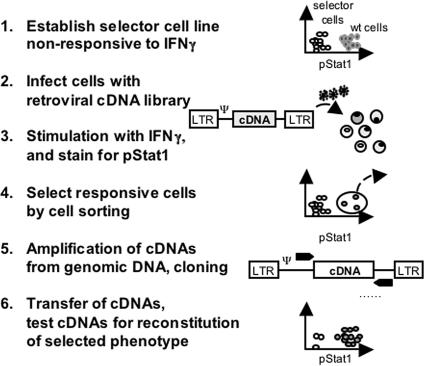
The genetic screen of retroviral libraries aimed at the identification of cDNAs altering intracellular phosphorylation events in signaling cascades, here the phosphorylation of Stat1 in response to IFN-γ stimulation, was performed as follows: (i) establishment of a suitable selector cell line not responding to IFN-γ, (ii) transduction of a cDNA library into the selector cells, (iii) stimulation with IFN-γ and subsequent staining for pStat1, (iv) selection of responding cells by cell sorting, (v) amplification of cDNAs from the genomic DNA of the selected cells and re-cloning into a retroviral vector for validation and (vi) analysis of the isolated cDNAs for the reconstitution of the selected phenotype upon expression in the selector cells (for schematic illustration of the experimental design see Figure 1).
Figure 1.
Schematic representation of the experimental design. Selector cells deficient in responsiveness toward a specific stimulation, here IFN-γ-induced phosphorylation of Stat1, are selected (step 1). After transduction of the retroviral cDNA library into the selector cells (step 2) stimulation and staining of the cells is performed (step 3). Responsive cells are selected by cell sorting (FACS) (step 4). The genomic DNA is isolated serving as a template for the specific amplification of inserted cDNAs that are re-inserted into a retroviral backbone (step 5). Retroviral transfer vectors harboring the selected cDNAs are transduced into naive selector cells and tested for their ability to mediate the reconstitution of the selected phenotype (step 6).
Establishment of a selector cell non-responsive to IFN-γ stimulation
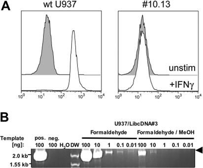
A successful genetic screen relies on the utilization of a selector cell exerting a minimal endogenous background for the phenotype desired to enrich upon the selection of a certain cDNA causing it. In this study, we were aiming to identify a cDNA involved in the phosphorylation of Stat1 induced by IFN-γ stimulation. Thus, we intended to establish a cell line that shows weak responsiveness toward the stimulus. G. Poli and co-workers (16) previously reported on a U937 cell clone (clone #10, plus) supporting HIV-1 replication in the presence of IFN-γ. The analysis of the phenomena revealed undetectable phosphorylation of Stat1 after exposure to IFN-γ. The mechanism by which this blockade to phosphorylation was caused is unknown. However, examination of the respective phosphorylation events using pStat-specific antibodies and FACS analysis demonstrated that ∼5% of the cell population responded to stimulation. Thus, the original U937 cell clone #10 we received most probably either contains genetic revertants, which were contaminated with wild-type cells, or possess an effect that was due to epigenetic variation. Therefore, we established subclones from U937 clone #10 by biological cloning. A total of 15 individual clones were analyzed for their induction of pStat1 after exposure to IFN-γ and subjected to a staining procedure using pStat1-specific antibodies conjugated with Alexa 647 as described previously (10,11,14,18). As shown in Figure 2A, clone #10.13 was demonstrated to respond very weakly in comparison with wt U937 cells indicating its suitability as a selector cell for the intended genetic screen. Revertants were not observed at high frequency; hence, we deemed that this subclone would be appropriate for use in the screen.
Figure 2.
Responsiveness of selector cells and selective amplification of cDNAs. (A) FACS analysis of IFN-γ-induced Stat1 phosphorylation of wt U937 cells and cell clone #10.13. Unstimulated cells (gray histogram) or after exposure to IFN-γ (open histogram) were fixed and permeabilized and subsequently stained with antibodies raised against pSTAT1 (Y701) conjugated with Alexa 647. (B) Detection limit of the PCR used to specifically amplifying an integrated cDNA. Genomic DNA isolated from U937 cells transduced with the retroviral library clone #3 harboring a cDNA (2 kb) after formaldehyde fixation and formaldehyde/methanol treatment were used as templates for PCR yielding ∼2 kb band (arrow). DNA of transduced and untransduced non-treated cells served as positive and negative controls, respectively. DW, DNA marker.
Detection limit of cDNA-specific amplification using PCR and genomic template DNA of formaldehyde- and methanol-treated cells
Since the screen outlined requires the fixation and permeabilization of cDNA-expressing cells to enable specific staining for pStat1 rather than the sorting of living cells, we first investigated the minimal amount of genomic DNA that allowed for the amplification of cDNAs using PCR. Therefore, U937 cells were transduced three times with one transfer vector from the retroviral cDNA (clone #3) harboring a cDNA of ∼2 kb spiked with 2% of an enhanced green fluorescence protein (EGFP)-encoding transfer vector (LibEGFP). Two days after the last transduction round, ∼1% of the targets cells were EGFP-positive indicating that about half of the cells were successfully transduced with the Lib cDNA#3. Subsequently, genomic DNA of untreated, formaldehyde- and formaldehyde/methanol-treated cells were isolated and used as the template in a PCR employing oligonucleotides allowing for the specific amplification of the integrated retroviral cDNA. Genomic DNA of untreated transduced and untransduced U937 cells served as positive and negative controls, respectively. As shown in Figure 2B, the PCR protocol described in Material and Methods allowed for the amplification of a 2 kb fragment from 100 ng of untreated and transduced cells but not from untransduced cells indicating the selective amplification of cDNA #3. Moreover, using decreasing amounts of template DNA revealed that >0.01 and >10 ng of genomic DNA of formaldehyde- and formaldehyde/methanol-treated cells were necessary to allow the successful amplification of integrated cDNA, respectively. Thus, the genomic DNA of ∼1 and 1000 treated cells, respectively, were sufficient for the amplification. However, when we first amplified 1 ng of genomic DNA with a non-specific amplification method using random hexamer oligonucleotides (Genomi-Phi kit; Amersham), we were able to detect the desired 2 kb band. This demonstrated that >100 cells with a respective integrated cDNA were sufficient to amplify, isolate and clone it, and thus, it was feasible to assume that the screen of a retroviral cDNA library could be performed with fixed and permeabilized cells and their selection by cell sorting.
cDNA library transfer and enrichment of cells responding to IFN-γ
To transduce the retroviral cDNA derived from human PBL, 3 × 10⁶ MLV-based Phoenix gp packaging cells were co-transfected with the plasmid library spiked with 5% of the vector pLibEGFP harboring the reporter gene EGFP and a VSV-G encoding construct. Two days post–transfection, 6 ml of cell-free supernatants containing MLV(VSV-G) pseudotype vectors were harvested and subsequently used to transduce 3 × 10⁶ U937 clone #10.13 cells. After 3 days of expansion, ∼1–2% of the cells expressed EGFP indicating that 20–40% of the cells were successfully transduced with cDNA-encoding retroviral transfer vectors. A total of 5 × 10⁶ of the transduced cells were stimulated with IFN-γ as described and stained using Alexa 647-conjugated pStat1-specific antibodies. Stimulated and unstimulated wt U937, and untransduced and transduced #10.13 cells served as controls.
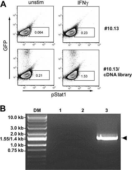
As shown in Figure 3A, ∼1% of the cDNA library expressing #10.13 cells revealed detectable pStat1 levels. The cells were sorted using FACS and their genomic DNA was isolated to serve as a template for unspecific amplification using the Genomi-Phi protocol. Subsequently, the pre-amplified DNA was subjected to cDNA-specific amplification using PCR. While no signal was obtained using control DNA of untransduced #10.13 cells, the use of genomic DNA of the selected cDNA-transduced cells as a template resulted in a single DNA fragment of ∼1.5 kb termed cDNA1.2. Although it is feasible to assume that more than one cDNA was present in the selected cell population, these were possibly not amplified as they were not enriched by the selection scheme applied and, therefore, represented at a much lower frequency in the genomic DNA used as a template. However, in two previous screens using the same experimental design a number of cDNAs were obtained of which one was shown to encode the IFNGR2. The other cDNAs were demonstrated not to reconstitute responsiveness to IFN-γ stimulation (data not shown).
Figure 3.
Cell sorting of cDNA expressing cells and amplification of cDNA. (A) pStat1 detected in unstimulated and IFN-γ-stimulated transduced selector cells. Boxes indicate the gates used to select pStat1-positive cells. Numbers within the boxes represent the percentage of cells within the gate. (B) Specific amplification of cDNAs. No DNA fragment was amplified using no template (1) or genomic DNA of untransduced #10.13 cells (2) serving as negative controls, respectively, using the oligonucleotides LIB5′MCSforw and LIB3′cDNAS/Xrev matching the flanking regions upstream and downstream of the cDNA insertion site in the retroviral backbone. Genomic DNA of transduced cells selected by FACS for the occurrence of pStat1 after IFN-γ stimulation (3) yielded one fragment termed cDNA1.2 shown to encode the IFNGR2 (arrow). DM, DNA marker.
This blunt end fragment was ligated into the vector pCR-Blunt II-TOPO and sequenced demonstrating that the coding region encoded human IFNGR2 (17). Thus, remarkably, after only one round of selection a single cDNA was enriched encoding a member of the IFN-γ-induced signaling cascade leading to the downstream phosphorylation of Stat1.
The selected cDNA reconstitutes responsiveness of the selector cells to IFN-γ stimulation
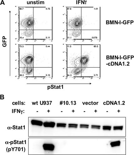
Next, we aimed to demonstrate that expression of the selected cDNA1.2 mediated the responsiveness to IFN-γ stimulation of U937 #10.13 cells. By using specific primers that introduced flanking NotI restriction sites and a standard PCR protocol, cDNA1.2 was amplified, digested with NotI and ligated into the retroviral vector BMN-I-GFP, which was previously digested with NotI and dephosphorylated with calf intestine phosphatase. In this construct, the MLV promoter in the 5′ long terminal repeat drives the expression of an inserted cDNA and an IRES-GFP cassette. The accuracy of the coding region and its right orientation of the inserted cDNA were analyzed by sequencing. MLV(VSV-G) vector particles packaging the mRNA transcripts of retroviral transfer vectors, BMN-I-GFP and BMN-I-GFP-cDNA1.2, were harvested from transiently transfected packaging cells as described and used to transduce U937 #10.13 cells. Within 2 weeks after transduction, the transduced cells expressing GFP were sorted twice using FACS analysis and expanded. As shown in Figure 4A, FACS analysis for the abundance of pStat1 of unstimulated and IFN-γ-stimulated cells using phospho-specific antibodies revealed that the cells transduced with the control vector BMN-I-GFP did not respond to IFN-γ. In contrast, cells expressing GFP and cDNA1.2 showed clear phosphorylation of Stat1 in response to IFN-γ. Notably, cells that were negative for GFP, and therefore did not contain the cDNA, did not show appreciable pStat1 levels. To exclude the possibility that this resulted in different amounts of Stat1-substrate proteins in the transduced cells, western-blot analysis of cell lysates was performed employing murine monoclonal antibodies directed against Stat1 and pStat1, respectively, and anti-murine IgG antibodies conjugated with HRP. Wild-type U937 cells expressed ∼2–3-fold more Stat1 compared with all #10.13-derived cell lines. Most importantly, besides wt U937 cells, only #10.13 cells transduced with BMN-I-GFP-cDNA1.2 responded toward IFN-γ stimulation by Stat1 phosphorylation in agreement with the observations obtained using FACS analysis. This showed that the recovery of the phenotype was mediated by cDNA1.2-encoded IFNGR2 expression rather than the different amount of Stat1 present in the cytoplasm.
Figure 4.
Reconstitution of IFN-γ responsiveness upon the expression of cDNA1.2. (A) Detection of pStat1 in unstimulated and IFN-γ-stimulated cells transduced with BMN-I-GFP and BMN-I-GFP-cDNA1.2, respectively. Numbers represent the percentage of cells within each quadrant. (B) Western-blot analysis of cell lysates of wt U937, naive U937 clone #10.13, BMN-I-GFP transduced #10.13 and BMN-I-GFP-cDNA1.2 transduced unstimulated cells (−) and after IFN-γ stimulation (+). Antibodies raised against Stat1 (upper panel) and pStat1 (pY701; lower panel).
CONCLUSION
We report a proof of concept that allows for the identification of cellular factors involved in the phosphorylation of proteins in signaling cascades following specific physiological stimuli by genetic screens of retroviral cDNA libraries. This new methodology utilizes the unique feature of retroviral libraries enabling the stable expression of only one cDNA in individual mammalian cells upon transduction and, therefore, facilitating the selection of a desired phenotype on a single cell level (2). In addition, the complementary use of a protocol that allows for the analysis of signaling events at the single cell level using flow cytometry was instrumental in the screening of such libraries (10–12).
First, we demonstrated that the rescue of cDNAs integrated in the genome of transduced cells following fixation and permeabilization with formaldehyde and methanol using highly sensitive and specific PCR with a detection limit of 10 ng of genomic DNA was sufficient to enable cDNA library screening. We chose the IFN-γ-induced phosphorylation of Stat1 in human promonocytic U937 cells as a model to screen for cDNAs that upon expression mediated the occurrence of pStat1. Therefore, we established a selector cell that was almost completely non-responsive to IFN-γ stimulation and allowed for a highly stringent genetic screen with high signal-to-noise ratios. After the transduction of the retroviral cDNA library into the selector cells and staining for pStat1 with fluorophore-conjugated antibodies, FACS was performed and genomic DNA of the selected cells was isolated and used as a template for a pre-amplification step using random hexamer primers and the Genomi-Phi protocol and subsequent cDNA-specific amplification using PCR. After only one round of selection, a single cDNA was obtained and demonstrated to encode the human IFNGR2 (17). Upon re-insertion of the cDNA into a retroviral transfer vector and subsequent transduction into selector cells, it was shown to reconstitute the selected phenotype exemplified in the IFN-γ-induced phosphorylation of Stat1. Thus, we demonstrated the successful and highly stringent genetic screen of a retroviral cDNA library for cellular factors mediating the phopshorylation of proteins in signaling cascades.
Interestingly, this cDNA reconstituted the phenotype of responsiveness, but did not led to an apparent increase in the expression of IFNGR2 at the cell surface (data not shown). Sequencing of the original mRNA of the IFNGR2 did not reveal any coding region mutations, but the original cell line does express IFNGR2 as revealed by western-blot analysis (data not shown). Thus, there must be another mutation in trafficking of the IFNGR2 to the cell surface. Clearly, though, this mutation must be a complex mutation—perhaps two or more genes—since we were not able to recover any obvious cDNAs that could account for this. However, IFNGR2 expression at higher levels through retroviral expression does reproducibly allow for sufficient restored responsiveness to induction. This interesting result once again demonstrates that in genetic screens you get what you ask for, but not always a predictable result.
We anticipate that this novel methodology will be instrumental in the characterization of a variety of signaling events, including the identification of malfunctioned or disturbed protein interaction in disease models such as cancer and of target proteins of pharmacological kinase inhibitors. The relatively high efficiency and rapid nature with which this genetic selection scheme can be conducted should accommodate a wide range of appliances facilitating rapid identification of protein–protein interactions or the selection of dominant effectors (2,19–21).
Acknowledgments
We want to acknowledge Guido Poli for the donation of U937 clone 10 (plus), Roland Wolkowicz for useful scientific discussions and Khoua Vang for administrative help. J.S. was supported by a European Molecular Biology Organization (EMBO) postdoctoral fellowship. P.O.K. was supported by a Howard Hughes Medical Institute predoctoral fellowship. G.P.N. was supported by NIH grant AI35304 R01 and the Juvenile Diabetes Foundation. Funding to pay the open access publication charges for this article was provided by NIH grant AI35304 R01.
REFERENCES
- 1.Yokota Y., Arai N., Lee F., Rennick D., Mosmann T. Use of a cDNA expression vector for isolation of mouse interleukin 2 cDNA clones: expression of T-cell growth-factor activity after transfection of monkey cells. Proc. Natl Acad. Sci. USA. 1985;82:68–72. doi: 10.1073/pnas.82.1.68. [DOI] [PMC free article] [PubMed] [Google Scholar]
- 2.Kitamura T., Onishi M., Kinoshita S., Shibuya A., Miyajima A., Nolan G.P. Efficient screening of retroviral cDNA expression libraries. Proc. Natl Acad. Sci. USA. 1995;92:9146–9150. doi: 10.1073/pnas.92.20.9146. [DOI] [PMC free article] [PubMed] [Google Scholar]
- 3.Tailor C.S., Nouri A., Zhao Y., Takeuchi Y., Kabat D. A sodium-dependent neutral-amino-acid transporter mediates infections of feline and baboon endogenous retroviruses and simian type D retroviruses. J. Virol. 1999;73:4470–4474. doi: 10.1128/jvi.73.5.4470-4474.1999. [DOI] [PMC free article] [PubMed] [Google Scholar]
- 4.Rasko J.E., Battini J.L., Gottschalk R.J., Mazo I., Miller A.D. The RD114/simian type D retrovirus receptor is a neutral amino acid transporter. Proc. Natl Acad. Sci. USA. 1999;96:2129–2134. doi: 10.1073/pnas.96.5.2129. [DOI] [PMC free article] [PubMed] [Google Scholar]
- 5.Shimohima M., Miyazawa T., Ikeda Y., McMonagle E.L., Haining H., Akashi H., Takeuchi Y., Hosie M.J., Willett B.J. Use of CD134 as a primary receptor by feline immunodeficiency virus. Science. 2004;303:1192–1195. doi: 10.1126/science.1092124. [DOI] [PubMed] [Google Scholar]
- 6.Stremlau M., Owens C.M., Perron M.J., Kiessling M., Autissier P., Sodorski J. The cytoplasmic body component TRIM5alpha restricts HIV-1 infection in old world monkey. Nature. 2004;427:848–853. doi: 10.1038/nature02343. [DOI] [PubMed] [Google Scholar]
- 7.Hitoshi Y., Lorenz J., Kitada S.I., Fisher J., LaBarge M., Ring H.Z., Francke U., Reed J.C., Kinoshita S., Nolan G.P. Toso, a cell surface, specific regulator of Fas-induced apoptosis in T cells. Immunity. 1998;8:461–471. doi: 10.1016/s1074-7613(00)80551-8. [DOI] [PubMed] [Google Scholar]
- 8.Perez O.D., Kinoshita S., Hitoshi Y., Payan D.G., Kitamura T., Nolan G.P., Lorenz J.B. Activation of the PKB/AKT pathway by ICAM-2. Immunity. 2002;16:51–65. doi: 10.1016/s1074-7613(02)00266-2. [DOI] [PubMed] [Google Scholar]
- 9.Perez O.D., Nolan G.P. Simultaneous measurement of multiple active kinase states using oplychromatic flow cytometry. Nat. Biotechnol. 2002;20:155–162. doi: 10.1038/nbt0202-155. [DOI] [PubMed] [Google Scholar]
- 10.Krutzik P.O., Nolan G.P. Intracellular phospho-protein staining techniques for flow cytometry: monitoring single cell signaling events. Cytometry. 2003;55A:61–70. doi: 10.1002/cyto.a.10072. [DOI] [PubMed] [Google Scholar]
- 11.Krutzik P.O., Irish J.M., Nolan G.P., Perez O.D. Analysis of protein phosphorylation and cellular signaling events by flow cytometry: techniques and clinical applications. Clin Immunol. 2004;110:206–221. doi: 10.1016/j.clim.2003.11.009. [DOI] [PubMed] [Google Scholar]
- 12.Perez O.D., Mitchell D., Jager G.C., South S., Murriel C., McBride J., Herzenberg L.A., Kinoshita S., Nolan G.P. Leukocyte functional antigen 1 lowers T cell activation threshold and signaling through cytohesin-1 and Jun-activating binding protein 1. Nature Immunol. 2003;4:1083–1093. doi: 10.1038/ni984. [DOI] [PubMed] [Google Scholar]
- 13.Perez O.D., Krutzik P.O., Nolan G.P. Flow cytometric analysis of kinase signaling cascades. Methods Mol. Biol. 2004;263:67–94. doi: 10.1385/1-59259-773-4:067. [DOI] [PubMed] [Google Scholar]
- 14.Irish J.M., Hovland R., Krutzik P.O., Perez O.D., Bruserud O., Gjertsen B.T., Nolan G.P. Single cell profiling of potentiated phospho-protein networks in cancer cells. Cell. 2004;118:217–228. doi: 10.1016/j.cell.2004.06.028. [DOI] [PubMed] [Google Scholar]
- 15.Kalvakolanu D.V. Alternate interferon signaling pathways. Pharmacol. Ther. 2003;100:1–29. doi: 10.1016/s0163-7258(03)00070-6. [DOI] [PubMed] [Google Scholar]
- 16.Bovolenta C., Lorini A.L., Mantelli B., Camorali L., Novelli F., Biswas P., Poli G. A selective defect of IFN-γ- but not of IFN-α-induced JAK/STAT pathway in a subset of U937 clones prevents the antiretroviral effect of IFN-γ against HIV-1. J. Immunol. 1999;162:323–330. [PubMed] [Google Scholar]
- 17.Soh J., Donnelly R.J., Kotenko S., Mariano T.M., Cook J.R., Wang N., Emauel S., Schwartz B., Miki T., Pestka S. Identification and sequence of an accessory factor required for activation of the human interferon gamma receptor. Cell. 1994;76:793–802. doi: 10.1016/0092-8674(94)90354-9. [DOI] [PubMed] [Google Scholar]
- 18.Stitz J., Mühlebach M.D., Blömer U., Scherr M., Selbert M., Wehner P., Steidl S., Schmitt I., König R., Schweizer M., Cichutek K. A novel vector derived from apathogenic Simian Immunodeficiency Virus. Virology. 2001;291:191–197. doi: 10.1006/viro.2001.1183. [DOI] [PubMed] [Google Scholar]
- 19.Xu X., Leo C., Jang Y., Chan E., Padilla D., Huang B.C., Lin T., Gururaja T., Hitoshi Y., Lorens J.B., et al. Dominant effector genetics in mammalian cells. Nature Genet. 2001;27:23–29. doi: 10.1038/83717. [DOI] [PubMed] [Google Scholar]
- 20.Kinsella T.M., Ohashi C.T., Harder A.G., Yam G.C., Li W., Peelle B., Pali E.S., Bennett M.K., Molineaux S.M., Anderson D.A., Masuda E.S., Payan D.G. Retrovirally delivered random cyclic peptide libraries yield inhibitors of interleukin-4 signaling in human B cells. J. Biol. Chem. 2002;277:37512–37518. doi: 10.1074/jbc.M206162200. [DOI] [PubMed] [Google Scholar]
- 21.Wiesner C., Hoeth M., Binder B.R., Martin R. A functional screening assay for the isolation of transcription factors. Nucleic Acids Res. 2002;30:e80. doi: 10.1093/nar/gnf079. [DOI] [PMC free article] [PubMed] [Google Scholar]


