Abstract
Correction to: The EMBO Journal (2005) 24, 261–269. doi:10.1038/sj.emboj.7600529
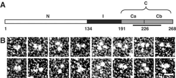
Due to a typesetting error, Figure 1 of the above article was published incorrectly in print. The correct figure is reproduced below.
Figure 1.

Molecular organization of human EB1. (A) Schematic representation of the domain organization of human EB1. Domains N, I, and C (subdivided in Ca and Cb) are depicted in white, black, and gray, respectively. Corresponding domain boundaries are indicated by residue positions. The unique EB1-like sequence motif is highlighted by a line. (B) High-magnification TEM gallery of glycerol sprayed/rotary metal shadowed EB1 specimens. Scale bar, 10 nm.
The Publisher would like to apologise for any inconvenience this may have caused.


