Abstract
INTRODUCTION:
Bone marrow examination is a useful investigative tool for the diagnosis of many hematological and nonhematological disorders. Bone marrow aspiration (BMA) provides information about the numerical and cytological features of marrow cells, whereas bone marrow trephine biopsies (BMB) provide excellent appreciation of spatial relationships between cells and of overall bone marrow structure. We conducted this study with the objective of comparing the accuracy of BMA with BMB in the diagnosis of various hematological disorders.
MATERIALS AND METHODS:
Both BMA and BMB were performed on a total of 130 cases and a comparative evaluation was performed in 100 cases to see the complementary role of both the procedures. However, 30 cases were excluded due to inadequate BMA, BMB, or both. Immunohistochemistry (IHC) was employed whenever required.
RESULTS:
In our study of 100 cases, 87% of cases were confirmed on bone marrow biopsy and in remaining 13% of cases final diagnosis was achieved with the help of other ancillary investigations. These cases were excluded for calculation of concordance rate between BMA and BMB. The concordance and disconcordance rate between BMA and BMB was 72.4% and 27.6%, respectively.
CONCLUSION:
BMA cytology and trephine biopsy histopathology complement each other and the superiority of one method over the other depended on the underlying disorder. Furthermore, application of ancillary techniques such as flow cytometery and IHC proved to be an additional advantage in further typing of various diseases.
Keywords: Aplastic anemia, bone marrow aspiration, bone marrow biopsy, megaloblastic anemia
Introduction
Bone marrow examination is a formidable weapon in the clinician's diagnostic armamentarium to hit an unsuspected diagnosis when other test results turn out to be noncontributory or inconclusive during the evaluation process.[1,2] It is a useful investigative tool for the diagnosis of many hematological and nonhematological disorders.[3]
Bone marrow examination may be performed by two methods: Aspiration and trephine biopsy. Bone marrow aspiration (BMA) is simple, reliable, and rapid method of marrow evaluation. It provides information about the numerical and cytological features of marrow cells. These cells are also well suited to further examination by cytogenetics, molecular and flow cytometric methods. However, BMA has low sensitivity in detecting solid tumor metastasis and lymphoma involvement.[4,5]
Bone marrow trephine biopsies (BMB) provide excellent appreciation of spatial relationships between cells and of overall bone marrow structure. It is required in conditions such as inadequate or failed aspirate, assessment of cellularity and bone marrow architecture, suspected focal lesion (for example, suspected granulomatous disease, or lymphoma) and bone marrow fibrosis.[2]
Nowadays, aspirate and trephine biopsy specimens are considered complementary and when both are obtained, they provide a comprehensive study of bone marrow. However, biopsy is a painful procedure and its processing takes at least 48–72 h. Hence, to perform trephine biopsies in all patients may not be cost effective in terms of clinician and laboratory personnel time, efforts, and patient discomfort.[6]
With advent of new technologies such as flow cytometry, immunohistochemistry (IHC) and molecular techniques combined analyses is useful in achieving more accurate and informative diagnostic data in some diagnostically challenging cases. We conducted this study with the objective of comparing the accuracy of BMA with BMB in the diagnosis of various hematological disorders.
Materials and Methods
The present study was conducted in the Department of Pathology, in a Tertiary Institute of North India. The study included 130 patients in whom both BMA and BMB were performed in years 2013–2014. A comparative evaluation was performed in 100 cases to see the complementary role of both the procedures. However, 30 cases were excluded due to inadequate BMA, BMB or both.
Aspirate smears were stained by Leishman's stain and Perl's Prussian stain for iron on smears. Cytochemical stain like Sudan black B was used wherever required. The aspirate smears examined for the adequacy of cellularity, presence of megakaryocytes, tumor cells and granulomas and a minimum of 500 nucleated and intact cells (myelogram) were evaluated.
Biopsies were performed on the posterior iliac crest and fixed by using 10% neutral buffered formalin. Decalcification was carried out by ethylenediamine tetraacetic acid solution for 24 h. About 4–6 μm thick sections were cut and stained with H and E. Gomori's, reticulin and Masson's trichrome were performed to grade marrow fibrosis. Ziehl Neelson stain was used for acid fast bacilli and periodic acid Schiff for glycogen and fungal hyphae.
Reporting protocol of trephine biopsy included: Specimen quality-size, integrity, overall cellularity, distribution, and maturation of granulocytes, erythrocytes, megakaryocytes, trabecular bone structure and pattern of stromal reticulin fibers were recorded. Distribution, composition and extent of any lymphoid, plasmacytic, granulomatous or foreign infiltrates, stromal changes such as edema, gelatinous change, collagen fibrosis, erythrophagocytosis or hemophagocytosis were noted. Additional information– clinical, imaging, serological, etc., if it contributed in addition to microscopy in achieving a final diagnosis were noted.
IHC was employed as per standard procedure. MPO, glycophorin, Tdt, CD10 were used routinely. In suspicious cases of marrow infiltration by lymphoma, panel of antibodies (CD45, CD20, CD15, CD30, CD3, CD5, λ and κ light chains) were employed for confirmation and further subtyping. To confirm nonhematopoietic marrow metastases in suspicious cases, antibodies against cytokeratin, neuron specific enolase, CD99, S-100 and epithelial membrane antigen were used wherever required.
Statistical analysis
All the data were compiled and entered in MS-excel. Statistical analysis was performed by using Cohen's test and software used was SPSS-20, Social Sciences Soft ware version 20 (SPSS, Armonk, NY: IBM Crop).
Results
In our study, out of 130 cases, 30 cases were excluded due to inadequacy of BMA, BMB, or both. Inadequate material was obtained more in BMA 70% (21/30) as compared to BMB 20% (6/30), whereas, 10% (3/30) cases were inadequate on both. Dry tap 66.6% (16/21) was the most common reason for failed aspiration followed by hemodilution (25%). Out of these 16 cases with dry tap, 5 (31.2%) cases diagnosed on BMB were of myelofibrosis (MF), 1 case (6.25%) of myeloproliferative disorder (MPD), 2 cases (12.5%) of acute leukemia and 3 cases (18.75%) cases of hypoplastic/aplastic anemia. However, one case of dry tap revealed myeloid hyperplasia on BMB. Four cases were inconclusive on trephine biopsy also. Two cases of poor quality smears of aspirate were diagnosed as Non-Hodgkin's lymphoma (NHL) B cell type and hypoplastic/aplastic anemia on trephine biopsy. In one hundred cases, comparison was made between BMA and bone marrow biopsy (BMB) [Figures 1 and 2].
Figure 1.
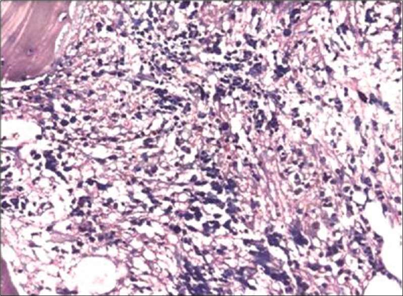
Bone marrow trephine biopsies section of myelofibrosis Grade II (H and E, ×100)
Figure 2.
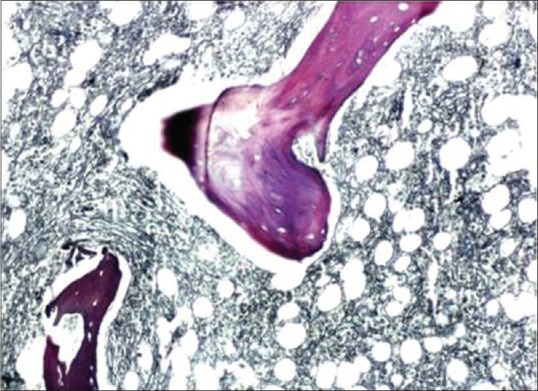
Bone marrow trephine biopsies section of myelofibrosis Grade III (Reticulin, ×100)
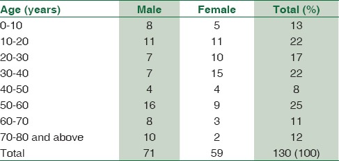
There were 71 (54.61%) males and 59 (45.38%) females with a male to female ratio of 1.2:1. The age of the subjects ranged from 1.5 to 88 years. Majority of the cases were in sixth decade [Table 1].
Table 1.
Age and sex distribution (n=130)
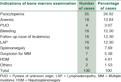
Pancytopenia (26.92%) and anemia (13.84%) were the most common clinical indications for performing a bone marrow examination. The other indications included were bleeding, hepatosplenomegaly, pyrexia of unknown origin, lymphadenopathy, etc., [Table 2].
Table 2.
Distribution of cases according to main clinical and laboratory indication of bone marrow examination (n=130)
On bone marrow examination, nutritional anemia (19%) was the most common benign disorder followed by hypoplastic/aplastic anemia (12%) and the most common malignant disorder was acute leukemia (21%) in our study. Out of 21 cases of acute leukemia, aspirates were diagnostic in 14 (8 acute myeloid leukemia [AML], 6 acute lymphocytic leukemia [ALL]) cases. However, in 5 cases, aspirate smears were unable in exact typing of acute leukemia, the definite typing was achieved by BMB and these cases were further categorized as 2 cases of AML (2/5) and 3 cases of ALL (3/5) and further subtyping of ALL cases was done with the use of IHC.
In this study, 7 cases of lymphomas, of which six cases of NHL along with one case of primary NHL of bone marrow and one case of Hodgkin disease were included. Three cases were picked up by both BMA and BMB. One case revealed presence of suspicious lymphoid cells on BMA, which came out to be reactive on BMB. Primary NHL of bone marrow revealed prominence of atypical lymphoid cells on aspirate smears, monoclonality of which was established on biopsy by IHC [Figures 3 and 4].
Figure 3.
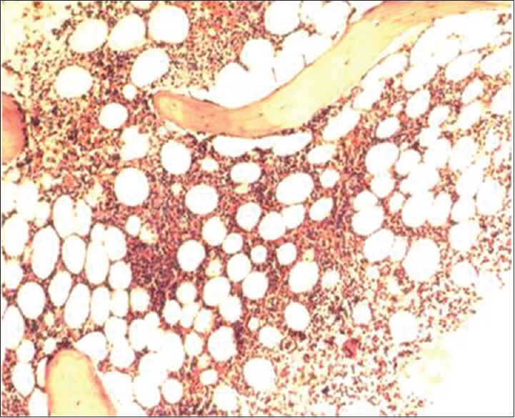
Bone marrow trephine biopsies section showing nodular and interstitial pattern (H and E, ×200)
Figure 4.
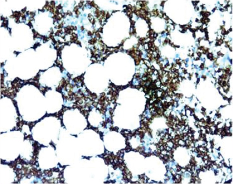
CD20 positive in Non-Hodgkin's lymphoma (IHC, ×200)
A case of anaplastic large cell lymphoma (confirmed on lymph node biopsy) was diagnosed as necrotizing granulomatous inflammation on BMB and showed atypical lymphoid cells (? viral induced) on aspirate smears. In this case, diagnosis could not be established on the basis of BMB. A case of Hodgkin lymphoma (diagnosed on lymph node biopsy) with suspicion for bone marrow involvement was negative for marrow infiltration on both aspiration and BMB.
Three cases required additional investigations for confirmation of diagnosis. One case showed reactive marrow as diagnosed on lymph node biopsy. One case each of nephrotic syndrome and light chain disease (diagnosed on renal biopsy) revealed reactive plasmacytosis and normal marrow study on BMA and BMB respectively [Table 3 and Figures 5–8].
Table 3.
Accuracy of diagnosis (n=100)
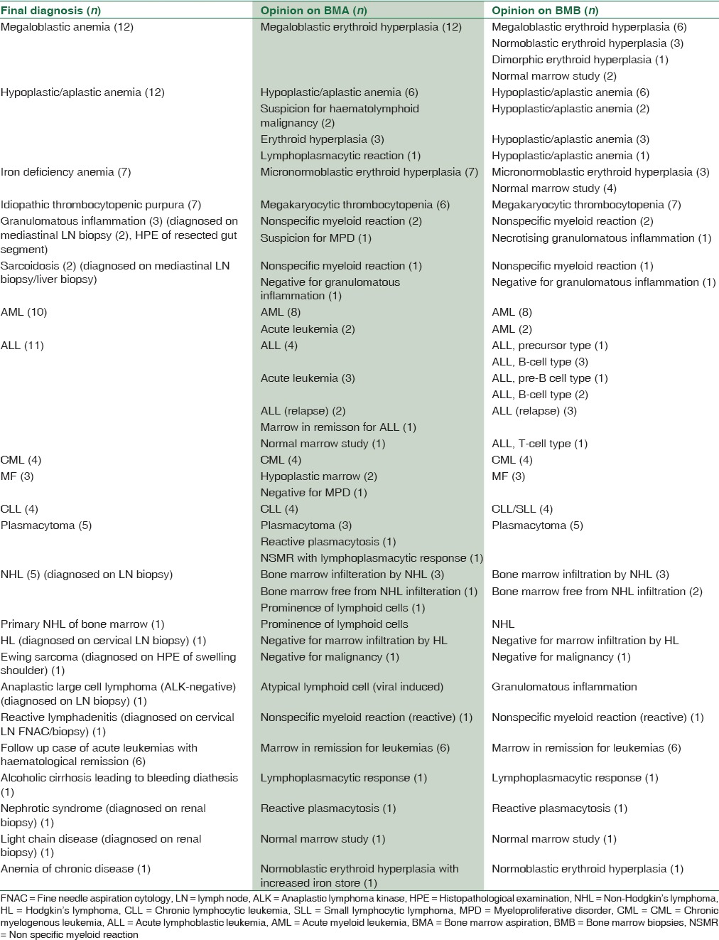
Figure 5.
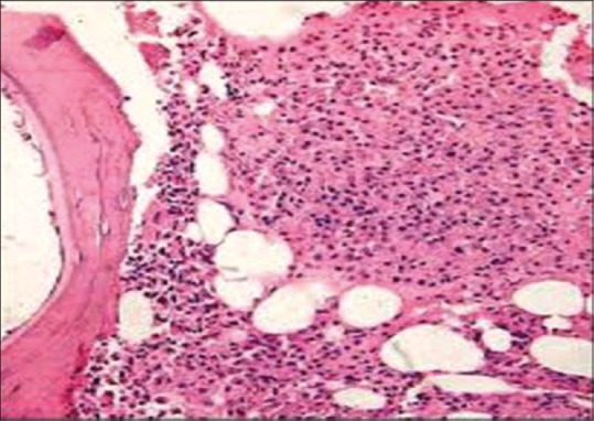
Bone marrow trephine biopsies section of plasmacytoma (H and E, ×100)
Figure 8.
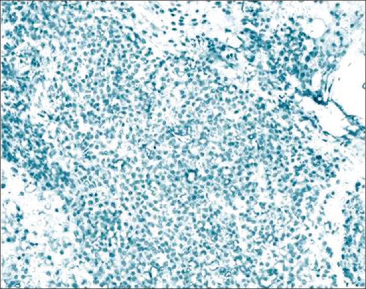
Lambda negativity in plasmacytoma (IHC, ×200)
Figure 6.
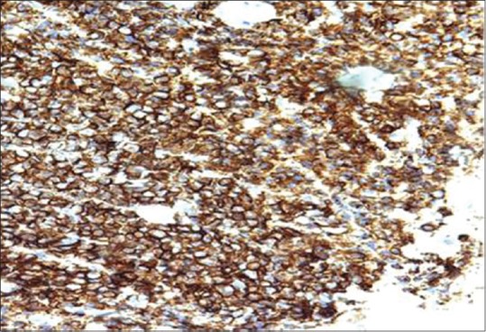
CD 138 positivity in plasmacytoma (IHC, ×200)
Figure 7.
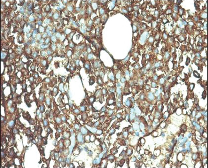
Kappa positivity in plasmacytoma (IHC, ×400)
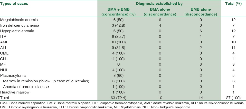
In our study of 100 cases, 87% of cases were confirmed on BMB and in remaining 13% of cases final diagnosis was achieved with the help of other ancillary investigations. These cases were excluded for calculation of concordance rate between BMA and BMB. The concordance and disconcordance rate between BMA and BMB was 72.4% and 27.6%, respectively. Of 87 cases, 10 (11.5%) cases were diagnosed on BMA alone and 14 (16.1%) cases were diagnosed on BMB alone [Table 4].
Table 4.
Concordance in the results of bone marrow aspiration and bone marrow biopsies in different types of haematological cases (n=87)
In our study, distribution of benign, malignant, and equivocal case on BMA and BMB (n = 100) were, out of 59 benign cases on BMA, 7 turned out to be malignant and 52 were benign on BMB. In 7 equivocal cases on BMA, 6 were confirmed as benign and 1 case as malignant on biopsy and the rest 34 cases were diagnosed as malignant on both [Table 5].
Table 5.
Distribution of benign, malignant and equivocal diagnosis made on bone marrow aspiration and bone marrow biopsies (n=100)

Discussion
BMA and BMB are tools for assessing health of marrow and a comparative evaluation is essential so that rapid and efficient method may be defined for early diagnosis of hematological disorders.
Out of 130 cases, 30 cases were excluded due to inadequacy. Dry tap was the most common reason for failed aspiration in our study. Extensive marrow fibrosis and hypercellularity lead to inaspirable marrow.[7] Biopsy was the only diagnostic method in MF cases in our study. Similar observations were reported by other authors and concluded that biopsy alone is diagnostic in all cases of MF.[8,9,10]
The commonest benign hematological disorder in the present study was anemia with 31% of cases belonging to this subset. Good sensitivity of BMA (100%) was found in diagnosing nutritional anemia as compared to BMB (42.8%). In rest of the cases, trephine biopsy revealed normal study. Our findings correlated with those of Khan et al. (94.4%)[11] and Nanda et al.[9] In iron deficiency anemia, aspiration was 100% diagnostic but iron stores could not be assessed properly on biopsy sections due to loss of iron during processing because of its solubility in acid during decalcification.[11,12] Due to prevalent malnutrition and infectious diseases, high incidence of nutritional anemia can be seen in tropical country as ours.[6]
Hypoplastic or aplastic anemia was the etiology in 12% of cases but aspirate was suggestive only in 50% of cases while BMB was diagnostic in all cases. This is in contrast to the study done by Mahajan et al. who found 87% of cases of hypoplastic anemia on BMA and additional 13% on BMB.[13] Trephine biopsy can provide information about number and distribution of megakaryocytes, lymphocytes, plasma cells, and blasts, these are prognostic markers required in follow up of aplastic anemia.[6]
The diagnostic sensitivity of aspiration was 86% in idiopathic thrombocytopenia (ITP) in our study. A positive correlation in cases of ITP was 85.7% in our study, which was consistent with the study, conducted by Khan et al. (83.3%).[11]
In diagnosing acute leukemia, the sensitivity and specificity of aspiration was 100% in our study, which in concordance with other studies.[14] BMB provided additional information over aspirate smears, about the bone marrow changes in AML and suggested that some of the features may have prognostic significance in addition to diagnostic importance. The important features were, presence or absence of inflammatory cells and were better depicted on core biopsy.[15]
In this study, there were 8 cases of chronic leukemias (chronic myeloid leukemia [CML] [n = 4], chronic lymphocytic leukemia [CLL] [n = 4]) which were concordant on aspiration and biopsy similar to Ghodasara and Gonsai[16] In CML cases, the BMB showed hypercellular marrow with granulocytic hyperplasia and loss of fat cells. The aspirates are better able to classify the phases of CML as compared to biopsy.[16] BMB provide additional information about the pattern of involvement in CLL and about prognosis that is nodular pattern over diffuse pattern.[10]
All the three cases of MF in our study were diagnosed on BMB. BMA does not have much role in diagnosis of MF because of diffuse osteomyelosclerosis, intrasinusoidal hematopoiesis and vascular proliferation, which are characteristic of fibrotic MF. Hence, BMB is more helpful in confirmation and grading.[6]
In our study, 60% of cases of multiple myeloma (MM) showed a positive correlation in BMA and BMB. The sensitivity was 88.5% in the study conducted by Goyal et al.[6] BMA is essential for appropriate evaluation of plasma cell differentiation. The diagnosis of MM in marrow biopsy depends on extent and pattern of plasma cell infiltration and cytological features of plasma cells. BMB in this disorder is an essential and important investigation for comparison with repeated biopsies during follow up.[17]
Bone marrow involvement in lymphoproliferative disease is a frequent finding and can be detected by morphological examination of bone marrow biopsies and aspirate smears, flowcytometric analysis of aspirate samples and IHC of tissue samples for B and T cell markers and molecular genetic analysis using polymerase chain reaction.[18]
In the present study, aspiration had 80% sensitivity in diagnosing NHL. These results were comparable with those in the literature, in which the sensitivity of BMA for bone marrow involvement in different malignancies ranges from 69% to 82%.[19] BMB provides valuable information regarding spatial distribution, extent of infiltrate, cellularity, and fibrosis in NHL which cannot be determined from aspiration. It is more useful in postchemotherapy patients to assess the residual tumor cell burden and degree of chemotherapy response.[6] The combined procedure of aspiration and biopsy gives a higher yield and is essential in patients with suspected carcinoma, NHL and Hodgkin's disease.[20]
In this study, a case of shoulder swelling was included which was diagnosed as Ewing's sarcoma on histopathological examination. However, both aspirate and trephine biopsy was negative for metastases. BMA and BMB should both be performed in patient with proven/suspected malignancies because staging may affect the management.[21]
Granulomatous inflammation was diagnosed in one case, on BMB; however, the aspirate smears revealed suspicion for MPD. Because of focal involvement of the marrow it is very difficult to detect granulomas on aspirate smears. Fibrosis in and around the granuloma leads to difficult marrow aspiration. Hence, trephine biopsy is a better tool to demonstrate granulomas because better preservation of morphology and more amount of tissue is available for study than aspirate smears.[22] Other studies have also observed detection of granulomas more on trephine biopsies than aspirates.[11] BMA and BMB of 4 cases revealed nonspecific myeloid reaction. However, later these patients had been diagnosed as granulomatous inflammation on lymph node biopsy (n = 2) and sarcoidosis on lymphnode biopsy and liver biopsy (n = 2).
BMB was confirmatory in 87% of cases while remaining 13% cases could not be diagnosed on BMB and required other ancillary investigations. On comparison of these 87% cases, 72.4% of cases were concordant on both BMA and BMB which was in agreement with the study published by Khan et al. and Ghodasara and Gonsai who reported a positive concordance of 73.8% in 443 patients studied and 73.9% in 73 patients studied, respectively.[11,16] Whereas Chandra and Chandrareported 78% positive correlation between the two procedures.[23] The disconcordance rate was 27.6%.
There were a few limitations of our study. We did not evaluate the cases by touch imprints which may increase the diagnostic accuracy. Furthermore, there were small numbers of cases in the subgroups during the given span of time. Further studies and more number of cases will provide better correlation of BMA and BMB.
Conclusion
Our study concludes that BMA cytology and trephine biopsy histopathology complement each other and the superiority of one method over the other depended on the underlying disorder. Among nonmalignant disorders BMA was superior to BMB in megaloblastic anemia, iron deficiency anemia, and ITP cases. BMB was the sole diagnostic tool in MF cases where BMA aspiration yielded diluted marrow or dry tap. BMB was more helpful in diagnosing MPDs, aplastic/hypoplastic anemia, granulomatous lesions, NHL, and acute leukemia. Furthermore, application of ancillary techniques such as flow cytometery and IHC proved to be an additional advantage in further typing of various diseases in our study.
Financial support and sponsorship
Nil.
Conflicts of interest
There are no conflicts of interest.
References
- 1.Bain BJ. Bone marrow aspiration. J Clin Pathol. 2001;54:657–63. doi: 10.1136/jcp.54.9.657. [DOI] [PMC free article] [PubMed] [Google Scholar]
- 2.Bain BJ. Bone marrow trephine biopsy. J Clin Pathol. 2001;54:737–42. doi: 10.1136/jcp.54.10.737. [DOI] [PMC free article] [PubMed] [Google Scholar]
- 3.Riley RS, Hogan TF, Pavot DR, Forysthe R, Massey D, Smith E, et al. A pathologist's perspective on bone marrow aspiration and biopsy: I. Performing a bone marrow examination. J Clin Lab Anal. 2004;18:70–90. doi: 10.1002/jcla.20008. [DOI] [PMC free article] [PubMed] [Google Scholar]
- 4.Bates I, Burthem J. Bone marrow biopsy. In: Bain BJ, Bates I, Laffan MA, Lewis SM, editors. Dacie and Lewis Practical Hematology. 11th ed. China: Elsevier, Churchill Livingstone; 2012. pp. 123–37. [Google Scholar]
- 5.Atla BL, Anem V, Dasari A. Prospective study of bone marrow in haematological disorders. Int J Res Med Sci. 2015;3:1917–21. [Google Scholar]
- 6.Goyal S, Singh UR, Rusia U. Comparative evaluation of bone marrow aspirate with trephine biopsy in hematological disorders and determination of optimum trephine length in lymphoma infiltration. Mediterr J Hematol Infect Dis. 2014;6:e2014002. doi: 10.4084/MJHID.2014.002. [DOI] [PMC free article] [PubMed] [Google Scholar]
- 7.Humphries JE. Dry tap bone marrow aspiration: Clinical significance. Am J Hematol. 1990;35:247–50. doi: 10.1002/ajh.2830350405. [DOI] [PubMed] [Google Scholar]
- 8.Sabharwal BD, Malhotra V, Aruna S, Grewal R. Comparative evaluation of bone marrow aspirate particle smears, imprints and biopsy sections. J Postgrad Med. 1990;36:194–8. [PubMed] [Google Scholar]
- 9.Nanda A, Basu S, Marwaha N. Bone marrow trephine biopsy as an adjunct to bone marrow aspiration. J Assoc Physicians India. 2002;50:893–5. [PubMed] [Google Scholar]
- 10.Varma N, Dash S, Sarode R, Marwaha N. Relative efficacy of bone marrow trephine biopsy sections as compared to trephine imprints and aspiration smears in routine haematological practice. Indian J Pathol Microbiol. 1993;36:215–26. [PubMed] [Google Scholar]
- 11.Khan TA, Khan IA, Mahmood K. Diagnostic role of bone marrow aspiration and trephine biopsy in haematological practice. J Postgrad Med Inst. 2014;28:217–21. [Google Scholar]
- 12.Sah SP, Matutes E, Wotherspoon AC, Morilla R, Catovsky D. A comparison of flow cytometry, bone marrow biopsy, and bone marrow aspirates in the detection of lymphoid infiltration in B cell disorders. J Clin Pathol. 2003;56:129–32. doi: 10.1136/jcp.56.2.129. [DOI] [PMC free article] [PubMed] [Google Scholar]
- 13.Mahajan V, Kaushal V, Thakur S, Kaushik R. A comparative study of bone marrow aspiration and bone marrow biopsy in haematological and non-haematological disorders – An institutional experience. J Indian Acad Clin Med. 2013;14:133–5. [Google Scholar]
- 14.Shastry SM, Kolte SS. Spectrum of hematological disorders observed in one-hundred and ten consecutive bone marrow aspirations and biopsies. Med J DY Patil Univ. 2015;5:118–21. [Google Scholar]
- 15.Younus U, Saba K, Aijaz J, Bukhari MH, Naeem S. Significance of bone marrow histology in the diagnosis of acute myeloid leukemia. Ann King Edward Med Univ. 2011;17:5–8. [Google Scholar]
- 16.Ghodasara J, Gonsai RN. Comparative evaluation of simultaneous bone marrow aspiration and bone marrow trephine biopsy – A tertiary care hospital based cross-sectional study. Int J Sci Res. 2014;10:2058–61. [Google Scholar]
- 17.Stifter S, Babarovic E, Valkovic T, Seili-Bekafigo I, Stemberger C, Nacinovic A, et al. Combined evaluation of bone marrow aspirate and biopsy is superior in the prognosis of multiple myeloma. Diagn Pathol. 2010;5:30. doi: 10.1186/1746-1596-5-30. [DOI] [PMC free article] [PubMed] [Google Scholar]
- 18.Bedu-Addo G, Ampem Amoako Y, Bates I. The role of bone marrow aspirate and trephine samples in haematological diagnoses in patients referred to a teaching hospital in Ghana. Ghana Med J. 2013;47:74–8. [PMC free article] [PubMed] [Google Scholar]
- 19.Musolino A, Guazzi A, Nizzoli R, Panebianco M, Mancini C, Ardizzoni A. Accuracy and relative value of bone marrow aspiration in the detection of lymphoid infiltration in non-Hodgkin lymphoma. Tumori. 2010;96:24–7. doi: 10.1177/030089161009600104. [DOI] [PubMed] [Google Scholar]
- 20.Bearden JD, Ratkin GA, Coltman CA. Comparison of the diagnostic value of bone marrow biopsy and bone marrow aspiration in neoplastic disease. J Clin Pathol. 1974;27:738–40. doi: 10.1136/jcp.27.9.738. [DOI] [PMC free article] [PubMed] [Google Scholar]
- 21.Moid F, DePalma L. Comparison of relative value of bone marrow aspirates and bone marrow trephine biopsies in the diagnosis of solid tumor metastasis and Hodgkin lymphoma: Institutional experience and literature review. Arch Pathol Lab Med. 2005;129:497–501. doi: 10.5858/2005-129-497-CORVOB. [DOI] [PubMed] [Google Scholar]
- 22.Jeevan SK, Paul-Tara R, Uppin S, Uppin M. Bone marrow granulomas: A retrospective study of 47 cases (A single center experience) Am J Int Med. 2014;2:90–4. [Google Scholar]
- 23.Chandra S, Chandra H. Comparison of bone marrow aspirate cytology, touch imprint cytology and trephine biopsy for bone marrow evaluation. Hematol Rep. 2011;3:65–8. doi: 10.4081/hr.2011.e22. [DOI] [PMC free article] [PubMed] [Google Scholar]


