Description
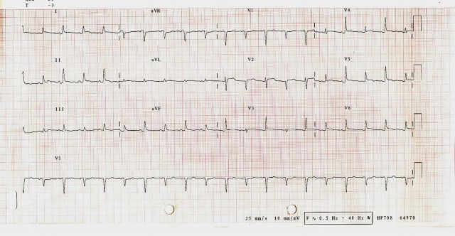
A 39-year-old woman with several weeks’ history of malaise and shortness of breath was referred because of acute deterioration. Examination revealed a regular tachycardia of 110 bpm, evidence of pulsus alternans and a blood pressure of 120/74, but was otherwise unremarkable. A routine admission 12-lead ECG (figure 1) demonstrated low voltage QRS alternans. As a result of this finding, an urgent transthoracic echocardiogram (TTE) was arranged.
Figure 1.
12-Lead ECG demonstrating QRS alternans best seen in leads V3 and V4.
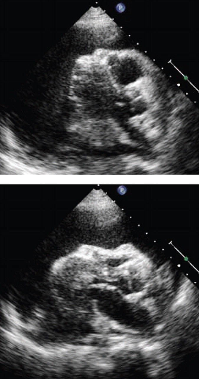
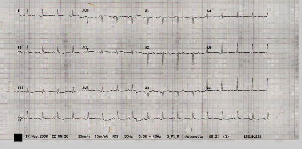
TTE (figure 2, video files) confirmed a large, circumferential pericardial effusion with signs of tamponade. Soon after, the patients condition declined and blood pressure dropped to 95/65 with pulsus paradoxus >10 mm Hg detected manually. Pericardiocentesis under echo-guidance was performed promptly and 900 ml of serosanguineous fluid was removed with immediate resolution in symptoms and an improvement in blood pressure to 139/87. Repeat ECG (figure 3) after drainage revealed resolution of electrical alternans.
Figure 2.

Parasternal views of the heart in mid-diastole. The position of the heart alternates within two consecutive heart beats, with the septum swinging along an antero-posterior axis. Anterior displacement of the septal wall results in the larger QRS amplitude (corresponding with chest leads V3 and V4 on ECG), whereas posterior displacement of the septum produces the smaller amplitude (almost negative) QRS.
Figure 3.
12-Lead ECG performed following drainage demonstrating resolution of QRS alternans.
Pericardial effusion should be considered likely when electrical alternans is seen in the presence of pulsus alternans, particularly when there is no evidence of explanatory arrhythmia on ECG. When present in this context, QRS alternans typically signifies a large, haemodynamically significant, effusion and urgent imaging to confirm the diagnosis and to guide treatment is important. In effusion-related QRS alternans, the heart swings once every second beat (2:1 swinging). This pendular cardiac motion is visible on echocardiography. True 2:1 QRS alternans occurs within a limited range of heart rates defined mathematically by the relative durations of diastole and systole.
Footnotes
Competing interests: None.
Patient consent: Obtained.


