

Abstract
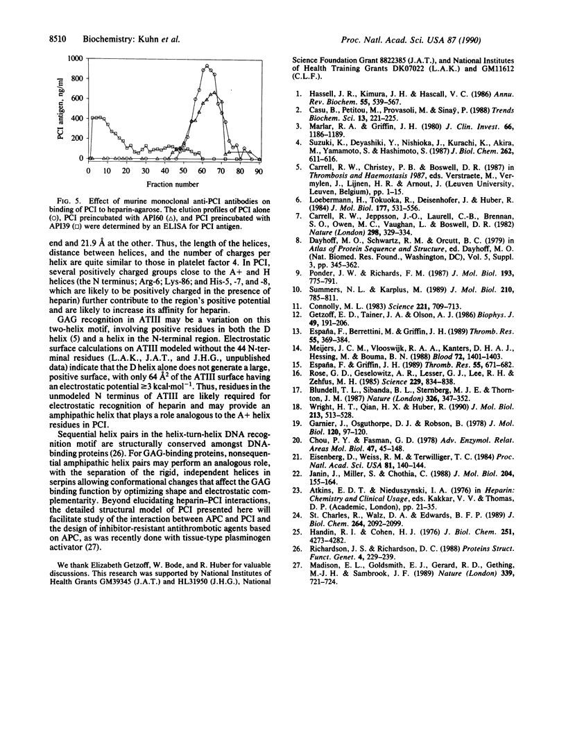
Glycosaminoglycans (GAGs) including heparin accelerate the inhibition of serine proteases by serine protease inhibitors (serpins), an essential process in regulating blood coagulation. to analyze the molecular basis for GAG recognition by the plasma serpin protein C inhibitor (PCI; also known as plasminogen activator inhibitor 3), we have constructed a complete, energy-minimized, three-dimensional model of PCI by using the structure of homologous alpha 1-antitrypsin as a template. Sequence analysis, hydrogen-bonding environment, and shape complementarity suggested that the N-terminal residues of PCI, which are not homologous to those of alpha 1-antitrypsin, form an amphipathic alpha-helix, here designated A+ since it precedes the alpha 1-antitrypsin A helix. Electrostatic calculations revealed a single, highly positive surface region arising from both the A+ and H helices, suggesting that this two-helix motif is required for GAG binding by PCI. The dominant role of electrostatic interactions in PCI-heparin binding was confirmed by the strong ionic strength dependence of heparin stimulation. The involvement of the A+ helix in heparin binding was verified by demonstrating that an anti-PCI antibody that specifically binds the A+ peptide blocks heparin binding.
Full text
PDF
Images in this article
Selected References
These references are in PubMed. This may not be the complete list of references from this article.
- Blundell T. L., Sibanda B. L., Sternberg M. J., Thornton J. M. Knowledge-based prediction of protein structures and the design of novel molecules. 1987 Mar 26-Apr 1Nature. 326(6111):347–352. doi: 10.1038/326347a0. [DOI] [PubMed] [Google Scholar]
- Carrell R. W., Jeppsson J. O., Laurell C. B., Brennan S. O., Owen M. C., Vaughan L., Boswell D. R. Structure and variation of human alpha 1-antitrypsin. Nature. 1982 Jul 22;298(5872):329–334. doi: 10.1038/298329a0. [DOI] [PubMed] [Google Scholar]
- Casu B., Petitou M., Provasoli M., Sinaÿ P. Conformational flexibility: a new concept for explaining binding and biological properties of iduronic acid-containing glycosaminoglycans. Trends Biochem Sci. 1988 Jun;13(6):221–225. doi: 10.1016/0968-0004(88)90088-6. [DOI] [PubMed] [Google Scholar]
- Chou P. Y., Fasman G. D. Prediction of the secondary structure of proteins from their amino acid sequence. Adv Enzymol Relat Areas Mol Biol. 1978;47:45–148. doi: 10.1002/9780470122921.ch2. [DOI] [PubMed] [Google Scholar]
- Connolly M. L. Solvent-accessible surfaces of proteins and nucleic acids. Science. 1983 Aug 19;221(4612):709–713. doi: 10.1126/science.6879170. [DOI] [PubMed] [Google Scholar]
- Eisenberg D., Weiss R. M., Terwilliger T. C. The hydrophobic moment detects periodicity in protein hydrophobicity. Proc Natl Acad Sci U S A. 1984 Jan;81(1):140–144. doi: 10.1073/pnas.81.1.140. [DOI] [PMC free article] [PubMed] [Google Scholar]
- España F., Berrettini M., Griffin J. H. Purification and characterization of plasma protein C inhibitor. Thromb Res. 1989 Aug 1;55(3):369–384. doi: 10.1016/0049-3848(89)90069-8. [DOI] [PubMed] [Google Scholar]
- España F., Griffin J. H. Determination of functional and antigenic protein C inhibitor and its complexes with activated protein C in plasma by ELISA's. Thromb Res. 1989 Sep 15;55(6):671–682. doi: 10.1016/0049-3848(89)90298-3. [DOI] [PubMed] [Google Scholar]
- Garnier J., Osguthorpe D. J., Robson B. Analysis of the accuracy and implications of simple methods for predicting the secondary structure of globular proteins. J Mol Biol. 1978 Mar 25;120(1):97–120. doi: 10.1016/0022-2836(78)90297-8. [DOI] [PubMed] [Google Scholar]
- Getzoff E. D., Tainer J. A., Olson A. J. Recognition and interactions controlling the assemblies of beta barrel domains. Biophys J. 1986 Jan;49(1):191–206. doi: 10.1016/S0006-3495(86)83634-7. [DOI] [PMC free article] [PubMed] [Google Scholar]
- Handin R. I., Cohen H. J. Purification and binding properties of human platelet factor four. J Biol Chem. 1976 Jul 25;251(14):4273–4282. [PubMed] [Google Scholar]
- Hassell J. R., Kimura J. H., Hascall V. C. Proteoglycan core protein families. Annu Rev Biochem. 1986;55:539–567. doi: 10.1146/annurev.bi.55.070186.002543. [DOI] [PubMed] [Google Scholar]
- Janin J., Miller S., Chothia C. Surface, subunit interfaces and interior of oligomeric proteins. J Mol Biol. 1988 Nov 5;204(1):155–164. doi: 10.1016/0022-2836(88)90606-7. [DOI] [PubMed] [Google Scholar]
- Loebermann H., Tokuoka R., Deisenhofer J., Huber R. Human alpha 1-proteinase inhibitor. Crystal structure analysis of two crystal modifications, molecular model and preliminary analysis of the implications for function. J Mol Biol. 1984 Aug 15;177(3):531–557. [PubMed] [Google Scholar]
- Madison E. L., Goldsmith E. J., Gerard R. D., Gething M. J., Sambrook J. F. Serpin-resistant mutants of human tissue-type plasminogen activator. Nature. 1989 Jun 29;339(6227):721–724. doi: 10.1038/339721a0. [DOI] [PubMed] [Google Scholar]
- Marlar R. A., Griffin J. H. Deficiency of protein C inhibitor in combined factor V/VIII deficiency disease. J Clin Invest. 1980 Nov;66(5):1186–1189. doi: 10.1172/JCI109952. [DOI] [PMC free article] [PubMed] [Google Scholar]
- Meijers J. C., Vlooswijk R. A., Kanters D. H., Hessing M., Bouma B. N. Identification of monoclonal antibodies that inhibit the function of protein C inhibitor. Evidence for heparin-independent inhibition of activated protein C in plasma. Blood. 1988 Oct;72(4):1401–1403. [PubMed] [Google Scholar]
- Ponder J. W., Richards F. M. Tertiary templates for proteins. Use of packing criteria in the enumeration of allowed sequences for different structural classes. J Mol Biol. 1987 Feb 20;193(4):775–791. doi: 10.1016/0022-2836(87)90358-5. [DOI] [PubMed] [Google Scholar]
- Richardson J. S., Richardson D. C. Helix lap-joints as ion-binding sites: DNA-binding motifs and Ca-binding "EF hands" are related by charge and sequence reversal. Proteins. 1988;4(4):229–239. doi: 10.1002/prot.340040402. [DOI] [PubMed] [Google Scholar]
- Rose G. D., Geselowitz A. R., Lesser G. J., Lee R. H., Zehfus M. H. Hydrophobicity of amino acid residues in globular proteins. Science. 1985 Aug 30;229(4716):834–838. doi: 10.1126/science.4023714. [DOI] [PubMed] [Google Scholar]
- St Charles R., Walz D. A., Edwards B. F. The three-dimensional structure of bovine platelet factor 4 at 3.0-A resolution. J Biol Chem. 1989 Feb 5;264(4):2092–2099. [PubMed] [Google Scholar]
- Summers N. L., Karplus M. Construction of side-chains in homology modelling. Application to the C-terminal lobe of rhizopuspepsin. J Mol Biol. 1989 Dec 20;210(4):785–811. doi: 10.1016/0022-2836(89)90109-5. [DOI] [PubMed] [Google Scholar]
- Suzuki K., Deyashiki Y., Nishioka J., Kurachi K., Akira M., Yamamoto S., Hashimoto S. Characterization of a cDNA for human protein C inhibitor. A new member of the plasma serine protease inhibitor superfamily. J Biol Chem. 1987 Jan 15;262(2):611–616. [PubMed] [Google Scholar]
- Wright H. T., Qian H. X., Huber R. Crystal structure of plakalbumin, a proteolytically nicked form of ovalbumin. Its relationship to the structure of cleaved alpha-1-proteinase inhibitor. J Mol Biol. 1990 Jun 5;213(3):513–528. doi: 10.1016/s0022-2836(05)80212-8. [DOI] [PubMed] [Google Scholar]