Abstract
Background and study aims
Endosurgical devices with injection function have been reported to decrease endoscopic submucosal dissection (ESD) operation times for experts, but the efficacy of these devices for inexperienced endoscopists is unclear. The aim of this study was to evaluate the feasibility of ESD using a novel ESD knife (DN-D2718B).
Patients and methods
This is a single-center prospective pilot clinical feasibility study. Patients diagnosed with superficial gastrointestinal neoplasms were enrolled. A pre-specified group of ESD trainees with ESD experience on a porcine gastric model and fewer than 30 cases of ESD in their selected fields performed ESD under expert supervision, using the DN-D2718B. En bloc resection rates, R0 resection rates, procedure times, and incidence of intra-operational/post-operational adverse events were assessed.
Results
Between June 2015 and January 2016, 13 esophageal, 27 gastric, and 14 colorectal ESD cases were performed per-protocol with mean resection speeds of 10.2, 12.0, and 15.5 mm 2 /min, respectively. There were no intra-operational complications.
Conclusion
ESD with this novel knife is feasible even when performed by non-experts.
Introduction
Endoscopic submucosal dissection (ESD) has become the gold standard for endoscopic treatment of superficial gastrointestinal neoplasms 1 2 . However, the procedure requires advanced training and is associated with a risk of adverse events (AEs) 3 . Endosurgical devices with injection function have been reported to decrease operation time for experts 4 . However, non-experts require longer procedure times and have a higher risk of AEs 5 , and therefore evaluation of the efficacy of use of these devices by non-experts may be clinically important.
In collaboration with Pentax Medical, HOYA Corporation, we have developed a second-generation endosurgical knife (DN-D2618B; HOYA Corporation, Pentax, Tokyo, Japan), and previously performed an animal feasibility study on the efficacy of this prototype 6 . The aim of this prospective pilot clinical feasibility study was to evaluate the efficacy of clinical ESD using this novel knife.
Patients and methods
Endosurgical device DN-D2718A/B
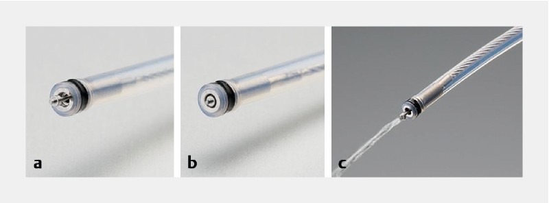
The endosurgical device (DN-D2718B; HOYA Corporation, Pentax, Tokyo, Japan) is a needle-type device 2.0 mm in length, with injection function, designed for uniform use in all gastrointestinal organs ( Fig. 1 ). Its unique characteristic is the 0.8-mm metal disk, which enables effective incision of the mucous layer and stabilizes the device during dissection. When retracted, electrical current spreads to the surrounding metal sheath, 1.8 mm in diameter, enabling effective hemostasis over a wide area. The wide injection shaft enables an effective elevation effect without the need for increasing injection pressure ( Supplementary Video ).
Fig. 1.

Endosurgical device DN-D2718B. a When the knife is extended, the disk enables effective incision and stability during dissection. b When the tip is retracted, the entire metal surface enables coagulation. c Effective injection can be achieved with this device.
Optimum electrocautery settings in humans
A team of expert endoscopists, each with experience of over 100 cases of gastrointestinal ESD, performed preliminary ESD on 10 patients to determine optimum settings with a high-frequency generator VIO 300 D (ERBE Elektromedizin GmbH, Tübingen, Germany). Settings with Soft Coag, Swift Coag, Forced Coag, and Endocut were each tested for incision, dissection and hemostasis. Optimum electrocautery settings for esophageal and colorectal ESD were unanimously determined to be EndoCut I (Effect 2, Duration 2, Interval 2) for incision and dissection, and Forced Coag (Effect 3, 30 W) for hemostasis. Similarly, optimum settings for gastric ESD were determined to be EndoCut I (Effect 3, Duration 3, Interval 3) and Forced Coag (Effect 3, 30 W).
Pilot prospective clinical feasibility study
This pilot prospective clinical feasibility study was begun after approval from the research ethics committee in our institution, and trial registration (UMIN 000017575) in May 2015. Patients referred to our institute with a diagnosis of superficial gastrointestinal neoplasms were enrolled. Written forms of informed consent were obtained from all patients.
ESD trainees
Two trainees each were selected for evaluation of esophageal, gastric, and colorectal ESD. All trainees had over 1000 cases of gastroscopy experience 7 8 , and ESD experience of 1 porcine gastric model. The selected trainees for gastric ESD had experience of 25, 26 gastric ESD each, while trainees for esophageal and colorectal ESD had attained competency in gastric ESD after over 30 cases, but had no experience in their specified field.
ESD procedure
All ESD procedures were performed under oral supervision by experts with over 6 years of ESD training. An expert took over only when a trainee could not accomplish ESD, due to inability to continue the procedure as judged by the expert or due to complications 7 8 . The selected trainees performed ESD as previously described 9 . In brief, the lesion margin was marked using the DN-D2718B with Forced Coag. Initial submucosal injection before incision was performed using a two-fold diluted solution of 0.4 % hyaluronic acid (Mucoup; Johnson and Johnson K.K., Tokyo, Japan), followed by incision and dissection with the DN-D2718B. Injections of normal saline with the device were repeated as required during the procedure, followed immediately by dissection of the elevated area. In cases of esophageal ESD, polyglycolic acid sheets and fibrin glue were prophylactically administered when lesions extended to over half the circumference of the esophagus 10 . In cases where stricture could not be prevented, balloon dilation was performed as required. Cases in which concurrent use of other ESD devices was required were predetermined to be excluded from analysis.
Study endpoints
Endpoints were set to assess the efficacy of the device. Procedure time, resection speed, en bloc resection rate, R0 resection rate, incidence of intra-operational AEs and postoperative AEs were assessed. R0 resection was defined as en bloc resection with histologically confirmed tumor-free horizontal and vertical margins. Perforation was defined as an endoscopically confirmed defect in the serosa, or free air detected by abdominal X-ray or CT. Intra-operational hemorrhage was defined as a decrease in hemoglobin of > 2 g/dL the day after ESD.
Sample size
As a pilot clinical study, 50 cases of ESD were set as the target for analysis. Assuming that all patients would have a single lesion, and with a conservative estimate that approximately 5 % of patients would not be able to complete per-protocol treatment and therefore be excluded, the sample size was predetermined to be 52 patients.
Results
Pilot prospective clinical feasibility study
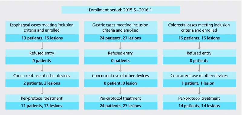
Between June 2015 and January 2016, a total of 52 patients composed of 13 with esophageal, 24 with gastric, and 15 with colorectal neoplasms were enrolled in this study (see Fig. 2 for details). After exclusion of cases as predetermined, a total of 49 patients and 54 cases of ESD were analyzed ( Table 1 ).
Fig. 2.

Flow diagram of the study patients.
Table 1. Results of ESD training with DN-D2718B.
| Esophagus | Stomach | Colon | ||||
| Trainee A | Trainee B | Trainee C | Trainee D | Trainee E | Trainee F | |
| Pre-study training | ||||||
|
1 | 1 | 1 | 1 | 1 | 1 |
|
0 | 0 | 26 | 25 | 0 | 0 |
|
Yes | Yes | No | No | Yes | Yes |
| Results of ESD with DN-D2718B | ||||||
|
10 | 3 | 13 | 14 | 9 | 5 |
| Patient Background | ||||||
|
10/0 | 3/0 | 9/4 | 12/2 | 4/5 | 2/3 |
|
70.0 ± 6.0 | 77.3 ± 10.0 | 70.6 ± 9.0 | 69.5 ± 6.9 | 69.2 ± 12.1 | 75.8 ± 11.7 |
| Tumor Background | ||||||
|
10/0/0 | 3/0/0 | 12/0/1 | 13/0/1 | 7/1/1 | 5/0/0 |
|
24.8 ± 15.4 | 30.0 ± 13.1 | 16.3 ± 8.9 | 13.2 ± 8.5 | 32.6 ± 11.2 | 23.8 ± 19.4 |
|
33.0 ± 13.8 | 43.3 ± 33.3 | 42.1 ± 9.4 | 39.2 ± 12.8 | 38.4 ± 9.0 | 31.5 ± 15.8 |
|
672.6 ± 465.7 | 1204.2 ± 131.7 | 1183.6 ± 593.0 | 1031.9 ± 588.3 | 1226.2 ± 451.2 | 932.5 ± 857.6 |
| Results of ESD | ||||||
|
10/10 (100) | 3/3 (100) | 10/13 (76.9) | 13/14 (92.9) | 9/9 (100) | 5/5 (100) |
|
69.8 ± 43.1 | 84.7 ± 43.6 | 122.9 ± 65.2 | 113.8 ± 61.5 | 73.7 ± 34.3 | 75.5 ± 26.3 |
|
9.4 ± 3.9 | 13.4 ± 4.6 | 11.1 ± 7.2 | 12.2 ± 5.9 | 16.7 ± 6.6 | 15.6 ± 9.1 |
|
10/10 (100) | 3/3 (100) | 13/13 (100) | 14/14 (100) | 9/9 (100) | 5/5 (100) |
|
7/10 (70.0) | 2/3 (66.7) | 13/13 (100) | 14/14 (100) | 6/9 (66.7) | 5/5 (100) |
| Adverse events | ||||||
|
0/10 (0) | 0/3 (0) | 0/13 (0) | 0/14 (0) | 0/9 (0) | 0/5 (0) |
|
2/10 (20.0) | 1/3 (33.3) | 0/13 (0) | 2/14 (14.3) | 0/9 (0) | 0/5 (0) |
ESD, endoscopic submucosal dissection; M, mucosal, SM, submucosal
All postoperative adverse events after esophageal ESD were stricture, and after gastric ESD were delayed hemorrhage.
En bloc resection was achieved in all cases. R0 resection rates were 69.2 %, 100 %, and 78.6 %, while resection speeds were 10.2 ± 4.5, 12.0 ± 6.7, 15.5 ± 7.3 mm 2 /min for esophageal, gastric and colorectal ESD, respectively.
There were no intraoperative AEs. Postoperative AEs comprised 3 cases (23.1 %) of postoperative stricture after esophageal ESD and 2 cases (7.4 %) of delayed hemorrhage after gastric ESD.
Discussion
While the feasibility of use of novel ESD devices by experts has often been assessed 11 12 , feasibility of use by non-experts, while difficult to demonstrate, may be more clinically important. Through this first pilot prospective clinical feasibility study, we have demonstrated that ESD with the DN-D2718B is effective even in the hands of non-experts.
Procedure times and resection speeds with this device were comparable to or better than previous reports on ESD 13 14 15 . The R0 resection rate for esophageal and colorectal ESD, although comparable to or higher than previous reports 13 14 15 , were lower than for gastric ESD. Histological evaluation of the resected specimens demonstrated a wide area of cauterization near the edges of the ESD specimen. While a large disk enables effective incision and hemostasis, resection with a wider margin may be required for histological evaluation of R0 resection, and finding the best balance will be necessary in future devices.
There were several limitations to our study. First, cases requiring use of other devices were excluded from analysis, which may be a cause of bias. The ITknife nano (KD-612L; Olympus Co.) was used in all of these cases, in circumstances where it became difficult to approach the target with the endoscope during the procedure. However, these circumstances may have been avoided in the hands of experts. Second, this was a single-center non-randomized pilot study with only a limited number of endoscopists and patients.
Conclusion
Although further studies are required for confirmation of the results in this study, effective ESD with this novel knife seems to be feasible.
Footnotes
Competing interests T. Y. and F. M. receive collaborative research funding form Pentax and honoraria from Olympus, CSL Behring, and GUNZE Medical.
References
- 1.Tanaka S, Kashida H, Saito Y et al. JGES guidelines for colorectal endoscopic submucosal dissection/endoscopic mucosal resection. Dig Endosc. 2015;27:417–434. doi: 10.1111/den.12456. [DOI] [PubMed] [Google Scholar]
- 2.Pimentel-Nunes P, Dinis-Ribeiro M, Ponchon T et al. Endoscopic submucosal dissection: European Society of Gastrointestinal Endoscopy (ESGE) Guideline. Endoscopy. 2015;47:829–854. doi: 10.1055/s-0034-1392882. [DOI] [PubMed] [Google Scholar]
- 3.Saito Y, Uraoka T, Yamaguchi Y et al. A prospective, multicenter study of 1111 colorectal endoscopic submucosal dissections (with video) Gastrointest Endosc. 2010;72:1217–1225. doi: 10.1016/j.gie.2010.08.004. [DOI] [PubMed] [Google Scholar]
- 4.Ciocîrlan M, Pioche M, Lepilliez V et al. The ENKI-2 water-jet system versus Dual Knife for endoscopic submucosal dissection of colorectal lesions: a randomized comparative animal study. Endoscopy. 2014;46:139–143. doi: 10.1055/s-0033-1344892. [DOI] [PubMed] [Google Scholar]
- 5.Imai K, Hotta K, Yamaguchi Y et al. Preoperative indicators of failure of en bloc resection or perforation in colorectal endoscopic submucosal dissection: implications for lesion stratification by technical difficulties during stepwise training. Gastrointest Endosc. 2016;83:954–962. doi: 10.1016/j.gie.2015.08.024. [DOI] [PubMed] [Google Scholar]
- 6.Fujishiro M, Sugita N. Animal feasibility study of an innovated splash-needle for endoscopic submucosal dissection in the upper gastrointestinal tract. Dig Endosc. 2013;25:7–12. doi: 10.1111/j.1443-1661.2012.01327.x. [DOI] [PubMed] [Google Scholar]
- 7.Tsuji Y, Ohata K, Sekiguchi M et al. An effective training system for endoscopic submucosal dissection of gastric neoplasm. Endoscopy. 2011;43:1033–1038. doi: 10.1055/s-0031-1291383. [DOI] [PubMed] [Google Scholar]
- 8.Yoshida M, Kakushima N, Mori K et al. Learning curve and clinical outcome of gastric endoscopic submucosal dissection performed by trainee operators. Surg Endosc. 2016 doi: 10.1007/s00464-016-5393-9. [DOI] [PubMed] [Google Scholar]
- 9.Ono S, Fujishiro M, Koike K. Endoscopic submucosal dissection for superficial esophageal neoplasms. World J Gastrointest Endosc. 2012;4:162–166. doi: 10.4253/wjge.v4.i5.162. [DOI] [PMC free article] [PubMed] [Google Scholar]
- 10.Sakaguchi Y, Tsuji Y, Ono S et al. Polyglycolic acid sheets with fibrin glue can prevent esophageal stricture after endoscopic submucosal dissection. Endoscopy. 2015;47:336–340. doi: 10.1055/s-0034-1390787. [DOI] [PubMed] [Google Scholar]
- 11.Okamoto K, Kitamura S, Muguruma N et al. Mucosectom2-short blade for safe and efficient endoscopic submucosal dissection of colorectal tumors. Endoscopy. 2013;45:928–930. doi: 10.1055/s-0033-1344644. [DOI] [PubMed] [Google Scholar]
- 12.Kawahara Y, Hori K, Takenaka R et al. Endoscopic submucosal dissection of esophageal cancer using the Mucosectom2 device: a feasibility study. Endoscopy. 2013;45:869–875. doi: 10.1055/s-0033-1344229. [DOI] [PubMed] [Google Scholar]
- 13.Park Y M, Cho E, Kang H Y et al. The effectiveness and safety of endoscopic submucosal dissection compared with endoscopic mucosal resection for early gastric cancer: a systematic review and metaanalysis. Surg Endosc. 2011;25:2666–2677. doi: 10.1007/s00464-011-1627-z. [DOI] [PubMed] [Google Scholar]
- 14.Arezzo A, Passera R, Marchese N et al. Systematic review and meta-analysis of endoscopic submucosal dissection vs endoscopic mucosal resection for colorectal lesions. United European Gastroenterol J. 2016;4:18–29. doi: 10.1177/2050640615585470. [DOI] [PMC free article] [PubMed] [Google Scholar]
- 15.Kim J S, Kim B W, Shin I S. Efficacy and safety of endoscopic submucosal dissection for superficial squamous esophageal neoplasia: a meta-analysis. Dig Dis Sci. 2014;59:1862–1869. doi: 10.1007/s10620-014-3098-2. [DOI] [PubMed] [Google Scholar]


